ITL-549ba5fd1e4b
Juan Li, MD · Updated November 17, 2026
Watch the video
Key points
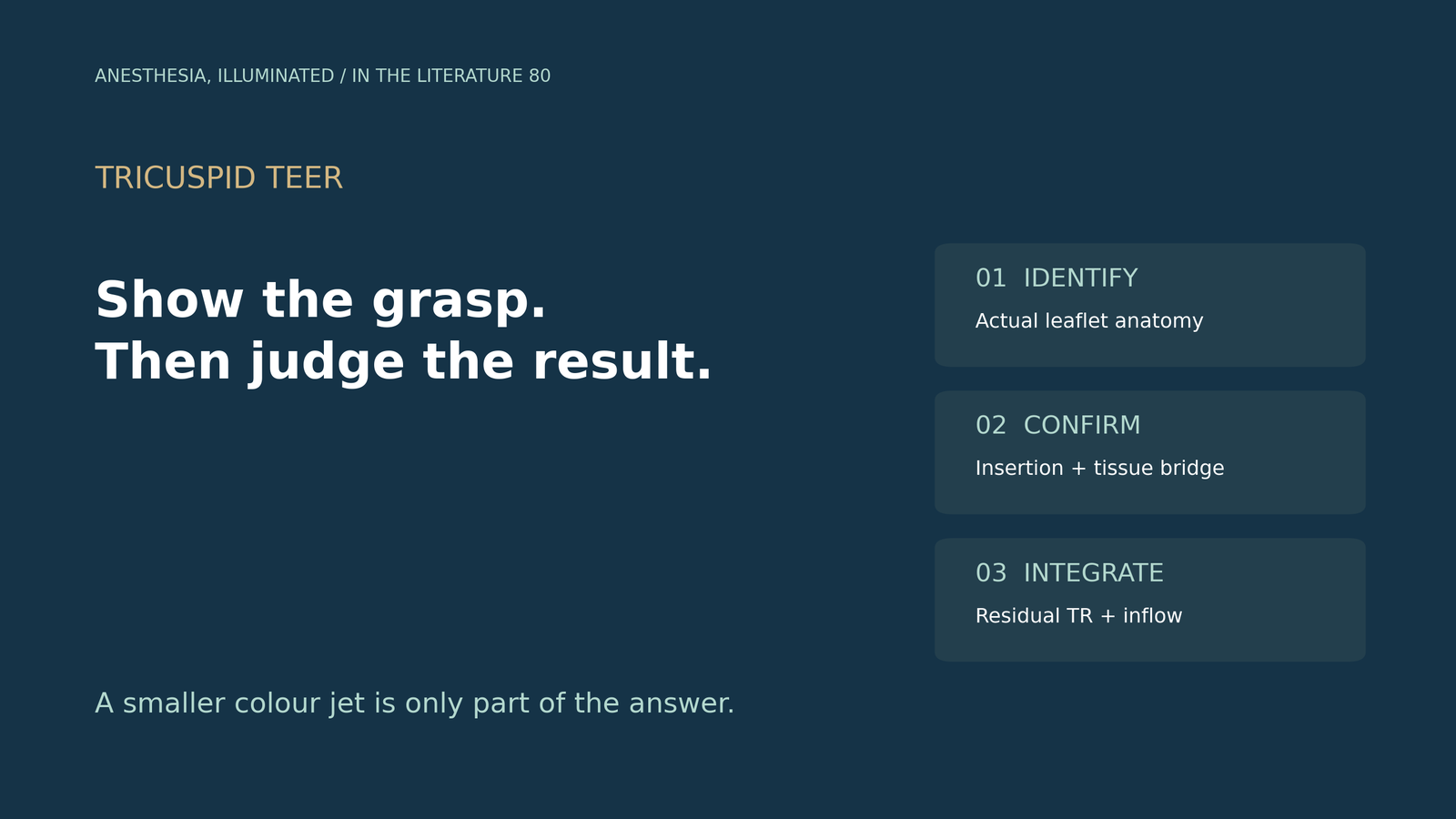
- Use complementary views: en-face imaging establishes spatial orientation; orthogonal views show the device–leaflet relationship.1
- Identify the patient's actual leaflets and target coaptation line; “tricuspid” does not guarantee three leaflets.2
- Three-dimensional TEE can improve morphological assessment, but an image-quality score is not a validated release criterion.3
- Confirm actual leaflet insertion and tissue bridging before release; a smaller colour jet alone cannot establish a secure grasp.4
- When a critical leaflet is obscured, change the imaging approach; selected cases may benefit from complementary intracardiac echocardiography (ICE).5
The device is closed and the colour jet looks smaller, but one leaflet disappears behind a shadow. Has it been captured, or is the image simply unable to answer? This is the central imaging question during tricuspid transcatheter edge-to-edge repair (TEER): the team needs evidence of the tissue–device relationship, alongside evidence that the valve result is acceptable.
Establish leaflet identity before choosing a grasping plane
A familiar probe angle does not reliably name the same leaflet in every patient. The practical approach is to relate the two-dimensional sections to the full valve: identify the septal attachment and surrounding landmarks, follow the coaptation lines, and then localize the intended leaflet pair. Mid/deep esophageal, transgastric short-axis and three-dimensional views contribute different information.1
In the bRIGHT anatomical analysis, an independent core laboratory evaluated 135 patients with available transgastric short-axis images. Leaflet configurations ranged from two to five; 28% were not trileaflet. This was a selected postmarket TEER cohort, not a prevalence survey of all patients with TR.2
The same study measured gaps at several locations along the valve. The largest measured gap and the gap at the intended grasping site can differ. Accordingly, an isolated “gap size” without its view and location is an incomplete description of the target. These observations support anatomical mapping; they do not create a new universal eligibility cutoff.2
Three-dimensional imaging helps place the sections in context. In a prospective single-center study of 54 patients with severe TR, Sobieraj and colleagues found that a study-defined detailed-morphology endpoint was achieved in 37/54 with 3D TEE versus 23/54 with 2D TEE (P=0.012). The comparison used a standardized midesophageal acquisition protocol. Its scoring thresholds were not validated against procedural outcomes, so the result supports better anatomical assessment—not release based on a score.3
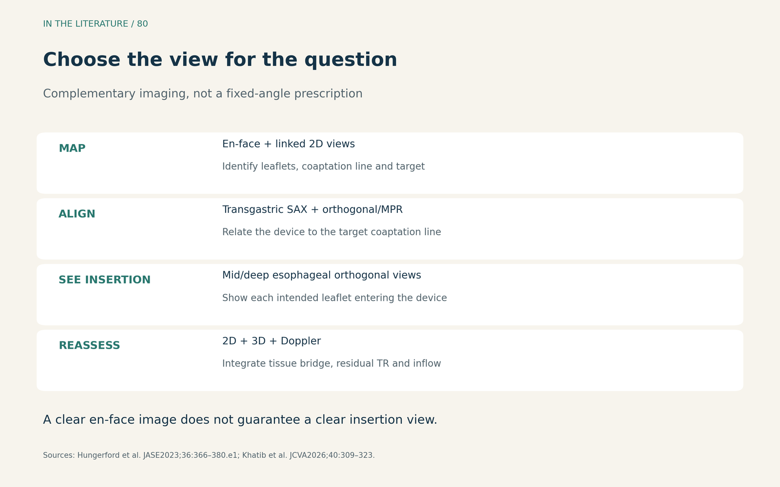
Figure 1. Select the view for the question being answered. A clear en-face image does not automatically provide a clear insertion view.1,4 View larger

Distinguish correct orientation from confirmed insertion
The device can appear correctly positioned over the jet while the imaging plane misses one leaflet. Orthogonal imaging or live 3D multiplanar reconstruction helps align the device with the actual target coaptation line. For clip orientation, the arms should be assessed perpendicular to that line; the screen's apparent angle is not the anatomical reference.14
Grasp confirmation requires a further step: visualize each intended leaflet entering the device. After closure, combine the observed insertion and leaflet motion on 2D imaging with the tissue bridge on 3D imaging. These are complementary findings; a suggestive short-axis configuration is not a guarantee of durable attachment.14
Khatib and colleagues' procedural review separates the tasks explicitly: orientation, leaflet insertion and bridge formation, then regurgitation and complication assessment. Clinically, that separation matters. Reduced colour flow is a hemodynamic observation; it cannot replace direct assessment of captured tissue.4
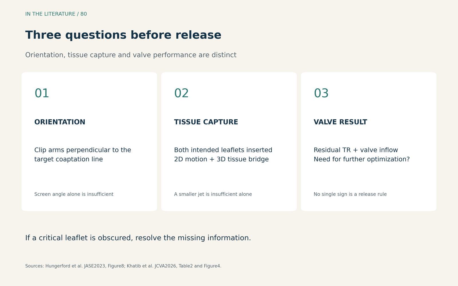
Figure 2. Orientation, tissue capture and valve performance answer different questions. No single sign is presented as an independent release rule.1,4 View larger

When shadowing hides the answer, obtain a different view
If insertion remains unclear, repeating the same obscured view does not resolve the uncertainty. Adjust probe depth and plane, use orthogonal or reconstructed views, and reassess which window displays the tissue. Device-related shadowing also matters: with PASCAL, the central spacer can obscure insertion. A transgastric view may improve orientation yet still fail to show insertion; nonstandard mid/deep esophageal windows may be needed.4
ICE offers another acoustic vantage point. Biroli and colleagues compared TEE and 4D-ICE in the same ten patients, using postprocedural image-quality ratings across eight procedural steps. For leaflet insertion, ICE received nine excellent, one good and no poor ratings; TEE received three excellent, three good and four poor ratings.5
This is a small, complex cohort: six patients had prior valve interventions, and reviewers could recognize the imaging modality. The results support ICE as a complementary option when TEE leaves a critical question unanswered. They do not establish superior clinical outcomes, noninferiority, or a requirement to use ICE for every procedure.5
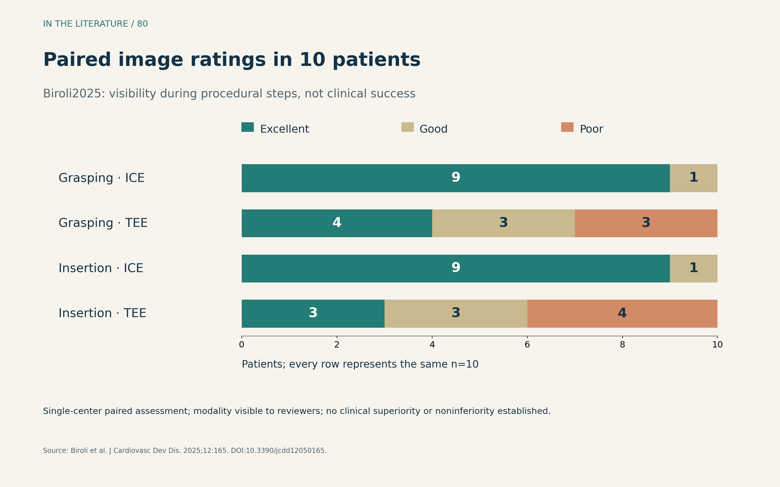
Figure 3. Original display of reported ratings from Biroli et al. Each row represents the same ten patients, not a separate treatment group. Ratings describe visibility, not clinical success.5 View larger

Before release, bring the tissue and hemodynamics together
Once capture is demonstrated, assess residual TR and the tricuspid inflow gradient before release. The decision about repositioning or another device must balance additional regurgitation reduction against inflow obstruction, using the actual anatomy and procedure conditions.14
The useful team handoff is therefore specific: which leaflets were captured, which views demonstrate insertion and bridging, and what are the residual regurgitation and inflow findings? Any remaining uncertainty about a critical leaflet should be stated explicitly. This is an integrated assessment, not a single image or an automatic numerical permission to release.14
Three takeaways
- Map before grasping: identify the actual leaflet configuration and describe the target gap by location and view.
- Demonstrate capture: combine visible insertion of both intended leaflets with complementary 2D and 3D findings; a smaller jet is insufficient by itself.
- Resolve the missing view, then assess the whole result: adapt the window or add ICE when appropriate, and integrate residual TR and inflow assessment before release.
References
- Hungerford SL, Rye EE, Hansen PS, et al. Key Echocardiographic Considerations for Tricuspid Valve Transcatheter Edge-to-Edge Repair. J Am Soc Echocardiogr. 2023;36(4):366-380.e1. doi:10.1016/j.echo.2023.01.013
- Donal E, Sitges M, Panis V, et al. Characterization of Tricuspid Valve Anatomy and Coaptation Gap in Subjects Receiving Tricuspid Transcatheter Edge-To-Edge Repair: Observations From the bRIGHT TriClip Study. J Am Soc Echocardiogr. 2024;37(4):397-404. doi:10.1016/j.echo.2023.12.002
- Sobieraj J, Rdzanek A, Kapłon-Cieślicka A, et al. Heart 3D: echocardiographic and anatomical features of the tricuspid valve in a heterogeneous population with severe regurgitation—implications for edge-to-edge procedure suitability. Front Cardiovasc Med. 2025;12:1637158. doi:10.3389/fcvm.2025.1637158
- Khatib D, Sheu R, Belani K, et al. Transcatheter Tricuspid Valve Interventions for Tricuspid Regurgitation: A Review of Percutaneous Annuloplasty, Edge-to-Edge Repair, and Valve Replacement Devices. J Cardiothorac Vasc Anesth. 2026;40(1):309–323. doi:10.1053/j.jvca.2025.10.015
- Biroli M, Fazzari F, Cannata F, et al. Early Experience with Acuson AcuNav 4D-ICE to Guide Transcatheter Tricuspid Edge-to-Edge Repair: 4D Intracardiac Echocardiography Compared to Transesophageal Echocardiography. J Cardiovasc Dev Dis. 2025;12(5):165. doi:10.3390/jcdd12050165