ITL-e0f99458499d
Juan Li, MD · Updated November 17, 2026
Watch the video
Key points
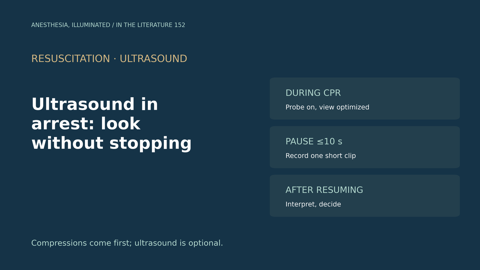
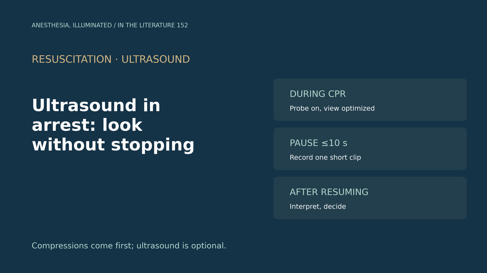
- Compressions come first. Guidelines allow ultrasound only if it does not interfere with the resuscitation protocol, note that it is associated with longer interruptions, and limit pulse checks to 10 s.[1]
- Prepare during compressions. Position the probe during CPR, record images during the break, and review them while compressions continue.[2]
- Fit the look into the pulse check. Ultrasound should be performed within the 10-s window, with one question per cycle, and the team leader should not scan.[3]
- Done well, it can be fast: ultrasound carotid pulse checks took a median of 1.62 s, rarely exceeded 5 s and never exceeded 10 s.[4]
- Transesophageal echo provides continuous images and shorter pulse checks, while transthoracic loop quality is often overestimated by the operator.[5], [6]
During a cardiac arrest in the operating room, the rhythm is pulseless electrical activity, and a colleague reaches for the ultrasound probe. How can the team look for a reversible cause without lengthening the pause in compressions?[1], [3]
What the guidelines allow
The 2020 American Heart Association guideline states that ultrasound may be considered if an experienced sonographer is present and its use does not interfere with the standard protocol. Cardiac ultrasound is associated with longer interruptions in compressions, a single small randomized trial found no improvement in outcomes, pulse checks should take no more than 10 s, and the chest compression fraction should be at least 60%.[1]
A workflow that protects compressions
The international SHoC consensus requires the core cardiac view to be obtained during the rhythm-check pause: position the probe during CPR to set up the view, record images during breaks, and review them during CPR. Absence of organized left ventricular activity suggests a poor outcome.[2] Anto and colleagues add that ultrasound should be performed within the 10-s pulse-check window, that the team leader should not perform it, that images should be acquired during the pause and interpreted during compressions, and that a single assessment per cycle is preferable. In a cited study, cardiac activity carried a 52% rate of return of circulation versus 12% without, but current evidence shows no outcome improvement from ultrasound during arrest.[3]
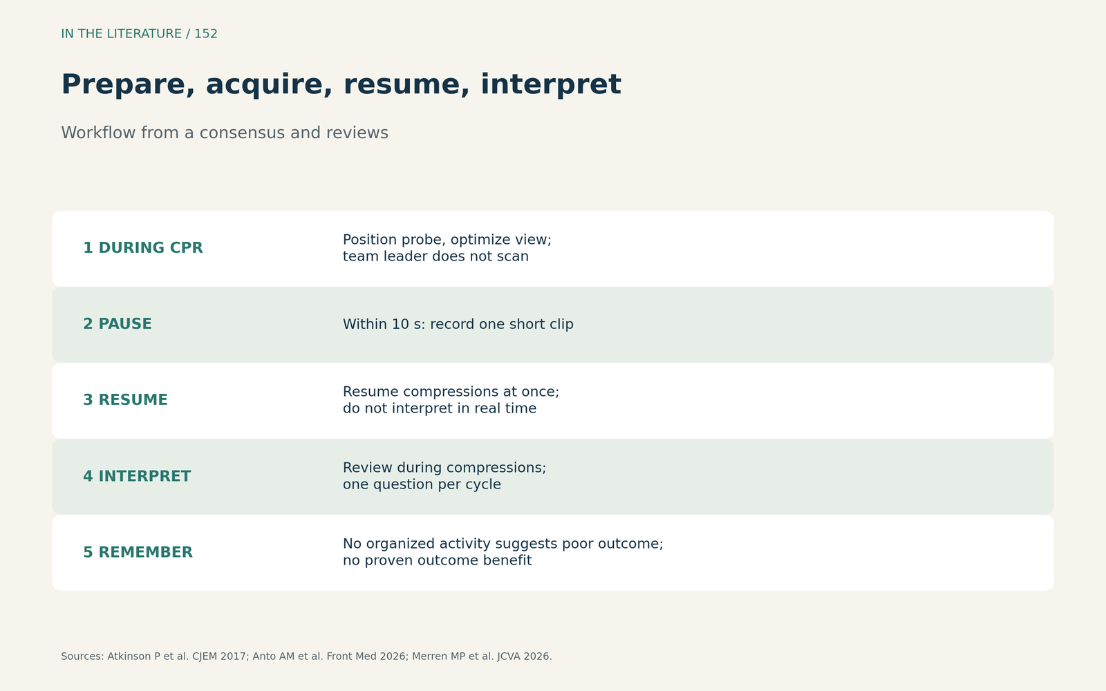
Figure 1. Prepare, acquire, resume, interpret. Synthesized from the SHoC consensus, Anto et al. and Merren et al.[2], [3], [4] View larger.

Merren and colleagues stress that ultrasound must not delay compressions and that the image should be optimized during ongoing compressions before the pause begins. In a cited study of carotid pulse checks, ultrasound took a median of 1.62 s versus 3.50 s by palpation, rarely exceeded 5 s and never exceeded 10 s.[4]
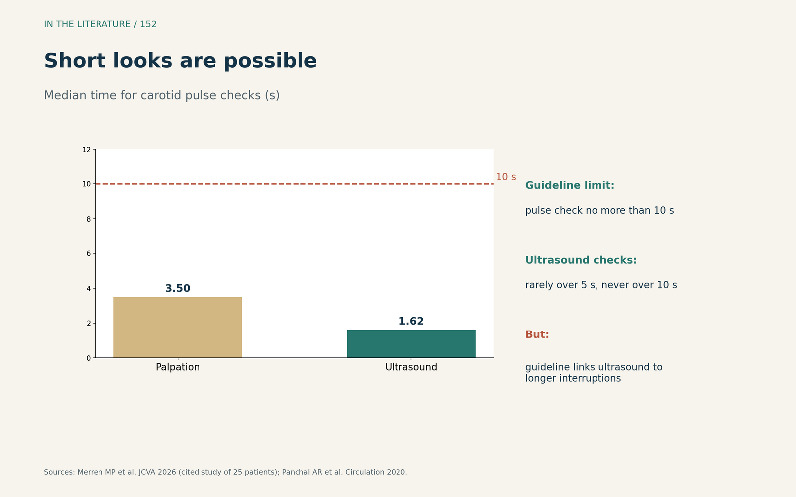
Figure 2. Short looks are possible. Redrawn from Merren et al. and the American Heart Association guideline.[4], [1] View larger.

Transesophageal echo and image reliability
Teran and colleagues describe transesophageal echo as providing a continuous image of myocardial activity, with feedback on compression quality and shorter pulse checks than transthoracic imaging.[5] It requires expertise, and in one study complications reached 4.3%.[4] For transthoracic loops recorded during out-of-hospital arrest, Papin and colleagues found that experts excluded 16 and 9 of 52 loops for poor quality, and that operators tended to overestimate loop quality compared with experts.[6]
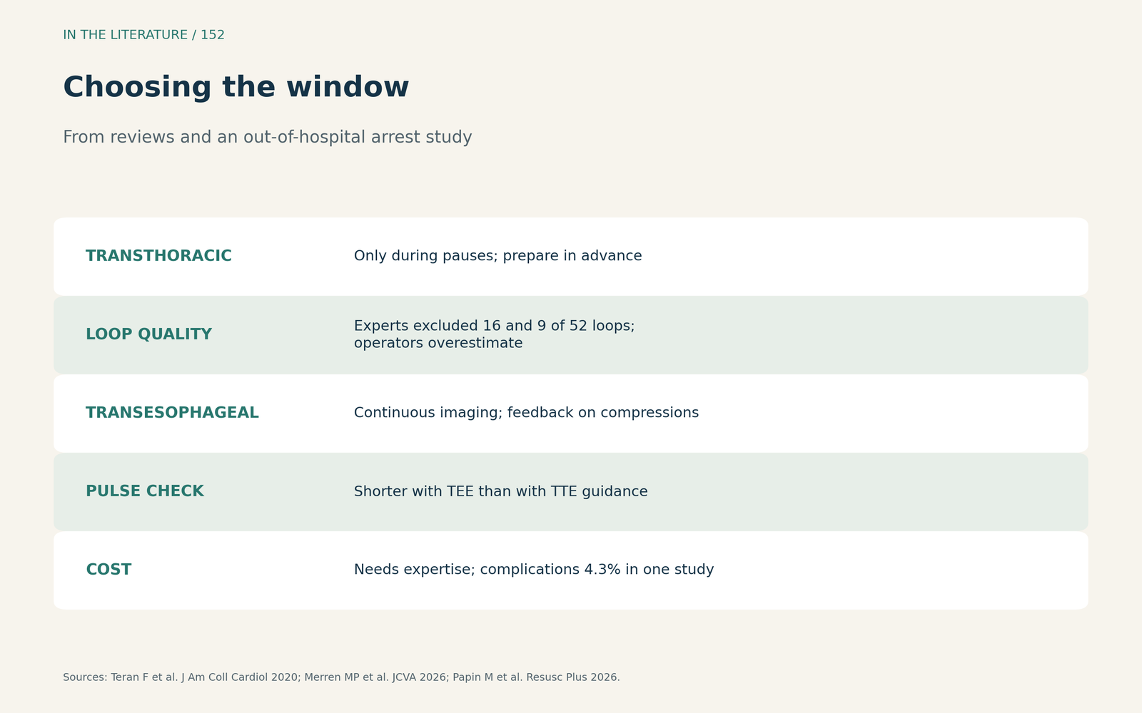
Figure 3. Choosing the window. Synthesized from Teran et al., Merren et al. and Papin et al.[5], [4], [6] View larger.

Three takeaways
- Never trade compressions for images. Pulse checks stay under 10 s, and ultrasound is optional.[1]
- Set up early, interpret late. Probe on during CPR, record in the pause, read after compressions resume.[2], [3]
- Consider TEE when available. Continuous imaging avoids pauses, but expertise and complications matter.[5], [4]
References
- Panchal AR, Bartos JA, Cabañas JG, et al. Part 3: adult basic and advanced life support: 2020 American Heart Association guidelines for cardiopulmonary resuscitation and emergency cardiovascular care. Circulation. 2020;142(16 Suppl 2):S366–S468. doi:10.1161/CIR.0000000000000916
- Atkinson P, Bowra J, Milne J, et al. International Federation for Emergency Medicine consensus statement: sonography in hypotension and cardiac arrest (SHoC): an international consensus on the use of point of care ultrasound for undifferentiated hypotension and during cardiac arrest. CJEM. 2017;19(6):459–470. doi:10.1017/cem.2016.394
- Anto AM, Javed N, Nallapareddy RR, et al. Point-of-care-ultrasound in cardiac arrest: a useful tool for resuscitation. Front Med (Lausanne). 2026;13:1795346. doi:10.3389/fmed.2026.1795346
- Merren MP, Augustin K, Ripoll JG, et al. Resuscitative point-of-care ultrasound in cardiac arrest: current evidence and clinical applications. J Cardiothorac Vasc Anesth. 2026;40(11):3829–3835. doi:10.1053/j.jvca.2026.07.002
- Teran F, Prats MI, Nelson BP, et al. Focused transesophageal echocardiography during cardiac arrest resuscitation: JACC review topic of the week. J Am Coll Cardiol. 2020;76(6):745–754. doi:10.1016/j.jacc.2020.05.074
- Papin M, Markarian T, Le Bastard Q, et al. Inter-rater reliability of point-of-care ultrasound during out-of-hospital cardiac arrest: an ancillary analysis of the observational prospective ACE trial. Resusc Plus. 2026;27:101191. doi:10.1016/j.resplu.2025.101191