ITL-04c77b47b2c9
Juan Li, MD · Updated November 18, 2026
Key points
- Rare but real. Unileaflet mitral valve prevalence was estimated at 1:8,800 in one German population, and it is commonly due to a severely hypoplastic posterior leaflet.[1], [2]
- Look for one working leaflet. A reported sign is an elongated anterior leaflet coapting with a ridge of tissue where the posterior leaflet should be, often with a single papillary muscle.[1]
- Dysplastic valves mimic other disease. Valves with one undifferentiated papillary muscle may masquerade as hypertrophic cardiomyopathy or rheumatic mitral disease.[3]
- Go 3D. TEE, 3D echocardiography or cardiac MRI help show whether the posterior leaflet is hypoplastic or absent.[2], [4]
- Search beyond the valve. A parachute mitral valve should prompt evaluation for other left-sided obstructive lesions.[5]
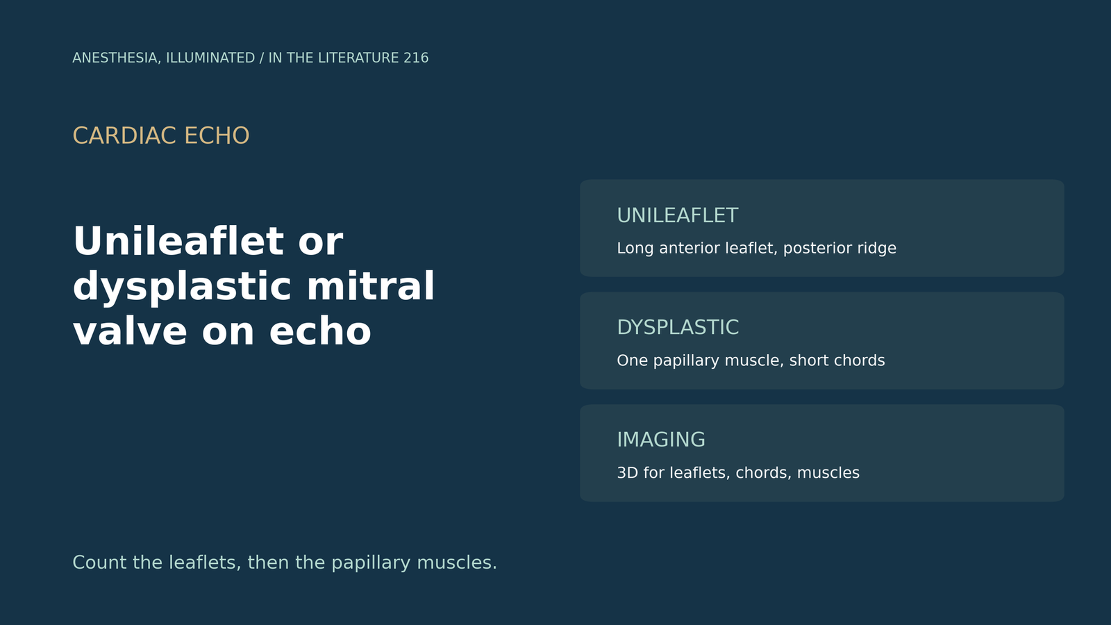
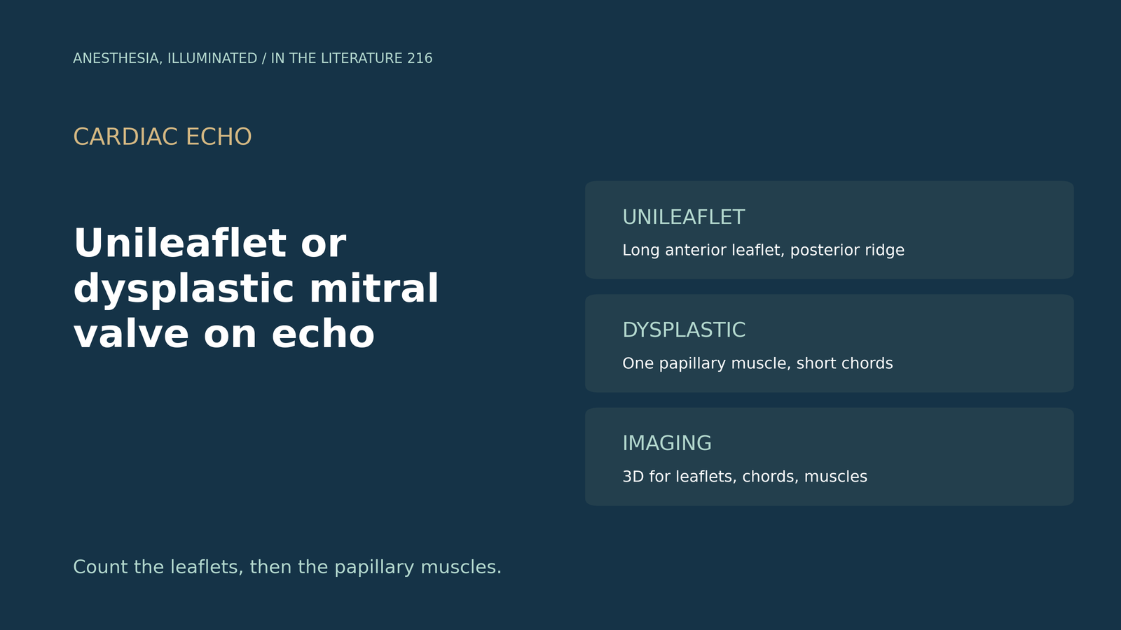
During a pre-bypass TEE, the mitral valve seems to have only one moving leaflet, with a thick, short subvalvular apparatus. Which echo signs separate a unileaflet valve, a dysplastic valve and a parachute valve?[1], [3]
Signs of a unileaflet valve
Arasaratnam et al. (CASE 2020) describe an elongated anterior leaflet coapting with a ridge of tissue where the posterior leaflet should be, and a single papillary muscle on the parasternal short-axis view. In their review of published cases, clinically significant MR was reported in 12 of 21, and imaging can show rudimentary valvular structures that inform repair.[1] Grandez and Skenderi (CASE 2023) add that the apical four-chamber view shows a markedly elongated, redundant anterior leaflet, and that M-mode shows the missing posterior leaflet with loss of the E-F slope.[2] Liu et al. (Eur Heart J Case Rep 2023) call the unileaflet valve the rarest congenital mitral anomaly; 3D TEE showed the posterior leaflet remnant en face in diastole, and a unileaflet valve with a single papillary muscle seems distinct from parachute valves.[4]
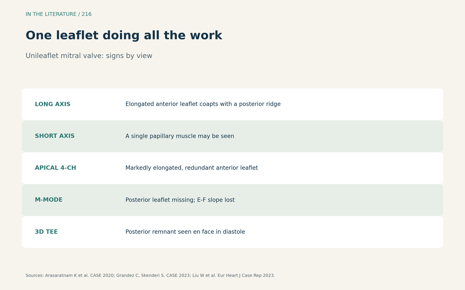
Figure 1. One leaflet doing all the work. Synthesized from Arasaratnam et al., Grandez and Skenderi, and Liu et al.[1], [2], [4] View larger.

Dysplastic and parachute-like valves
Mohan et al. (Indian Heart J 2016) found a solitary undifferentiated papillary muscle in six of 9600 consecutive echocardiograms. Clues were thickened leaflets without commissural fusion and short chords, and associated lesions could create a funnel rather than parachute-like appearance. Such valves may masquerade as hypertrophic cardiomyopathy or rheumatic disease. In true parachute valves, the chordae insert into one central papillary muscle, and parachute valves are associated with other congenital anomalies in over 95% of cases.[3] Mitral arcade, in which leaflets attach almost directly to the papillary muscles, can escape everyone: in a two-patient series (Dokollari et al., J Cardiothorac Vasc Anesth 2021), none of the preoperative imaging tools detected it; additional imaging may help when the mechanism of mitral stenosis is unclear, and MRI offers higher spatial resolution.[6]
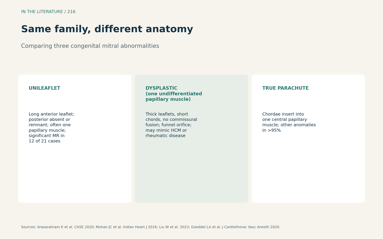
Figure 2. Same family, different anatomy. Synthesized from Mohan et al., Liu et al. and Goeddel et al.[3], [4], [5] View larger.

How to image and what to check
Simpson et al. (EACVI/ASE consensus 2017) recommend 3D echocardiography for valvar lesions and complex abnormalities, and to visualize valves, papillary muscles and chordal support for surgical planning; valve quantitation software often assumes normal morphology.[7] TEE, 3D echocardiography or cardiac MRI help separate a hypoplastic from an absent posterior leaflet.[2] Goeddel et al. (J Cardiothorac Vasc Anesth 2020) note that a parachute valve should prompt a search for other left-sided obstruction and that hemodynamics under general anesthesia with positive pressure ventilation may not reflect the awake state.[5] The evidence is case reports, a small series and expert consensus.
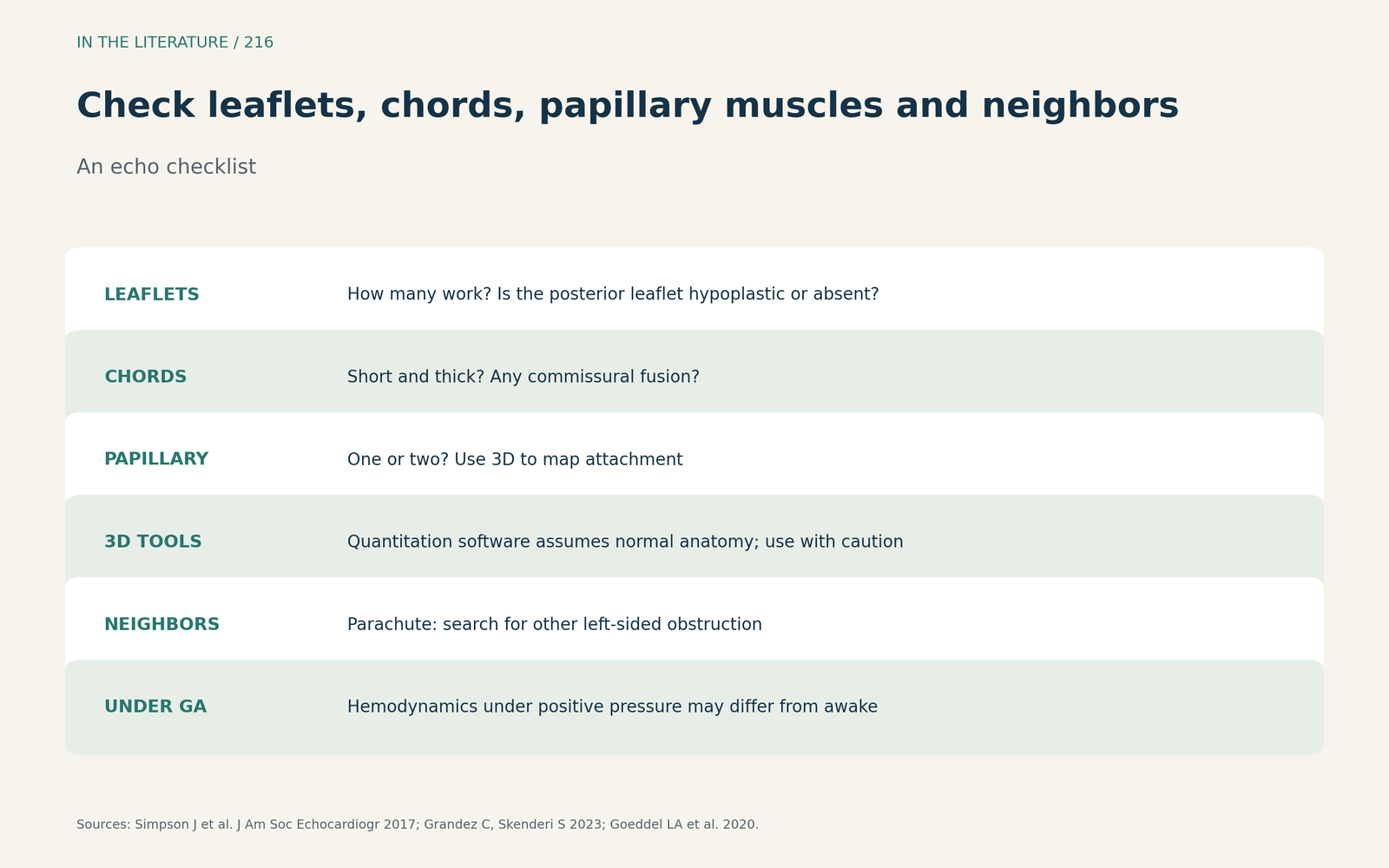
Figure 3. Check leaflets, chords, papillary muscles and neighbors. Synthesized from Simpson et al., Grandez and Skenderi, and Goeddel et al.[7], [2], [5] View larger.

Three takeaways
- Suspect a unileaflet valve when an elongated anterior leaflet coapts with a posterior ridge and a single papillary muscle is seen.[1], [2]
- Thick leaflets, short chords and no commissural fusion around one papillary muscle suggest a dysplastic valve, not HCM or rheumatic disease.[3]
- Use 3D imaging for leaflets, chords and papillary muscles, and look for other left-sided obstruction.[7], [5]
References
- Arasaratnam K, Tomlinson S, Dahiya A, et al. Surgical repair of a unileaflet mitral valve: a rare congenital abnormality and a novel surgical approach. CASE (Phila). 2020;4(5):420–428. doi:10.1016/j.case.2020.04.009
- Grandez C, Skenderi S. Unileaflet mitral valve in patient with marfanoid habitus. CASE (Phila). 2023;7(1):10–13. doi:10.1016/j.case.2022.07.005
- Mohan JC, Shukla M, Mohan V, et al. Spectrum of congenital mitral valve abnormalities associated with solitary undifferentiated papillary muscle in adults. Indian Heart J. 2016;68(5):639–645. doi:10.1016/j.ihj.2015.12.014
- Liu W, Ma L, Tuo Y, et al. Unileaflet mitral valve with one single papillary muscle: a rare congenital anomaly characterized by two- and three-dimensional echocardiography. Eur Heart J Case Rep. 2023;7(1):ytac484. doi:10.1093/ehjcr/ytac484
- Goeddel LA, Jung YH, Patel P, et al. Analysis of the 2018 American Heart Association/American College of Cardiology guidelines for the management of adults with congenital heart disease: implications for the cardiovascular anesthesiologist. J Cardiothorac Vasc Anesth. 2020;34(5):1348–1365. doi:10.1053/j.jvca.2019.08.004
- Dokollari A, Cameli M, Bisleri G, et al. Mitral arcades unexpectedly encountered during cardiac surgery. J Cardiothorac Vasc Anesth. 2021;35(3):914–916. doi:10.1053/j.jvca.2020.10.018
- Simpson J, Lopez L, Acar P, et al. Three-dimensional echocardiography in congenital heart disease: an expert consensus document from the European Association of Cardiovascular Imaging and the American Society of Echocardiography. J Am Soc Echocardiogr. 2017;30(1):1–27. doi:10.1016/j.echo.2016.08.022