
ITL-7153e1870e71
Juan Li, MD · Updated November 18, 2026
Key points
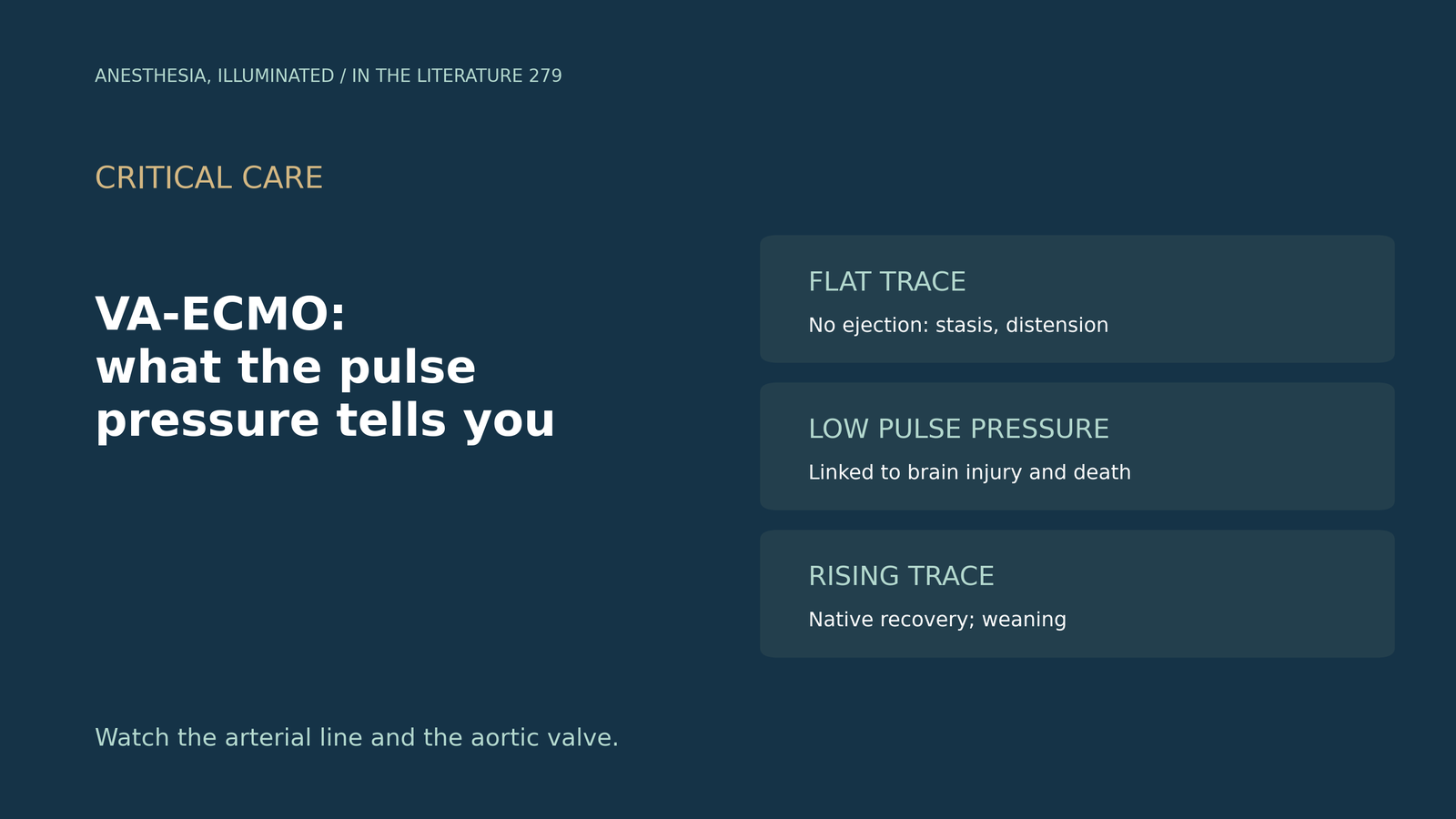
- Pulsatility shows the heart is ejecting. It is a marker of LV contraction and native output; a flat trace means the aortic valve has stopped opening.[1]
- A closed valve is not benign. VA-ECMO raises LV workload, and a non-ejecting LV risks distension, stasis and thrombus.[2], [3]
- Low pulse pressure tracks harm. Pulse pressure of 10 mmHg or less at 24 hours was linked to brain injury in the ELSO Registry.[4]
- Loss of pulsatility after ECPR was common and linked to death. It occurred in 49%, with higher odds for each extra hour.[5]
- Trends matter for weaning and for surprises. A rising pulse pressure accompanied successful weaning,[6] and a sudden waveform change can reveal new aortic regurgitation.[7]
A patient on peripheral VA-ECMO for cardiogenic shock has an arterial trace that has gone almost flat. Is that a problem, and what should be done?
What pulsatility and aortic valve opening mean
Pulsatility on VA-ECMO is a marker of LV contraction and native cardiac output. A flat arterial waveform signals excessive afterload, minimal LV preload or contractility, and loss of aortic valve opening. If LV ejection and pulsatility stay low for long, ventricular or aortic-root thrombus and embolic stroke may follow; a native output of about 20% of total flow, corresponding to a pulse pressure of at least 10 mmHg, may reduce these risks (Hoyler et al., J Cardiothorac Vasc Anesth 2020). This target is expert-based. Besides decreased pulse pressure, signs of LV distention include a dilated hypokinetic LV, arrhythmias, raised wedge pressure and pulmonary oedema.[1]
VA-ECMO increases LV workload and can lead to progressive distension and loss of aortic valve opening; an editorial in Anesthesiology calls for research on proactive LV unloading and synchronized pulsatile extracorporeal flow (Shekar et al. 2020).[2] With high flows and poor contractility, the aortic valve may stay closed even in systole, causing stasis and thrombus (Loforte et al., J Cardiothorac Vasc Anesth 2022).[3]
Low pulse pressure and outcomes
In 9,807 ELSO patients on peripheral VA-ECMO (Kalra et al., ASAIO J 2025), 85% had a pulse pressure above 10 mmHg at 24 hours. Acute brain injury occurred in 15% with pulse pressure of 10 mmHg or less vs 11% above it. After adjustment, low pulse pressure was associated with acute brain injury (aOR 1.25) but not with intracranial haemorrhage (aOR 1.03). Pulse pressure was a single 24-hour value and may mark a sicker population; the authors suggest that aiming for a pulse pressure above 10 mmHg may be desirable.[4]
After ECPR at a high-volume Australian centre (Nanjayya et al., Resusc Plus 2026), loss of pulsatility occurred in 108 patients (49%), 38% of them at ECMO initiation. It was independently associated with death (aOR 2.55), and each extra hour raised the odds by 17%. Mortality was 38.7%, 65.8% and 74.1% with normal, low and absent pulsatility. The authors see loss of pulsatility as a marker of injury severity rather than a direct cause.[5]
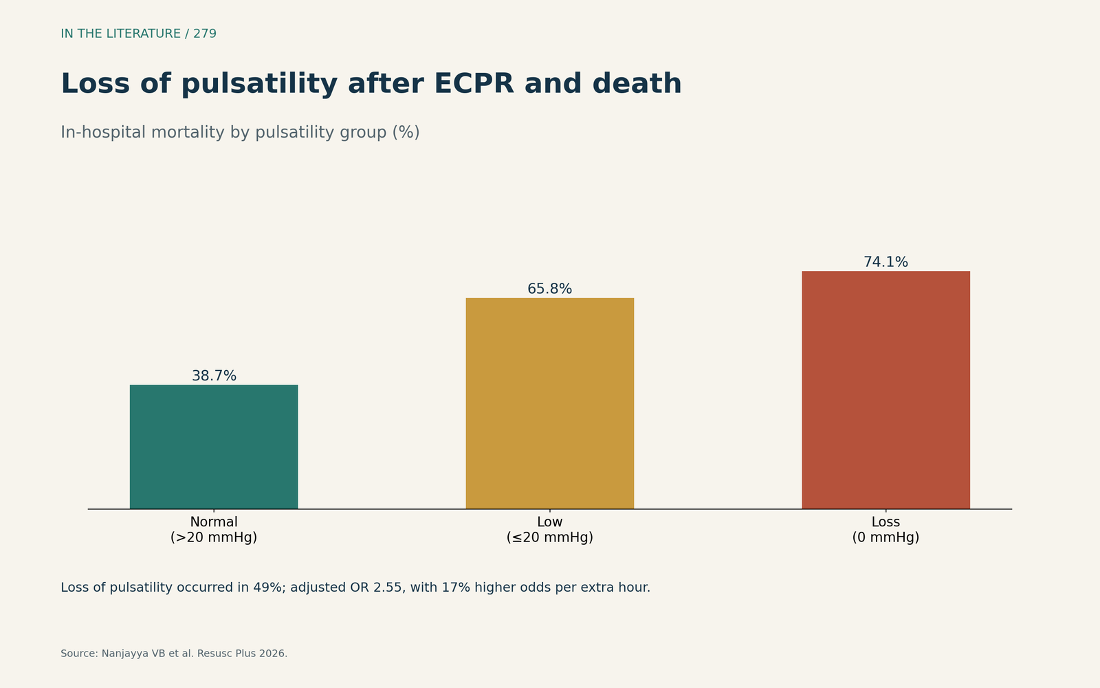
Figure 1. Mortality rose stepwise from normal to low to absent pulsatility. Data from Nanjayya et al.[5] View larger.

A rising pulse pressure and weaning
In 96 VA-ECMO patients (Hermens et al., Perfusion 2026), pulse pressure was consistently higher from 12 hours onward in those who were weaned (45.8%). Pulse pressure at 36 hours was the only independent predictor of weaning success after adjustment for flow and pump speed; the authors describe the finding as hypothesis-generating.[6] Expert reviews list a pulsatile waveform for at least 24 hours, with MAP above 60 mmHg on no or low vasoactive drugs, among weaning prerequisites,[3] and advise halting a wean if arterial pulse pressure fails to rise.[1]
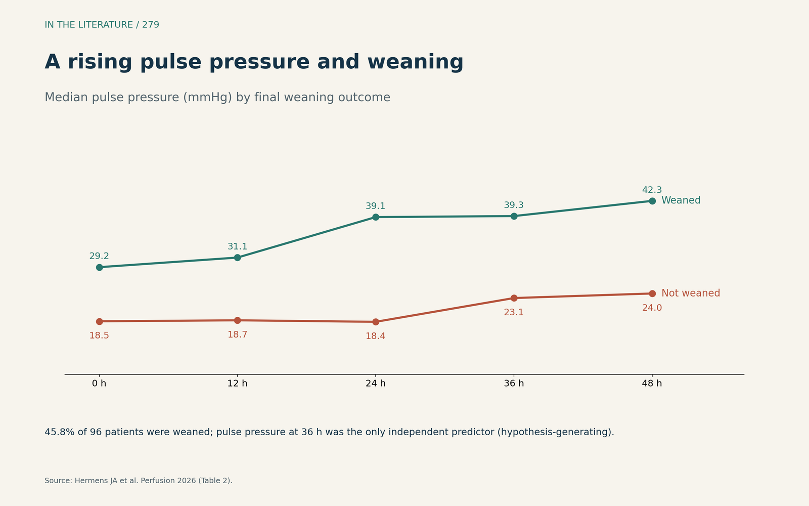
Figure 2. Patients who were weaned had a higher and rising pulse pressure. Data from Hermens et al.[6] View larger.

When the waveform changes suddenly
A new bisferiens arterial trace on peripheral VA-ECMO signalled severe aortic regurgitation, confirmed by TEE; the team lowered ECMO flow and weaned vasopressin to favour forward flow (Janjua et al., Anesthesiology 2021).[7] A wider pulse pressure is not always good news.
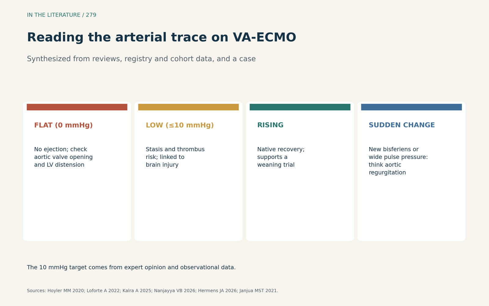
Figure 3. Flat, low, rising and suddenly changed traces each prompt a different question. Synthesized from Hoyler et al., Loforte et al., Kalra et al., Nanjayya et al., Hermens et al. and Janjua et al.[1], [3], [4], [5], [6], [7] View larger.

Three takeaways
- Treat a flat or very low arterial trace as a sign that the LV may not be ejecting; check aortic valve opening and LV distension.[1], [3]
- Low or absent pulse pressure was linked to brain injury and death, but may mark severity rather than cause harm.[4], [5]
- Follow the trend: a rising pulse pressure supports a weaning trial, while a sudden change in shape deserves an echo.[6], [7]
References
- Hoyler MM, Flynn B, Iannacone EM, et al. Clinical management of venoarterial extracorporeal membrane oxygenation. J Cardiothorac Vasc Anesth. 2020;34:2776–2792. doi:10.1053/j.jvca.2019.12.047
- Shekar K, Donker DW, Brodie D. Venoarterial extracorporeal membrane oxygenation: if you cannot measure it, you cannot improve it. Anesthesiology. 2020;133:708–710. doi:10.1097/ALN.0000000000003487
- Loforte A, Comentale G, Botta L, et al. How would the authors treat their own temporary left ventricular failure with mechanical circulatory support? J Cardiothorac Vasc Anesth. 2022;36:1238–1250. doi:10.1053/j.jvca.2021.10.020
- Kalra A, Kang JK, Wilcox C, et al. Pulse pressure and acute brain injury in venoarterial extracorporeal membrane oxygenation: an Extracorporeal Life Support Organization registry analysis. ASAIO J. 2025;71(2):99–108. doi:10.1097/MAT.0000000000002294
- Nanjayya VB, Nichol A, Kaye DM, et al. Incidence and impact of loss of pulsatility on outcomes after extracorporeal cardiopulmonary resuscitation: a retrospective cohort study. Resusc Plus. 2026;31:101429. doi:10.1016/j.resplu.2026.101429
- Hermens JA, Fennema A, van der Tak K, et al. Temporal evolution of arterial pulse pressure trajectories as early predictor of weaning success in venoarterial extracorporeal membrane oxygenation. Perfusion. 2026;41(8):1442–1452. doi:10.1177/02676591261439578
- Janjua MST, Shukla S, Tantawy H. Pulsus bisferiens on extracorporeal membrane oxygenation. Anesthesiology. 2021;134:628. doi:10.1097/ALN.0000000000003697