
ITL-cb64eb0dac1e
Juan Li, MD · Updated November 18, 2026
Key points
- Image only when it helps. Echo should be done only if expected to provide clinical benefit, and TEE carries extra risk.[1]
- Rank requests. Active or recent cardiovascular symptoms and therapy safety monitoring sit in the highest-priority tier.[2], [3]
- Triage can defer most studies safely. One centre performed 30% of requests, with no apparent harm from deferral.[4]
- Scan short. A limited tablet protocol was diagnostic in all cases and cut disinfection time, and TEE was kept for intubated patients with poor TTE windows.[5], [6]
- Start at the bedside, and look at the RV. Expert POCUS protocols begin with focused cardiac ultrasound, and RV dilatation carried prognostic weight.[7], [8]
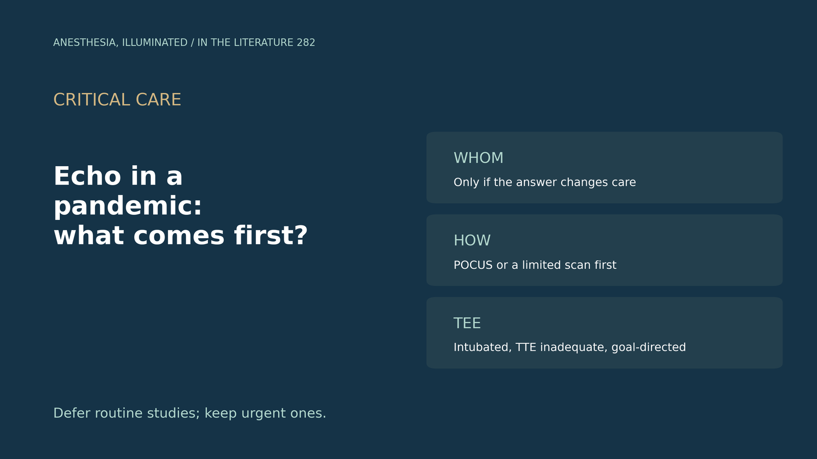
During a surge, the echo lab receives more requests than it can safely perform. Which studies should go first, and how should they be done?
Society frameworks for whom to image
Early in COVID-19, the ASE stated that TTE, stress echo and TEE should be performed only if expected to provide clinical benefit (Kirkpatrick et al., J Am Soc Echocardiogr 2020). TEE carries a heightened risk of viral spread, and contrast CT and MRI have emerged as alternatives. In crisis settings, triage by indication may be necessary, and studies should be as focused as necessary but complete enough to avoid a return visit.[1]
When services reopened, scheduling was to be prioritized, and when urgency is unclear, consultation with the referring physician is critical (Hung et al., J Am Soc Echocardiogr 2020). The highest-priority tier includes an active or recent change in cardiovascular symptoms, and safety monitoring for therapy, even if asymptomatic.[2]
The ASE update for future pandemics (Kirkpatrick et al., J Am Soc Echocardiogr 2023) follows a disaster-response model of conventional, contingency and crisis standards. About half of patients evaluated with TTE had an abnormality, yet even appropriate requests should be deferred under certain circumstances. Dobutamine stress echo may carry the least exposure risk of any stress test, and if cardiac POCUS or critical care echo answers the question, a confirmatory formal echo is usually not needed.[3]
Triage and limited protocols in practice
Of 145 inpatient TTE requests at one centre, 44 (30%) were performed after physician triage (Hennessey et al., J Am Soc Echocardiogr 2020). Of deferred patients who tested negative, 95% had TTE within 24 hours, and no adverse cardiovascular outcomes attributable to deferral were apparent.[4]
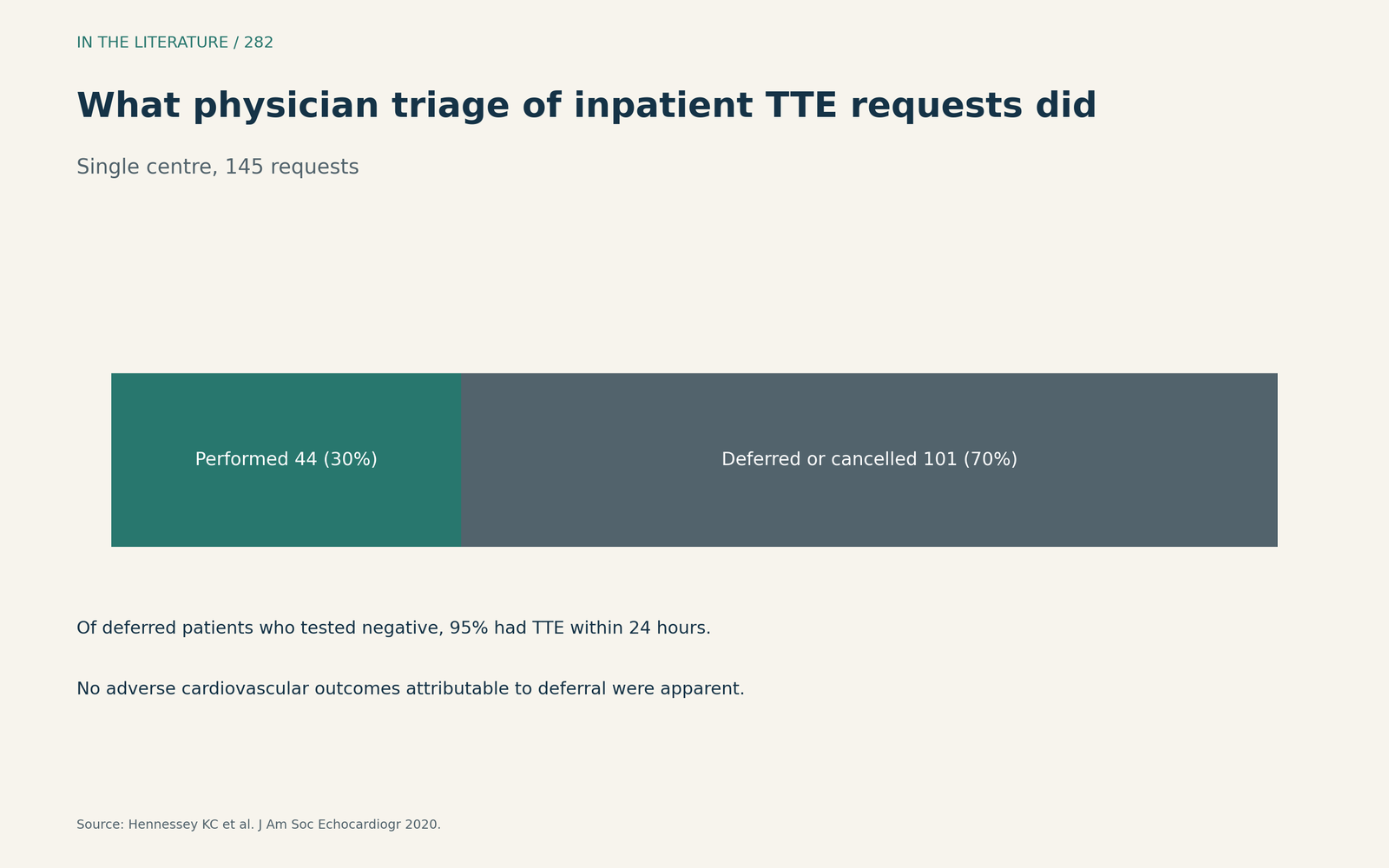
Figure 1. Most requests were deferred or cancelled, and deferred patients who tested negative were scanned quickly. Data from Hennessey et al.[4] View larger.

Of 91 appropriate requests in another centre, 90 (99%) were done with a handheld tablet and all were diagnostic (McMahon et al., J Am Soc Echocardiogr 2020). Disinfection time fell by 86% with the tablet, and only 12 patients (13%) undergoing tablet-based echo needed further imaging.[5]
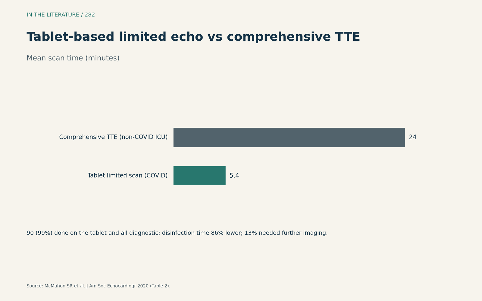
Figure 2. A limited tablet scan took a fraction of the time of a comprehensive TTE. Data from McMahon et al.[5] View larger.

TEE and POCUS-first protocols
In critically ill patients, TEE is performed only on intubated patients when TTE images are inadequate; such imaging is time sensitive, never elective and always goal directed (Teran et al., J Am Soc Echocardiogr 2020).[6]
A modified Delphi survey among 39 emergency imaging experts produced a pandemic POCUS protocol (Kwon et al., Medicina 2025). The protocol begins with focused cardiac ultrasound, and it is consensus-based and has not been prospectively tested.[7]
What to look for
In a Colombian cohort (Méndez-Toro et al., World J Methodol 2026), 147 studies (98.6%) were classed as high priority by ASE criteria. RV dilatation or overload, seen in 26.8%, was the only echo finding associated with in-hospital death.[8]
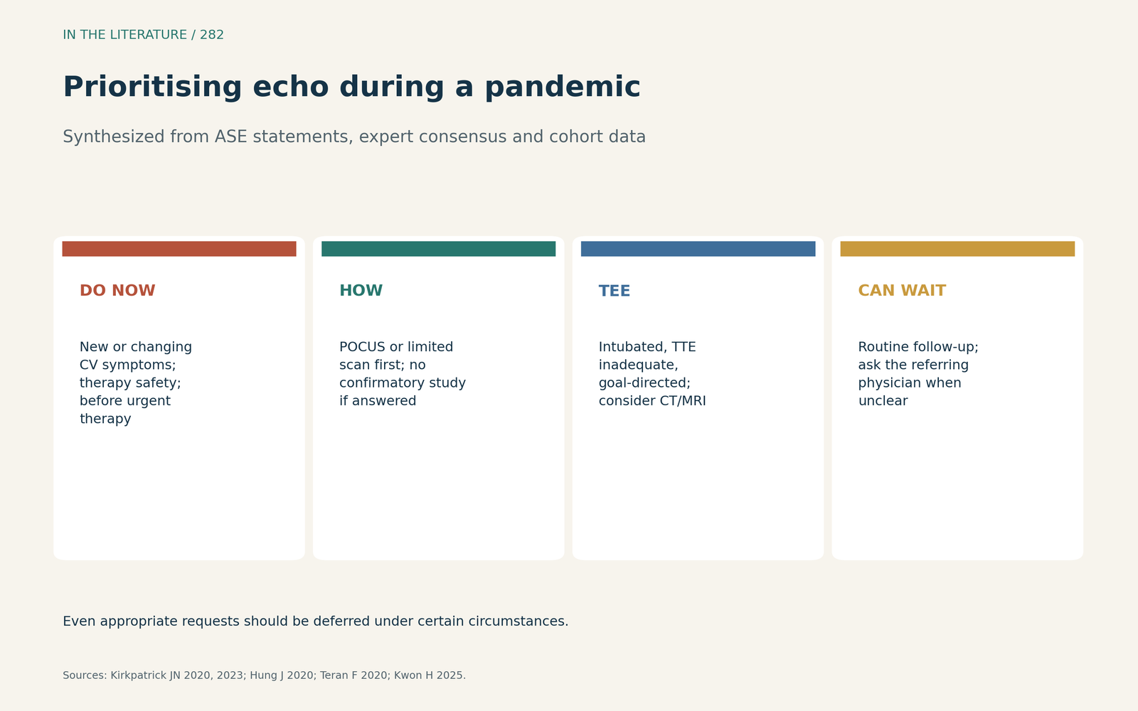
Figure 3. Do urgent studies now, start with POCUS or a limited scan, keep TEE goal-directed, and let routine studies wait. Synthesized from the ASE statements, Teran et al. and Kwon et al.[1], [2], [3], [6], [7] View larger.

Three takeaways
- Ask whether the result will change care; prioritise new symptoms, therapy safety and pre-procedure needs.[1], [2]
- Start with POCUS or a limited scan; if it answers the question, a formal study is usually unnecessary.[3], [5]
- Reserve TEE for intubated patients with inadequate TTE, and include the right heart in every focused study.[6], [8]
References
- Kirkpatrick JN, Mitchell C, Taub C, Kort S, Hung J, Swaminathan M. ASE statement on protection of patients and echocardiography service providers during the 2019 novel coronavirus outbreak: endorsed by the American College of Cardiology. J Am Soc Echocardiogr. 2020;33:648–653. doi:10.1016/j.echo.2020.04.001
- Hung J, Abraham TP, Cohen MS, et al. ASE statement on the reintroduction of echocardiographic services during the COVID-19 pandemic. J Am Soc Echocardiogr. 2020;33:1034–1039. doi:10.1016/j.echo.2020.05.019
- Kirkpatrick JN, Swaminathan M, Adedipe A, et al. American Society of Echocardiography COVID-19 statement update: lessons learned and preparation for future pandemics. J Am Soc Echocardiogr. 2023;36:1127–1139. doi:10.1016/j.echo.2023.08.020
- Hennessey KC, Shah N, Soufer A, et al. Inpatient transthoracic echocardiography during the COVID-19 pandemic: evaluating a new triage process. J Am Soc Echocardiogr. 2020;33:1418–1419. doi:10.1016/j.echo.2020.07.018
- McMahon SR, De Francis G, Schwartz S, et al. Tablet-based limited echocardiography to reduce sonographer scan and decontamination time during the COVID-19 pandemic. J Am Soc Echocardiogr. 2020;33:895–899. doi:10.1016/j.echo.2020.05.005
- Teran F, Burns KM, Narasimhan M, et al. Critical care transesophageal echocardiography in patients during the COVID-19 pandemic. J Am Soc Echocardiogr. 2020;33:1040–1047. doi:10.1016/j.echo.2020.05.022
- Kwon H, Lee JH, Suh D, You KM; PULSE Group. Developing a consensus-based POCUS protocol for critically ill patients during pandemics: a modified Delphi study. Medicina (Kaunas). 2025;61(8):1319. doi:10.3390/medicina61081319
- Méndez-Toro A, Rojas-Ruiz IT, Silva-DiazGranados LE, et al. Prognostic impact of right ventricular dilatation and echocardiography use criteria in hospitalized COVID-19 patients. World J Methodol. 2026;16(3):117845. doi:10.5662/wjm.v16.i3.117845