ITL-5e83b7280ca4
Juan Li, MD · Updated November 17, 2026
Watch the video
Key points
- MR severity is assessed in a physiological setting. Record the conditions when interpreting a change between examinations.1,2
- For stable secondary MR, assess severity after medical optimization, in a euvolemic and normotensive state.2
- A smaller MR measurement after induction of general anesthesia does not demonstrate improvement of the underlying valve disorder.3
- Exercise echocardiography can help when symptoms and resting findings disagree; assess MR together with pulmonary pressure and ventricular response.1,2
- Exercise can uncover important MR, but an observational association does not establish that intervening on exercise-induced MR improves outcomes.4
A patient has substantial MR before a planned procedure, yet the jet and calculated regurgitant volume look smaller after induction. Another patient describes exertional breathlessness despite a less impressive resting study. In both situations, the useful first question is: Were we examining the valve under comparable conditions?1–3
Compare the setting before comparing the grade
MR reflects the valve apparatus interacting with ventricular and atrial conditions. Changes in loading and ventricular geometry can alter coaptation and regurgitant flow, particularly in secondary MR. A changed measurement can therefore represent a real physiological change, an acquisition difference, or both. It need not mean that the structural lesion has improved or deteriorated.1,3
A practical comparison records blood pressure, heart rate and rhythm, sedation or anesthesia, ventilation and relevant vasoactive support. Review the mechanism, views and measurement method as well. This is a clinical synthesis of the imaging principles—not a validated checklist or a requirement to reproduce every variable exactly.1–3
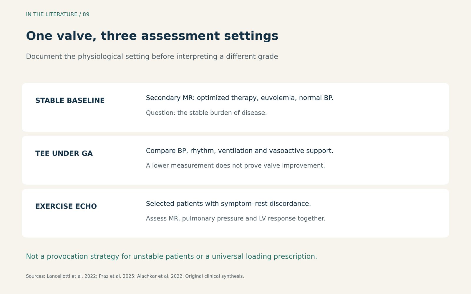
Figure 1. Different examinations answer different questions. An anesthetized measurement and an exercise measurement should be interpreted with their recorded physiological conditions.1–3 Enlarge

A smaller measurement under anesthesia is not evidence of repair
Alachkar and colleagues retrospectively compared TEE under conscious sedation with TEE after induction of general anesthesia in 50 patients undergoing transcatheter mitral repair. The anesthetized images were acquired before transseptal puncture, before treatment of the valve. This was neither an awake TTE-versus-TEE comparison nor a pre-repair/post-repair comparison.3
In the 39 patients with secondary MR, mean effective regurgitant orifice area decreased from 30 ± 11 to 24 ± 10 mm², and regurgitant volume from 47 ± 17 to 34 ± 13 mL/beat. These are group means ± standard deviations. They are not individual patient trajectories or a correction factor to apply to a new patient.3
Three-dimensional vena contracta area also decreased, from 66 ± 27 to 60 ± 29 mm². Direct three-dimensional measurement does not make the valve physiologically independent of loading. The small primary-MR subgroup did not show statistically significant changes, which does not establish that primary MR is immune to anesthesia.3
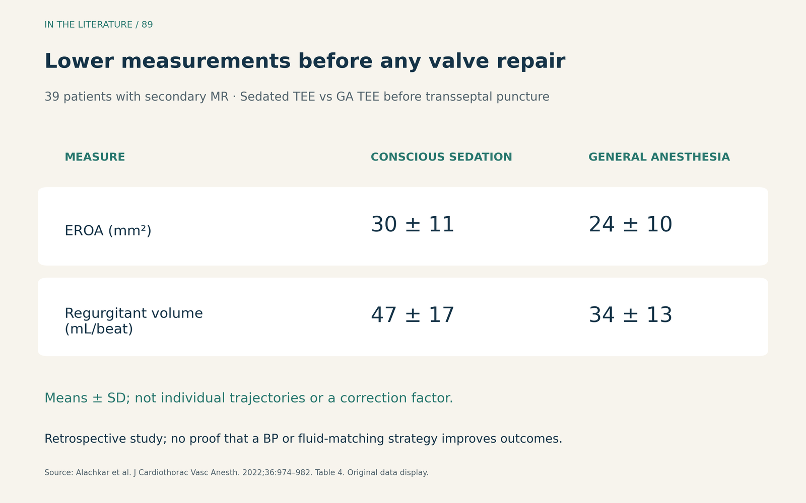
Figure 2. Original display of Table 4 data from Alachkar et al.: means ± SD in the same 39 patients. The comparison is conscious-sedation TEE versus general-anesthesia TEE before transseptal puncture.3 Enlarge

For the procedural team, the implication is to reconcile the intraoperative finding with the preprocedural assessment and current hemodynamics. Do not infer spontaneous disease improvement from a smaller jet after induction. Conversely, this study did not establish a universal blood-pressure target, fluid challenge or vasopressor dose for recreating baseline MR, and it did not demonstrate that a particular matching strategy improves procedural outcomes.3
Choose the test for the unresolved clinical question
Is this the patient's stable secondary MR burden? The 2025 ESC/EACTS guideline specifies assessment after medical optimization, in a euvolemic and normotensive state. A congested admission study and a later compensated study may legitimately differ. Describe that trajectory rather than treating one grade as a permanent property of the valve.2
Do symptoms appear out of proportion to resting MR? Exercise echocardiography can reveal changes that a resting acquisition misses. EACVI guidance supports this approach in relevant symptom–rest discordance, including secondary MR with unexplained recurrent pulmonary edema. Evaluate the MR signal, tricuspid-regurgitation Doppler for pulmonary-pressure estimation, and global and regional LV response; a larger colour jet alone is an incomplete stress assessment.1,2
This applies to an appropriately selected, clinically stable patient. It is not a reason to provoke symptoms during unstable acute pulmonary edema or to delay urgent evaluation. Handgrip and bicycle exercise also impose different physiological demands; results and thresholds should not be treated as interchangeable simply because both are called “stress.”1,4
Does quantitative discordance persist? CMR can help quantify regurgitant volume and fraction when echocardiographic measurements are inconclusive or discordant, as the guideline explicitly describes for primary MR. It answers a complementary quantitative question; a resting CMR examination does not recreate a transient exertional episode.2
What exercise can reveal—and what it cannot prove
In Spieker and colleagues' 2025 observational study of 133 patients with ischemic cardiomyopathy, handgrip reclassified 25 initially non-severe cases as severe MR, reported as 20% of those non-severe at rest. The study illustrates why a resting grade can miss dynamic disease in a selected ischemic population.4
The prognostic interpretation needs care. Clinicians were not blinded, and the composite endpoint included mitral interventions. After excluding those interventions, the remaining composite showed no statistically detected difference between exercise-severity groups (P=0.132). That is not proof of equivalence, and the study does not demonstrate benefit from intervention. Use the exercise result to inform assessment, not as a stand-alone procedural indication.4
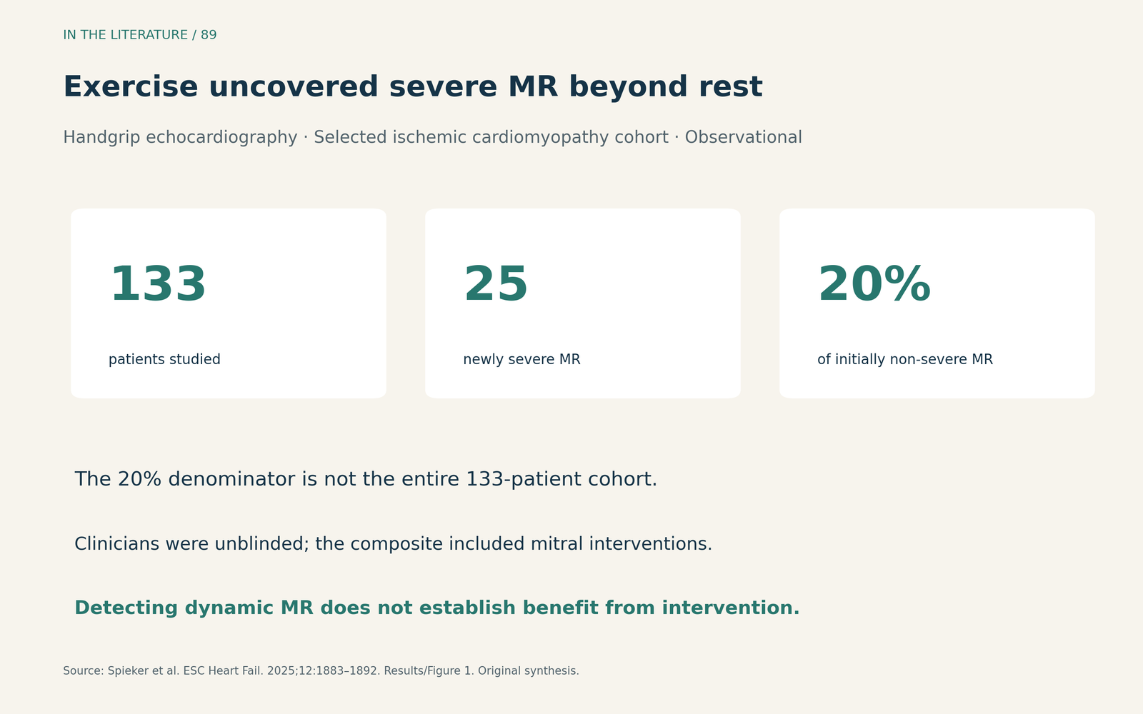
Figure 3. Original synthesis of the Spieker cohort. The reported 20% refers to patients with non-severe MR at rest, not the entire 133-patient cohort. The result cannot be assumed to apply to all primary or atrial MR.4 Enlarge

Three takeaways
- Attach the conditions to the grade. Compare mechanism, acquisition and hemodynamics before interpreting a change in MR.
- Choose the examination for the discrepancy. Optimize stable secondary MR assessment; consider exercise for symptom–rest discordance and CMR for unresolved quantification.
- Keep diagnosis separate from treatment benefit. A smaller anesthetized measurement does not prove improvement, and exercise-induced worsening alone does not prove that intervention will help.
References
- Lancellotti P, Pibarot P, Chambers J, et al. Multi-modality imaging assessment of native valvular regurgitation: an EACVI and ESC council of valvular heart disease position paper. Eur Heart J Cardiovasc Imaging. 2022;23(5):e171–e232. doi:10.1093/ehjci/jeab253
- Praz F, Borger MA, Lanz J, et al. 2025 ESC/EACTS Guidelines for the management of valvular heart disease. Eur J Cardiothorac Surg. 2025;67(8):ezaf276. doi:10.1093/ejcts/ezaf276
- Alachkar MN, Kirschfink A, Grebe J, et al. General Anesthesia Leads to Underestimation of Regurgitation Severity in Patients With Secondary Mitral Regurgitation Undergoing Transcatheter Mitral Valve Repair. J Cardiothorac Vasc Anesth. 2022;36(4):974–982. doi:10.1053/j.jvca.2021.10.024
- Spieker M, Sidabras J, Lagarden H, et al. Exercise-induced dynamic mitral regurgitation is associated with outcomes in patients with ischaemic cardiomyopathy. ESC Heart Fail. 2025;12(3):1883–1892. doi:10.1002/ehf2.15195