
Lower explained the leg veins of pregnancy by uterine pressure on the vena cava; in 1961 the same compression caused supine hypotension under epidural block.
Reading Library · Anesthesia Epochs · 中文版
Watch the education video (for clinicians)
1669–1961: Aortocaval Compression — Lower to Grennell · Anesthesia Epochs Education Series
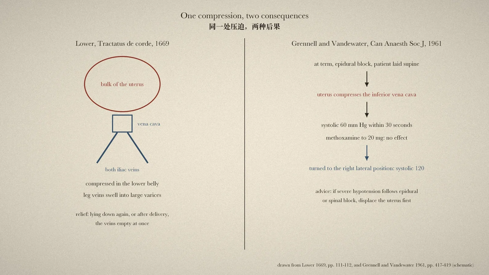
Every obstetric anaesthetist learns to tilt a patient at term, or push the uterus to the left, before trusting a blood pressure taken flat on the back. The reason is mechanical: the heavy uterus can sit on the inferior vena cava and choke the return of blood to the heart. The timeline of the Australian Society of Anaesthetists traces the idea back to 1669, to the Oxford physician Richard Lower and the Tractatus de corde, as "possibly the earliest description" of caval compression in pregnancy. This article reads Lower's passage in the 1669 printing, to see what it says and what it does not, and then reads one 1961 anaesthesia paper in which the same compression showed its dangerous side. [1][2][3]
What Lower wrote
The passage is in the second chapter, on the motion of the heart, pages 111 and 112. Lower is explaining why veins become dilated. The causes, the text says, are compression of some part, an excess of blood, or difficulty in the blood's return. "And therefore in pregnant women", in this article's translation, "because the vena cava and both iliac veins in the lowest part of the belly are compressed by the bulk of the uterus, and because of the increase of blood and humours and their difficult ascent from the lower parts", the veins of the legs are often stretched "into large varices". The blood, held back on its way home and not driven on with enough force from behind, lingers in the lower veins and distends them by its weight. [1]
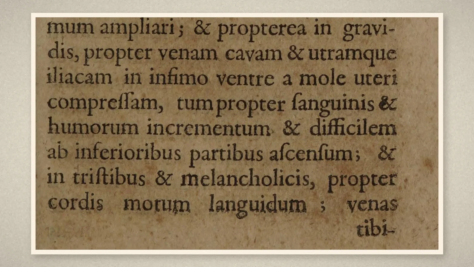
Then comes an observation that matters for what follows: "And so when they take to their beds again, or have delivered the child, because the blood returns more easily towards the heart, those veins at once empty." The next sentence moves to dilated veins in general. Such veins, Lower says, divert blood from the heart and the vena cava and interrupt the thread of the circulation, "whence weakness of the pulse and languor of strength" follow; and Lower describes a countryman of about forty, worn by long sadness, with a varix of the left leg swollen "like a great sausage" from ankle to knee. [1]
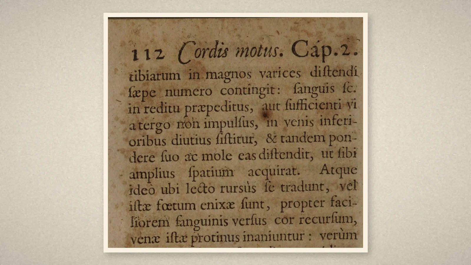
So the anatomy is right there: a heavy uterus pressing on the vena cava and the iliac veins. But the consequence Lower describes is swollen leg veins, and the relief comes from lying down. Nothing in the passage describes a pregnant woman fainting or losing blood pressure when laid on the back. Reading it as an early account of supine hypotension asks more of it than it says.
Kingston, 1961
The anaesthetic danger appears plainly in a short paper in the Canadian Anaesthetists' Society Journal of July 1961, by H. J. Grennell and S. L. Vandewater of the Department of Anaesthesia at Queen's University, Kingston, Ontario. The authors begin with the obstetric literature: Goodson and colleagues had found hypotension in the supine position in 11.2 per cent of women near term; Schmidt had proposed that the uterus falls backward and compresses the inferior vena cava, reducing venous return and cardiac output; and Kennedy had reported hypotension in 17.7 per cent of vaginal deliveries under spinal anaesthesia, with displacement of the uterus to the left restoring the pressure in 93.4 per cent of them. [2]
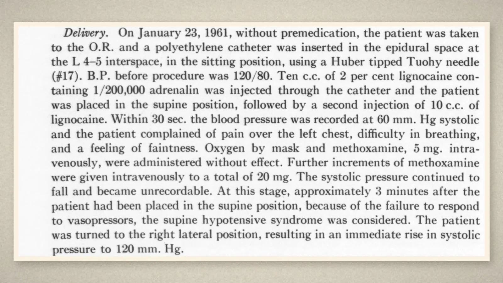
The first case is the reason the paper is remembered. On 23 January 1961, a patient at term came to the operating room for elective caesarean section. An epidural catheter was placed with a Huber-tipped Tuohy needle, lignocaine with adrenaline was injected, and the patient was laid supine. Within 30 seconds the systolic pressure was 60 mm Hg; the patient had chest pain, difficulty breathing and faintness. Oxygen and methoxamine had no effect, and the pressure became unrecordable. About three minutes after the patient had been laid flat, the supine hypotensive syndrome was considered and the patient was turned to the right lateral position: the systolic pressure rose at once to 120. Laid supine again, it fell to 40, and the operation was abandoned for that day. The next day, under general anaesthesia, the pressure was low until the uterus was emptied and then rose at once. [2]
In the second case, a vaginal delivery under epidural block, the pressure fell to 75/45 in the lithotomy position and rose to 95/55 whenever the uterus was pushed to the left by hand. Since the syndrome had been drawn to their attention, the authors wrote, eight of their last 100 consecutive vaginal deliveries under epidural anaesthesia had shown it, none of them before the block. They argued that sympathetic paralysis would exaggerate the effect of caval obstruction, that pressor drugs might fail where a change of position would save a life, and that collapse of this kind can mimic a total spinal; so when severe hypotension follows an epidural or spinal block in a patient lying supine, "uterine displacement might be carried out first". [2]
What the record shows
Lower saw the mechanism and drew a modest conclusion from it, about veins in the legs. Nearly three centuries later, regional anaesthesia removed the sympathetic tone that had been compensating for the same compression, and the consequence moved from the legs to the circulation as a whole. The practical lesson of 1961 is still taught: before reaching for more vasopressor in a pregnant patient who collapses lying flat after a block, move the uterus off the vena cava.
Sources and reading limits
Read: Lower's Tractatus de corde (London, 1669), chapter 2, pp. 111-113; Grennell and Vandewater's paper of 1961, pp. 417-420, in full; the 2024 ASA timeline entries.
Not covered: Howard, Goodson and Mengert's paper of 1953 that named the supine hypotensive syndrome (only the journal's index is open), and the papers of Schmidt, Kennedy and Holmes cited in 1961; later work on aortic compression; an English translation of Lower. Pressures are given as printed; doses are not reproduced. Translations from the Latin are this article's own. The article does not attribute the supine hypotensive syndrome to Lower.
Three illustrations are page crops from the 1669 book and the 1961 paper; one is drawn from them.
- Lower R. Tractatus de corde. Item de motu & colore sanguinis et chyli in eum transitu. London: Jo. Redmayne for J. Allestry; 1669. Cap. 2, pp. 111-113. Internet Archive
- Grennell HJ, Vandewater SL. The supine hypotensive syndrome during conduction anaesthesia for the near-term gravid patient: case reports. Can Anaesth Soc J. 1961;8(4):417-20. Internet Archive
- Australian Society of Anaesthetists. Anaesthesia history timeline. 2024. ASA
Continue exploring
Related reading connects shared questions and contexts; it does not by itself establish historical influence.
Anesthesia Epochs · The Source Room · About & Editorial Method
Published November 2, 2026. Sources and reading limits are stated above. Illustrations are labelled as original source images or AI-made. Historical accounts are educational; they are not current treatment instructions.