
A nine-page paper in Acta Radiologica, May 1953: a puncture needle, a flexible metal leader and a polyethylene catheter as wide as the needle, and 40 arterial catheterizations at Karolinska Sjukhuset.
Reading Library · Anesthesia Epochs · 中文版
Watch the education video (for clinicians)
1953: The Seldinger Technique — A Catheter Over a Leader · Anesthesia Epochs Education Series
The Australian Society of Anaesthetists' history timeline lists "the Seldinger technique" under 1953 and credits it with transforming vascular access. Its first description ran to nine pages and dealt with the arteries of radiology patients. In May 1953 Acta Radiologica printed "Catheter replacement of the needle in percutaneous arteriography: a new technique", by Sven Ivar Seldinger of the Roentgen Diagnostic Department at Karolinska Sjukhuset in Stockholm. The paper does not use the name "Seldinger technique", and it says nothing about anaesthesia except that its patients did not need a general anaesthetic.
The problem
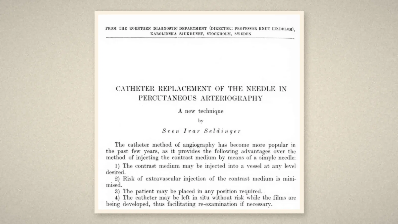
The paper opens with the case for catheters. The catheter method of angiography "has become more popular in the past few years": contrast could be injected at any level, the risk of extravascular injection was smaller, the patient could lie in any position, and the catheter could stay in place while the films were developed. [1, p. 368] What had held it back was "the lack of a suitable flexible thin-walled catheter which could be used percutaneously". [1, p. 368]
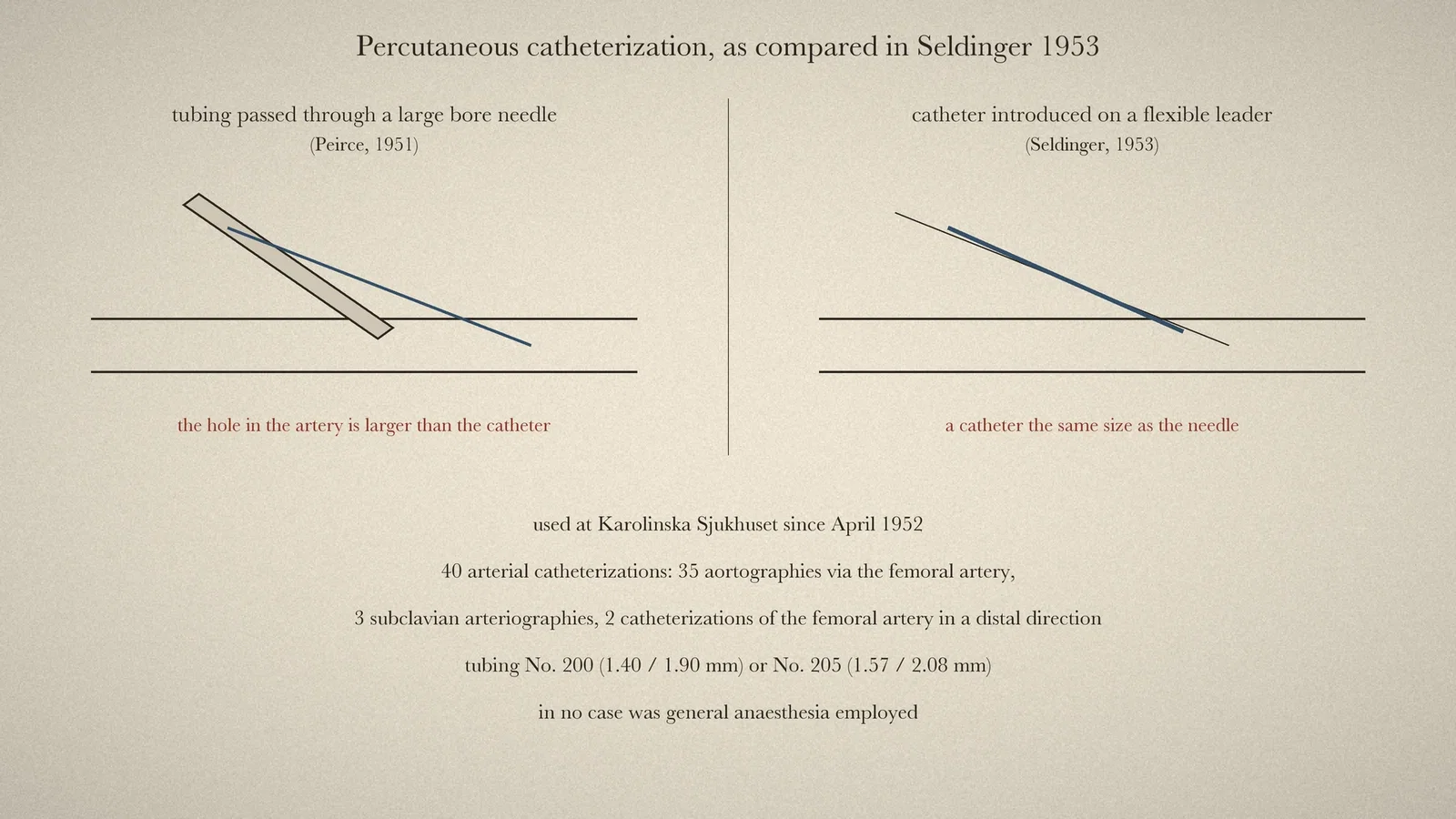
Seldinger listed the earlier attempts. Farinas in 1941 passed a urethral catheter into the aorta through a trocar in the exposed femoral artery; Radner in 1947 catheterized the exposed and ligated radial artery; Jönsson in 1949 guided a blunt cannula down from a puncture of the carotid artery. [1, p. 368] Exposing the artery was "time-consuming, troublesome" and carried risks. [1, p. 369] Polyethylene tubing made a percutaneous route possible, and Peirce in 1951 passed the tubing through a large bore needle. But a large needle made puncture harder and confined the method to large arteries, and because the hole in the artery was larger than the catheter, "haemorrhage after removal of the needle may be troublesome". [1, p. 369]
The method
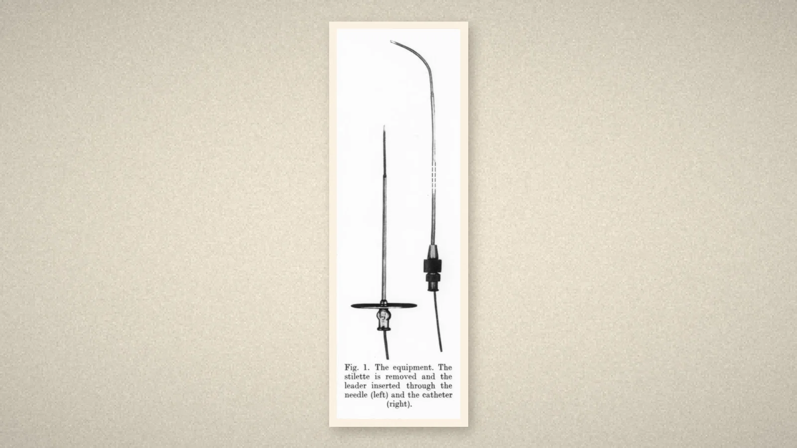
"There is a simple method, however, of using a catheter the same size as the needle", Seldinger wrote, one that had been used at Karolinska Sjukhuset since April 1952. Its main principle was that the catheter was introduced "on a flexible leader through the puncture hole after withdrawal of the puncture needle". [1, p. 369] The equipment, supplied by A. B. Stille-Werner of Stockholm, was a puncture needle with stilette, a flexible rounded-end metal leader whose distal 3 cm was extra flexible, and a polyethylene tube of the same diameter as the needle. [1, p. 369] The leader had to pass through both needle and catheter, fit the catheter's lumen closely, and be at least 8-9 cm longer than the catheter. [1, p. 370]
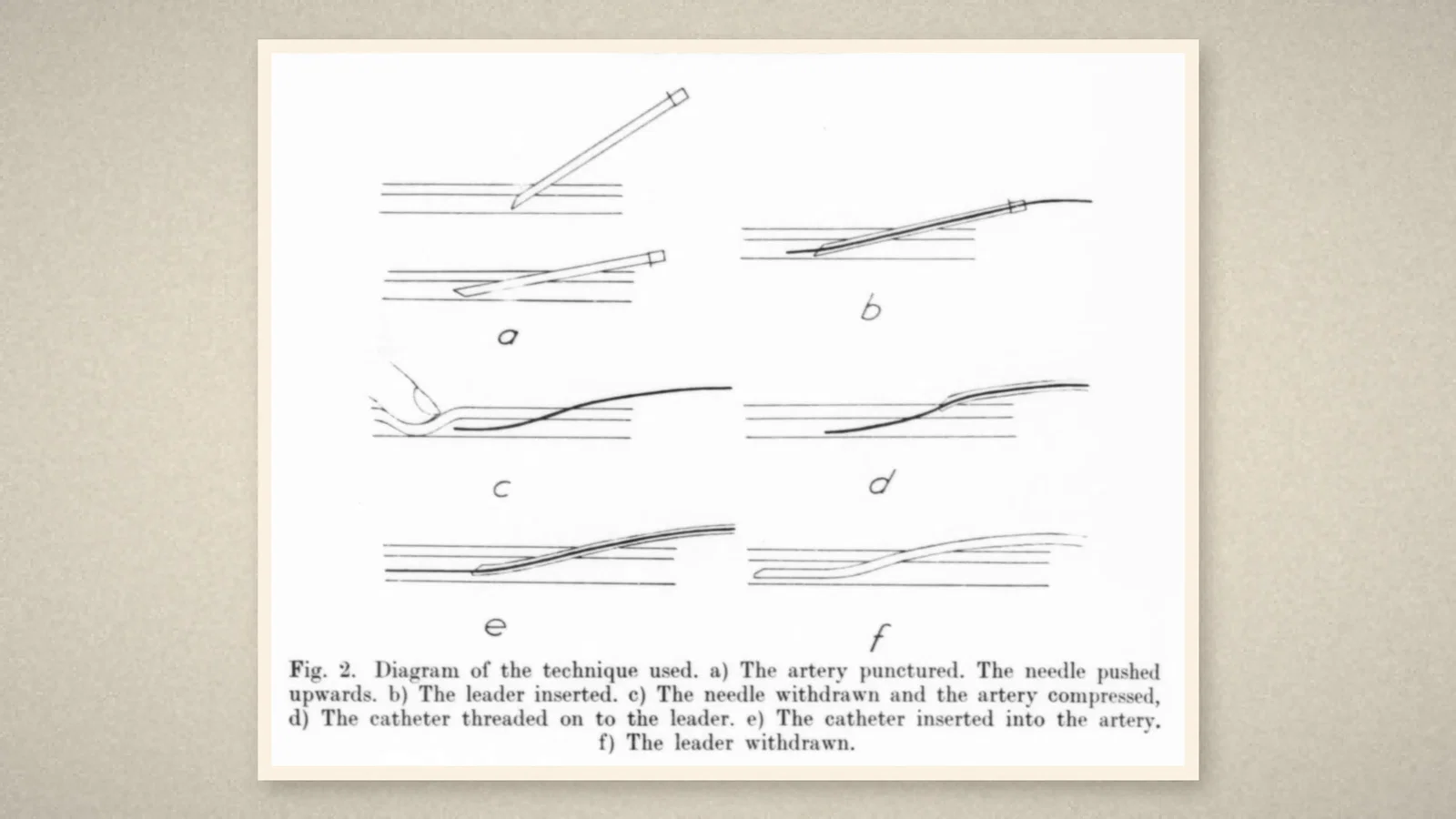
Figure 2 shows six steps. After local anaesthesia, the artery is punctured percutaneously at a small angle. The supple tip of the leader is passed a very short distance into the artery through the needle. The leader is held and the needle removed, with pressure on the artery above the puncture to control bleeding, since the leader is thinner than the hole. The catheter is threaded on to the leader and pushed through the skin into the artery. Then the leader is removed and the catheter directed to the level required. [1, p. 370-371] "This technique is simpler than appears on paper", the paper says, but if considerable resistance was met, force "must therefore never be applied". [1, p. 371-372]
The first forty
The paper reports 40 arterial catheterizations: 35 aortographies via the femoral artery, 3 subclavian arteriographies through the brachial artery, and 2 catheterizations of the femoral artery in a distal direction. "In no case was general anaesthesia employed." The tubing was No. 200 or No. 205, the latter apparently best for aortography. [1, p. 373] Seldinger performed 25; four other members of the department performed 15. One patient could not be catheterized despite five attempts. [1, p. 374] Bleeding at the puncture site was unimportant; "no haematoma of clinical consequence ever formed", and no thrombosis was seen. After local anaesthesia, the patients "felt nothing during the manipulations". [1, p. 375]
The argument for the method was arithmetic. With a catheter that did not have to slide through the needle, a larger catheter could pass through a needle of the same size, and by Poiseuille's law flow rises with "the 4th power of the radius of the tube". [1, p. 375] The one drawback the author admitted was the extra step with the leader, and a small risk that an unskilfully handled leader might pass through the back wall of the artery. [1, p. 376] The summary is one sentence: a method for inserting, after percutaneous puncture, "a catheter of the same size as the needle used into an artery". [1, p. 376]
Afterwards
The paper is about arteries and X-ray contrast. Its place in anaesthesia comes from later use, which this article knows only through a society timeline. The Australian Society of Anaesthetists' history timeline lists "the Seldinger technique" under 1953. It describes the technique as introduced to avoid trauma from large-bore cannulas, and says that it "revolutionized vascular access". [2, PDF p. 61] Under 1984 the same timeline lists an emergency cricothyrotomy kit "based only on the Seldinger technique". [2, PDF p. 79]
What the record shows
The 1953 paper changed the order of steps. The needle no longer had to be larger than the catheter: it only had to make a hole and carry a wire. The paper puts its own case in terms of hole size, bleeding and flow through a narrow tube, and supports it with forty cases done under local anaesthesia.
Sources and reading limits
Read: Seldinger's article in Acta Radiologica 1953;39(5):368-376, in full; the ASA timeline entries for 1953 and 1984.
Not covered: the papers Seldinger cites (Farinas, Radner, Jönsson, Peirce, Donald and colleagues) were not read and are described only as the article describes them. The Helsinki presentation of June 1952 was not found. Seldinger's later work and biography are not covered, and neither are accounts from that decade of the method's use for veins or by anaesthetists. Statements about the technique's later importance are the ASA timeline's. The radiographs in the article are not reproduced.
One illustration is a drawn comparison following the article's text; three are crops of the article's pages and figures.
- Seldinger SI. Catheter replacement of the needle in percutaneous arteriography; a new technique. Acta Radiol. 1953;39(5):368-76. Internet Archive
- Australian Society of Anaesthetists. Anaesthesia History Timeline. 2024. PDF
Continue exploring
Related reading connects shared questions and contexts; it does not by itself establish historical influence.
Anesthesia Epochs · The Source Room · About & Editorial Method
Published November 2, 2026. Sources and reading limits are stated above. Illustrations are labelled as original source images or AI-made. Historical accounts are educational; they are not current treatment instructions.