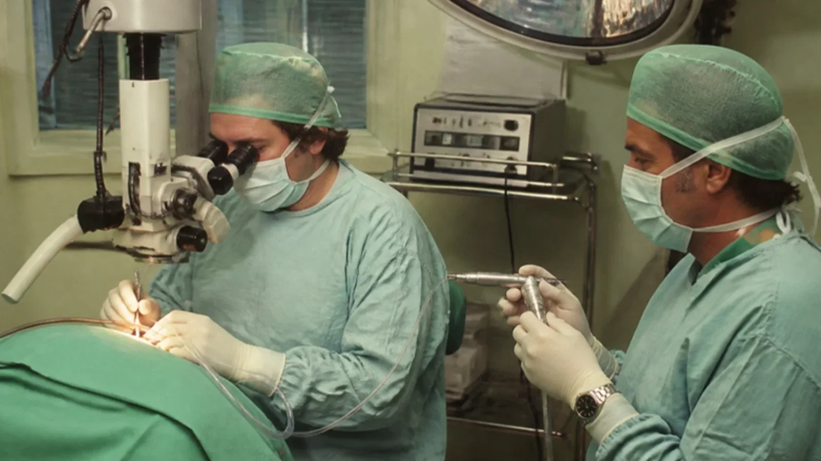
A thin plastic catheter with four soft petals, driven by a Venturi jet injector, gave ENT surgeons an unobstructed larynx and let them use the laser; it grew out of a decade of jet ventilation since Sanders' bronchoscope injector of 1967.
Reading Library · Anesthesia Epochs · 中文版
Watch the education video (for clinicians)
1977: The Benjamin Jet Tube — Microlaryngeal Surgery · Anesthesia Epochs Education Series
Operating on the vocal cords through a microscope leaves little room. The surgeon needs a clear view of the larynx, and the anaesthetist needs a way to ventilate the lungs through the same narrow passage. This article follows one Australian answer: a thin jet-ventilation tube designed in Sydney in 1977 by the ENT surgeon Bruce Benjamin and described with David Gronow in 1979. The 1979 paper itself could not be read. The account rests on its PubMed abstract and on the Australian Society of Anaesthetists' history timeline and its supplement, which quotes the paper's description of the tube.
A decade of jets
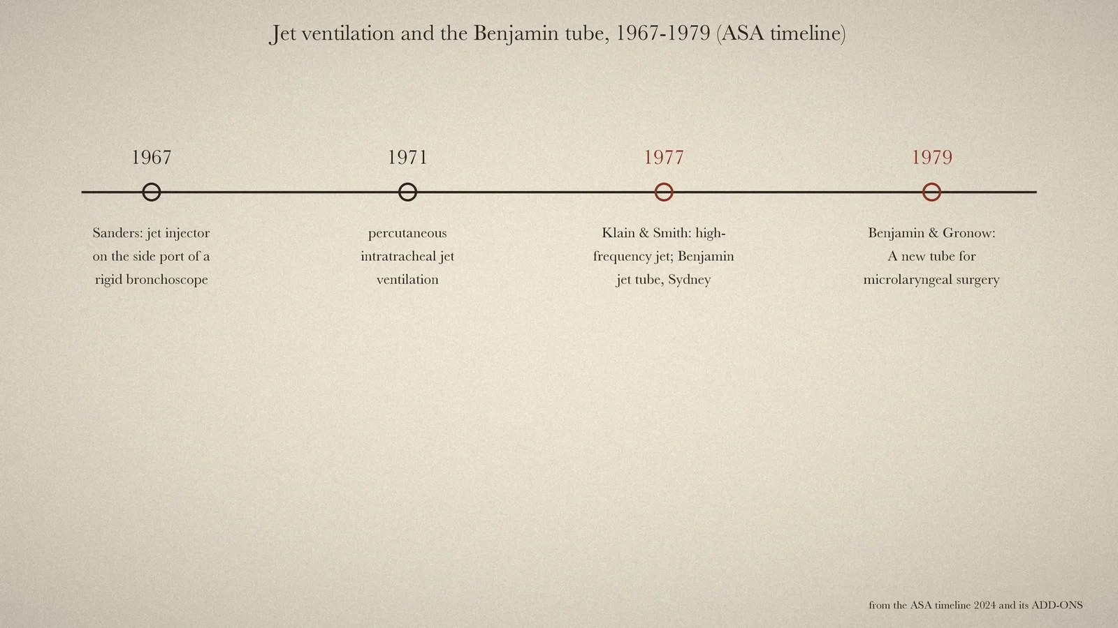
The ASA timeline lists the steps that made such a tube possible. In 1967 came the "Introduction of 'jet ventilation'" by Richard Douglas Sanders in Delaware. Sanders' injector was "attached to a side port of a rigid bronchoscope" and driven by wall oxygen through a pressure regulator and a hand-held valve. The bronchoscope had a second side port through which air or anaesthetic gases were entrained, and the adequacy of ventilation was judged by watching the chest move. [3, p. 70] In 1971 came the "Introduction of percutaneous intra-tracheal 'jet ventilation'". [3, p. 72] In 1977 came the "Introduction of high-frequency 'jet ventilation'", credited to Klain and Smith, whose paper on "High frequency percutaneous transtracheal jet ventilation" appeared that year. [3, p. 76; 4]
The Benjamin tube
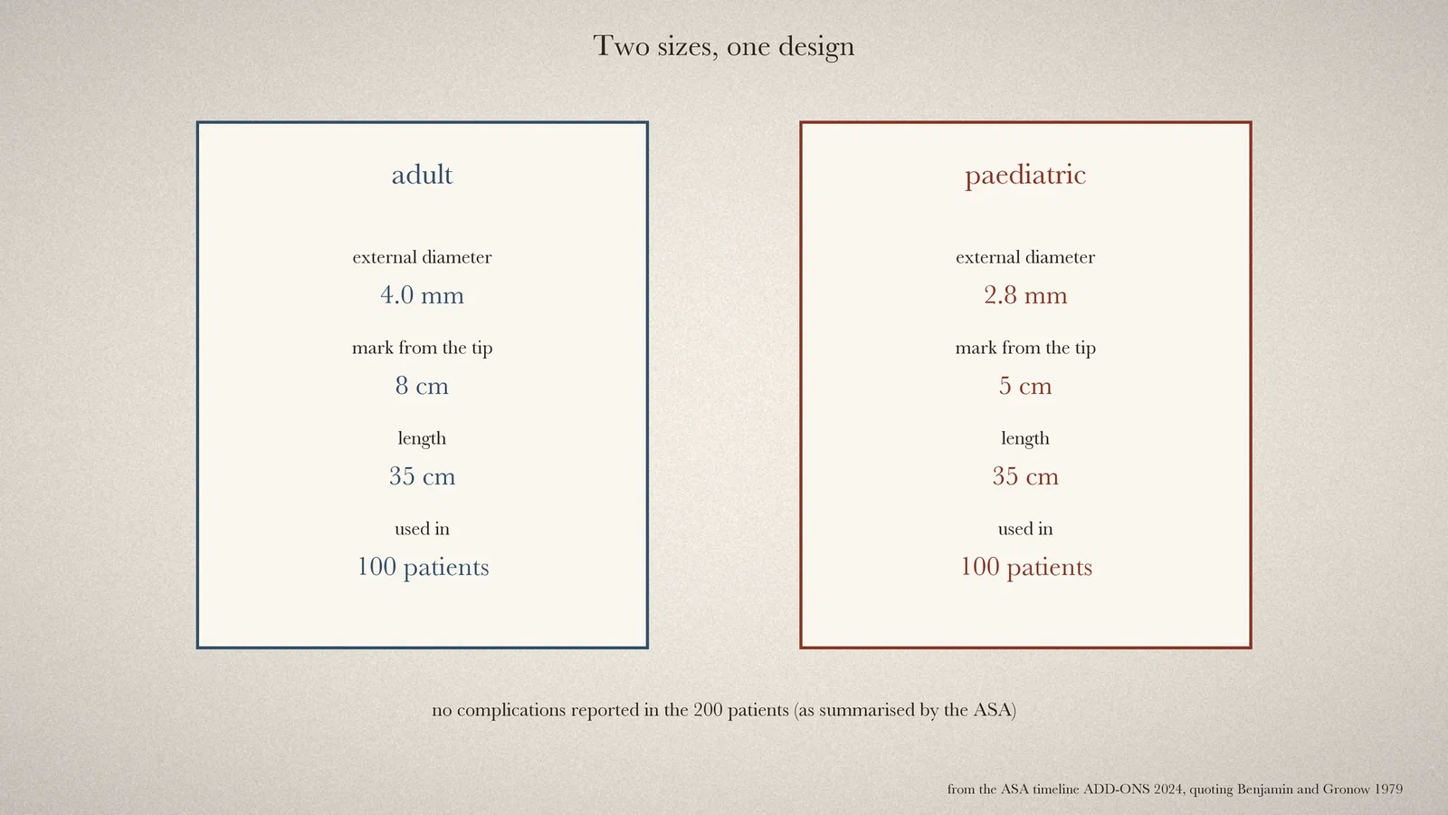
The ASA supplement records that in 1977 the "Benjamin Jet (BenJet) Anaesthesia Tube developed for use with microlaryngeal surgery". It was designed by the ENT surgeon "Bruce Neil Procter Benjamin (1913-2018)" of Sydney, "for use with a Venturi jet injector". [2, p. 2] Benjamin and Gronow published their results in Anaesthesia and Intensive Care in 1979 under the title "A new tube for microlaryngeal surgery". [1] By the supplement's account, they had used it in "100 adult and 100 paediatric patients without complication". [2, p. 2]
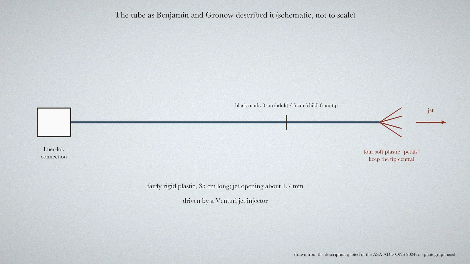
The supplement quotes the paper's description. "The tube is made of fairly rigid plastic", with a proximal Luer-lok connection. There are "four soft plastic 'petals' at the distal end", "designed to maintain it in a central position in the trachea", to prevent trauma to the tracheal wall and to distribute the gas evenly. "The tube is simple, cheap, sterile", easy to put in and take out, and cannot kink. [2, p. 2] The adult tube measured "4.0mm in external diameter with a black mark 8cm" from its tip, "the paediatric tube is 2.8mm in" diameter with a mark at 5 cm, and "Both tubes are 35cm long". [2, p. 2]
What it offered
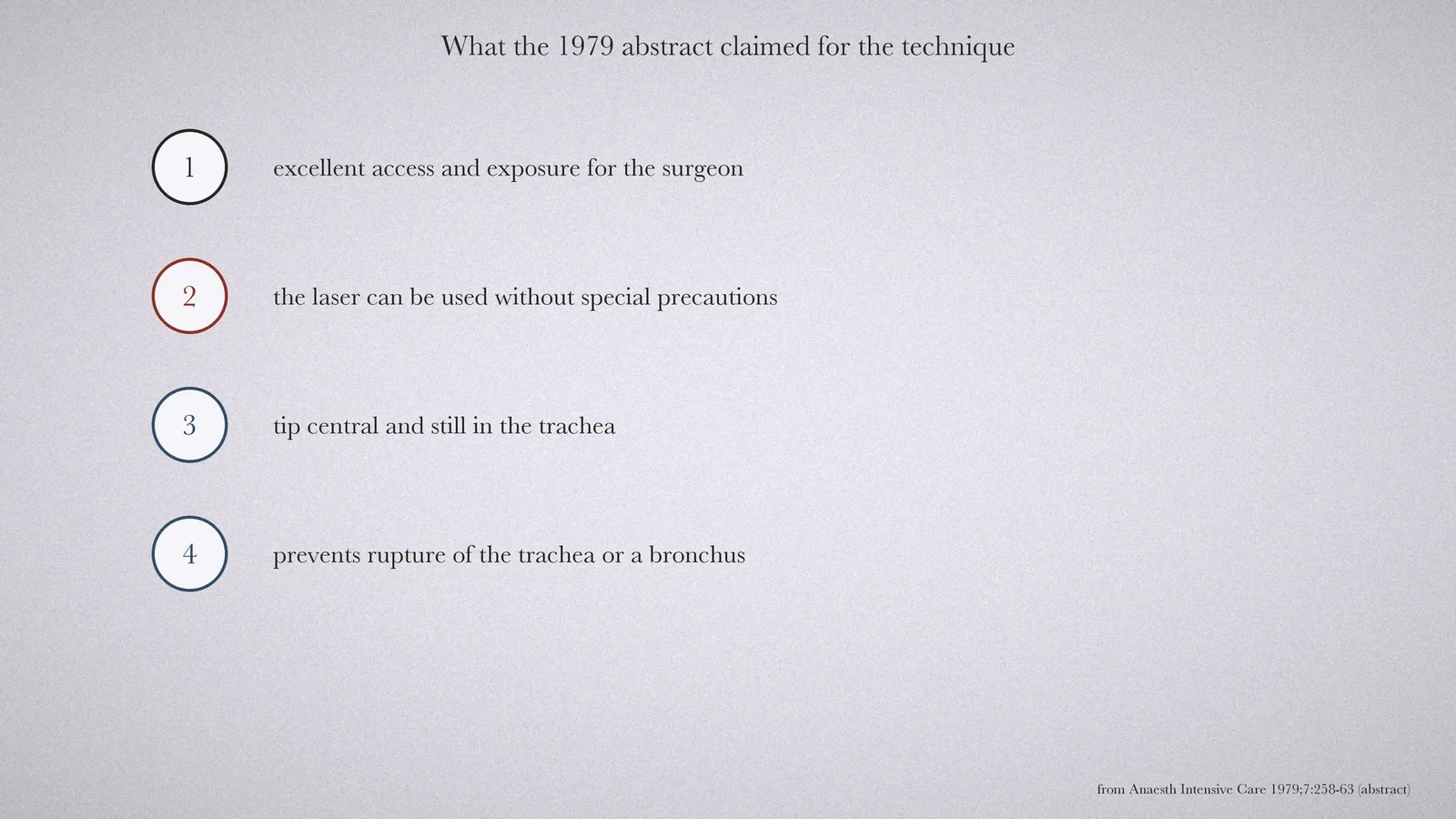
The 1979 abstract describes anaesthesia for microlaryngeal surgery using a "new catheter and the venturi jet injector". "Access and exposure for the surgeon are excellent", and "the laser can be used without special precautions". Because the tip sat centrally and did not move, the technique was meant to "prevent the potential complication of rupture of the trachea" or of a bronchus. [1]
The laser mattered to Benjamin in particular. The supplement notes that "He was the first person in Australia to use the CO2 laser". Benjamin later became the "Foundation President of the Australasian Society of Paediatric Otolaryngology" in 1993. The supplement adds ten years as a lecturer at Sydney University and work at several hospitals, including the Royal Alexandra Hospital for Children and the Royal North Shore Hospital in Sydney. [2, p. 2]
What the record shows
The Benjamin tube is a small example of a surgeon designing for the anaesthetist. A thin plastic line, centred by four petals and driven by a jet, kept the airway open while leaving the larynx clear for the microscope and the laser.
Sources and reading limits
Read: the PubMed abstract of Benjamin and Gronow 1979; the ASA timeline ADD-ONS 2024 entries on the Benjamin tube; the main ASA timeline entries on jet ventilation; the PubMed record of Klain and Smith 1977.
Not covered: the 1979 paper itself was not read. The tube's description, its sizes and the 200 patients come from the ASA supplement's quotation and summary. The supplement dates the tube to 1977 and the paper to 1979, and what happened in 1977 is not specified. Sanders' 1967 paper was not found. The schematic follows the quoted description only and is not drawn from a photograph of the device.
All four illustrations are drawn from these sources.
- Benjamin B, Gronow D. A new tube for microlaryngeal surgery. Anaesth Intensive Care. 1979;7(3):258-63. PubMed
- Australian Society of Anaesthetists. Anaesthesia History Timeline ADD-ONS 2024. PDF
- Australian Society of Anaesthetists. Anaesthesia History Timeline 2024. PDF
- Klain M, Smith RB. High frequency percutaneous transtracheal jet ventilation. Crit Care Med. 1977;5(6):280-7. PubMed
Continue exploring
Related reading connects shared questions and contexts; it does not by itself establish historical influence.
Anesthesia Epochs · The Source Room · About & Editorial Method
Published November 2, 2026. Sources and reading limits are stated above. Illustrations are labelled as original source images or AI-made. Historical accounts are educational; they are not current treatment instructions.