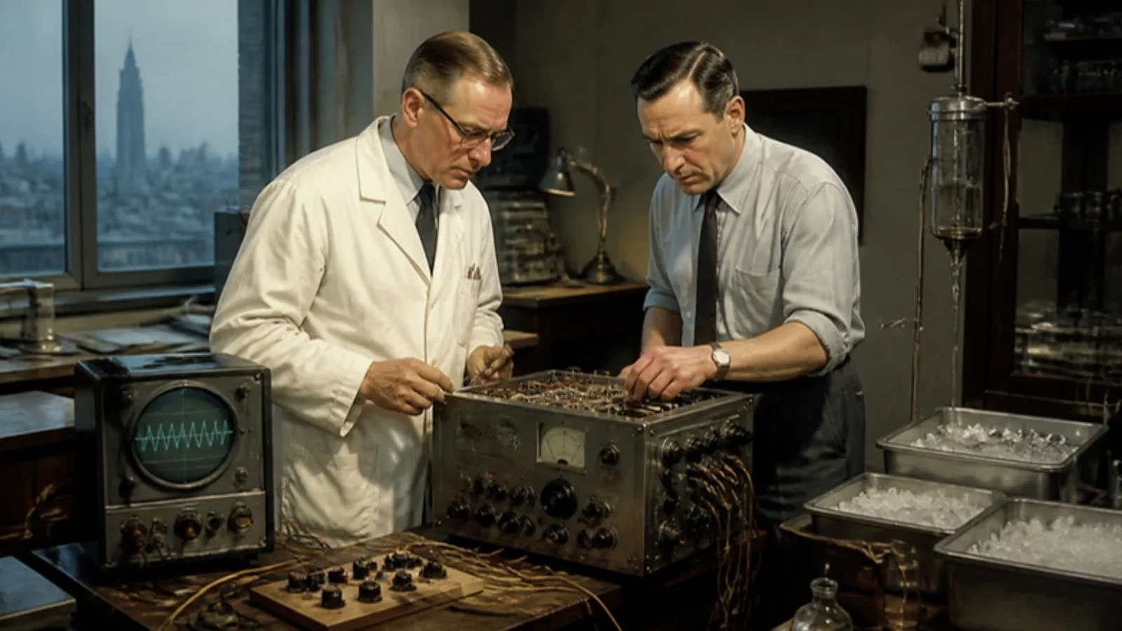
Bigelow, Callaghan and the engineer Hopps paced the S-A node region and restarted four arrested, cooled dog hearts.
Reading Library · Anesthesia Epochs · 中文版
Watch the education video (for clinicians)
1950: Bigelow's Pacemaker — Born in Hypothermia Experiments · Anesthesia Epochs Education Series
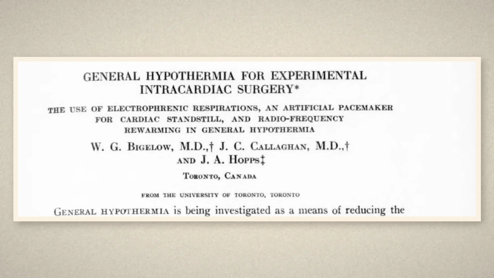
Temporary pacing wires on the heart after cardiac surgery are routine today. One root of the idea is a paper in the Annals of Surgery of September 1950, read before the American Surgical Association on 20 April that year: "General Hypothermia for Experimental Intracardiac Surgery", by W. G. Bigelow and J. C. Callaghan of the Department of Surgery, University of Toronto, and J. A. Hopps of the Radio and Electrical Engineering Division of the National Research Council of Canada. [1, p. 531] Its subtitle lists three techniques, and the second is "an artificial pacemaker for cardiac standstill".
Cooling dogs to operate inside the heart
The team was studying general hypothermia as a way to lower the body's oxygen needs enough to exclude the heart from the circulation, "thereby permitting intracardiac surgery under direct vision". [1, p. 531] "A total of 176 dogs have been cooled." Cooling to 20°C had become fairly safe, but "death at lower temperatures is usually due to ventricular fibrillation". [1, p. 531]
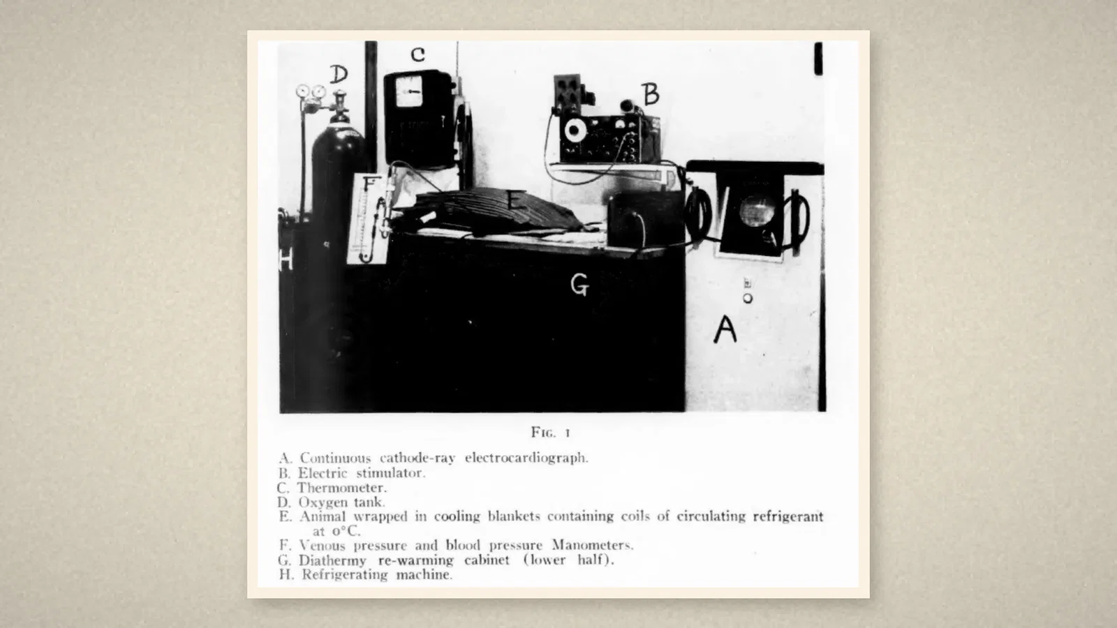
Several details concern anaesthetists directly. The dogs were given digoxin and procaine, and hypothermia was induced "with the aid of intravenous pentothal and curare to control shivering". Below 28°C the animal entered a state of "cold narcosis", in which "an anesthetic agent is no longer necessary to maintain unconsciousness and relaxation". At 20°C, oxygen consumption, cardiac output, blood pressure and heart rate were "about 15 per cent of normal". [1, p. 531] Because breathing failed in the cold, they ventilated the dogs by rhythmic electrical stimulation of the exposed phrenic nerve, "electrophrenic respiration", using a stimulator whose rate was set on a dial. [1, p. 532]
The heart was the weak point. Of the dogs that died while cold, "five experienced cardiac standstill, while the remaining 14 died from ventricular fibrillation". [1, p. 533] Defibrillation sometimes left the heart in standstill too.
"An artificial pacemaker"
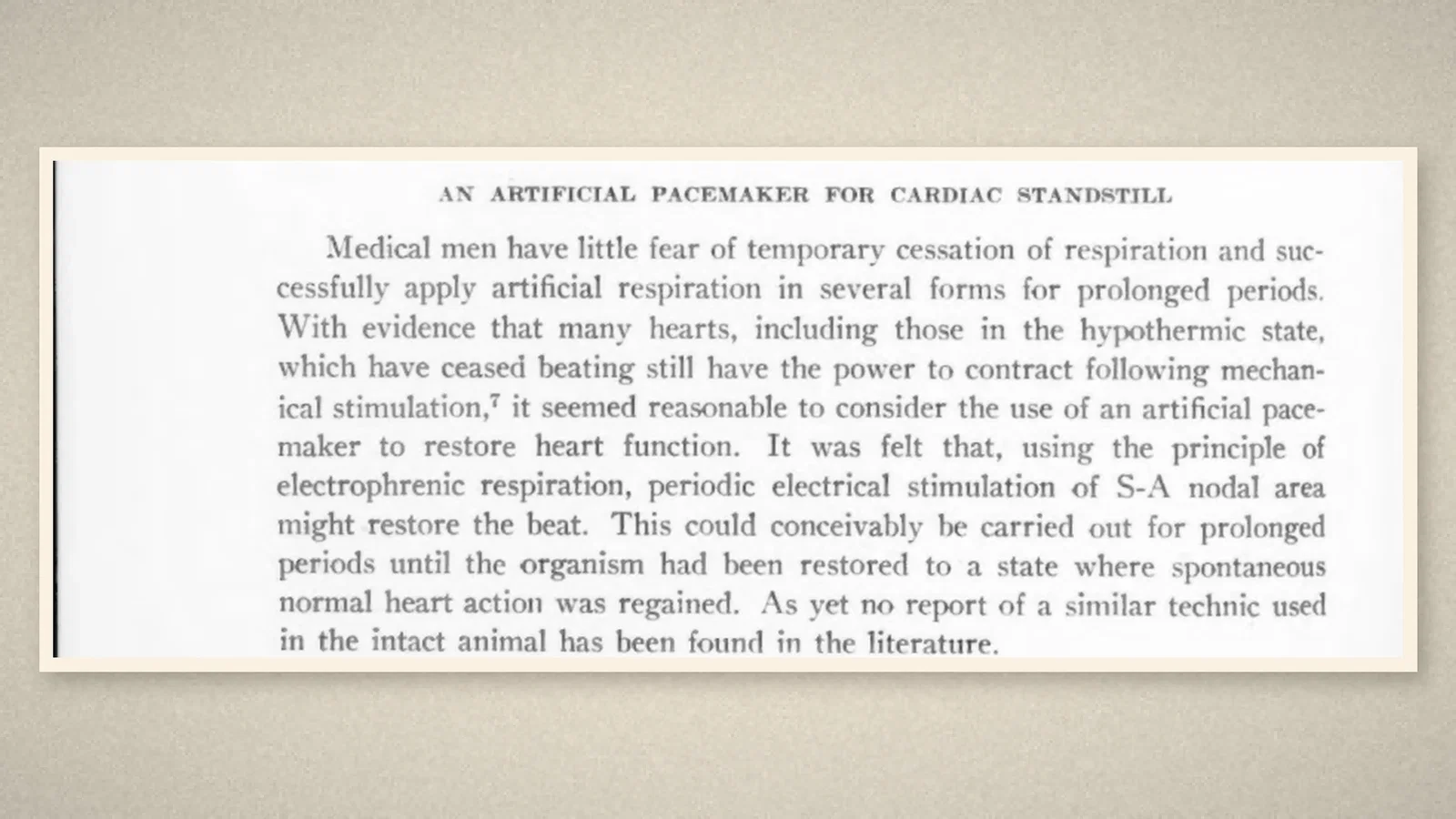
Their argument was an analogy with breathing. "Medical men have little fear of temporary cessation of respiration" and apply artificial respiration for long periods. Since many stopped hearts, including cold ones, can still contract when stimulated, "it seemed reasonable to consider the use of an artificial pacemaker to restore heart function". Using the principle of electrophrenic respiration, "periodic electrical stimulation of S-A nodal area might restore the beat". [1, p. 534] And: "As yet no report of a similar technic used in the intact animal has been found in the literature." [1, p. 534]
It was "a very early report", tried "in four cases of cardiac standstill, in each case with restoration of heart action". [1, p. 535] The same stimulator delivered impulses at any rate; "an indifferent electrode is clipped onto the chest wall and the stimulating electrode placed in the region of the S-A node", and "the heart rate is varied within limits by adjusting a dial". In two dogs, after 10 to 15 minutes of pacing, the heart "returned to standstill" when it was stopped; in the other two, "following electrical control of the heart beat for ten and 30 minutes, normal spontaneous heart beats returned". One of these, after cardiotomy, fibrillation, defibrillation and ten minutes of pacing, was rewarmed, "only to die several hours later". [1, p. 535] The authors saw the advantage of leaving the stimulating wire in place while closing the chest, and wrote that "its extension to other clinical conditions with cardiac arrest might be considered". [1, p. 535]
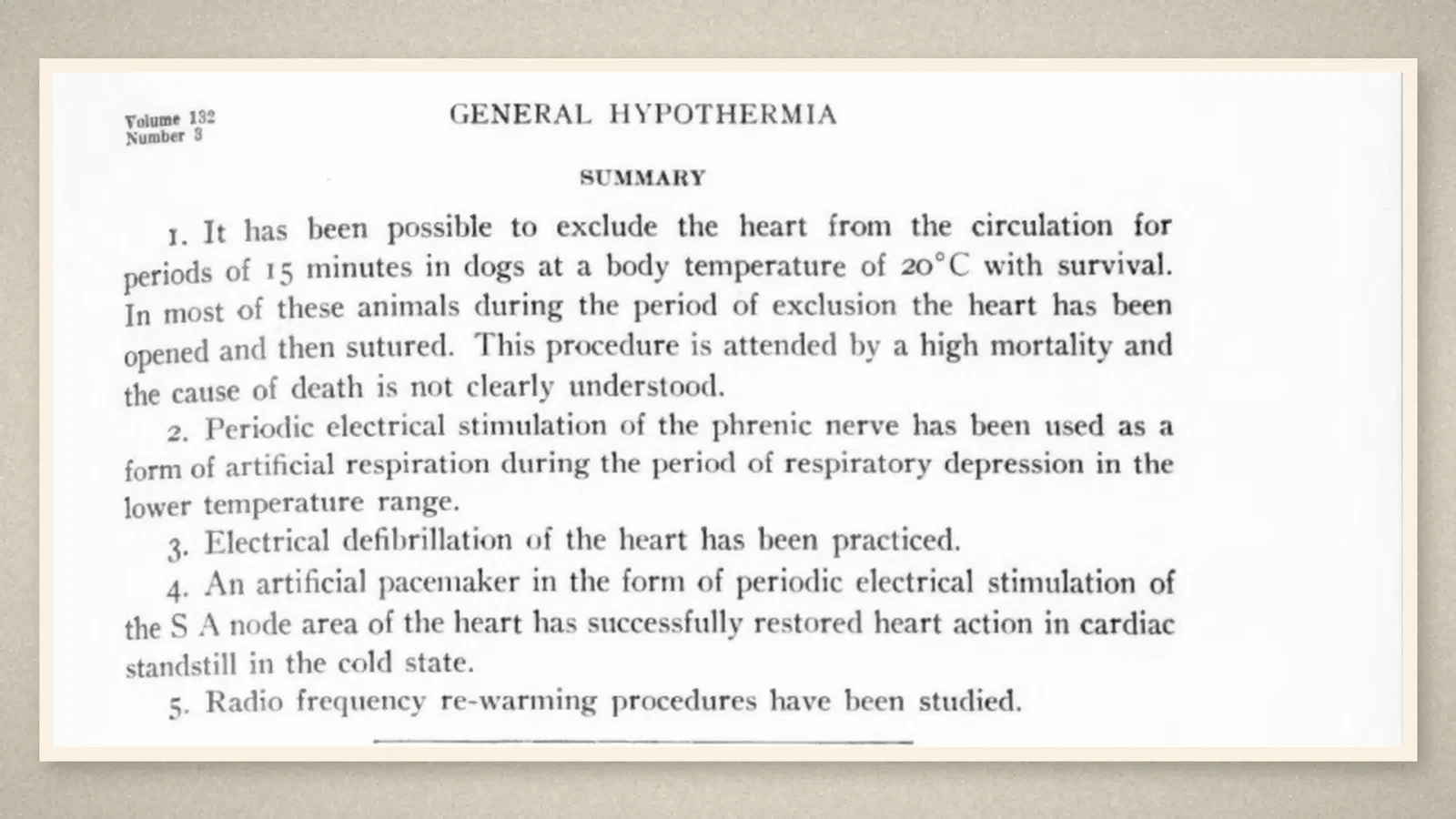
Before and after, according to a review
The Toronto team did not know of an earlier attempt on the other side of the world. According to Aquilina's history of pacing, the Australian anaesthetist Mark Lidwill used a needle inserted into the ventricle and intermittent electrical stimulation "in 1928 ... to save the life of a child born in cardiac arrest", and reported it to an Australian medical congress in 1929. [2] The same review describes Hopps's pacemaker as an external unit "driven by vacuum tubes" with a transvenous catheter to the atria [2]; the 1950 paper, however, describes an electrode on the S-A node region of the exposed heart. [1, p. 535] Eight years later, on 8 October 1958, the first fully implantable pacemaker, developed by the surgeon Åke Senning and the physician-inventor Rune Elmqvist, was implanted in Stockholm in Arne Larsson, a 43-year-old engineer whose Stokes-Adams attacks "required resuscitation many times daily". [2]
What the record shows
A device for pacing the heart came out of an attempt to make the heart safe to open under hypothermia, from a team of two surgeons and an electrical engineer, and from an analogy with artificial respiration that any anaesthetist would recognize. In 1950 it worked in four dogs, two of which needed it only for a while.
Sources and reading limits
Read: Bigelow, Callaghan and Hopps, Ann Surg 1950;132:531–539, in full with Fig. 1; Aquilina's open-access review (2006), as a secondary source.
Not covered: Lidwill's 1929 congress report and Booth's 1930 paper were not read; Lidwill appears here only through Aquilina, who gives 1928. Elmqvist and Senning's 1960 abstract (reported at a 1959 conference) and Greatbatch's work were not read; the 1958 implant is given from the review only.
All four illustrations are crops of the 1950 paper.
- Bigelow WG, Callaghan JC, Hopps JA. General hypothermia for experimental intracardiac surgery: the use of electrophrenic respirations, an artificial pacemaker for cardiac standstill, and radio-frequency rewarming in general hypothermia. Ann Surg. 1950;132(3):531-9. Internet Archive
- Aquilina O. A brief history of cardiac pacing. Images Paediatr Cardiol. 2006;8(2):17-81. Europe PMC
Continue exploring
Related reading connects shared questions and contexts; it does not by itself establish historical influence.
Anesthesia Epochs · The Source Room · About & Editorial Method
Published November 2, 2026. Sources and reading limits are stated above. Illustrations are labelled as original source images or AI-made. Historical accounts are educational; they are not current treatment instructions.