ITL-7ae6153625de
Juan Li, MD · Updated November 17, 2026
Watch the video
Key points
- Bubbles in the left heart prove a right-to-left shunt, not its location. Timing alone cannot separate a patent foramen ovale (PFO) from an intrapulmonary shunt.[1]
- The strongest clue is where the bubbles enter: across the interatrial septum or out of a pulmonary vein. Transesophageal echocardiography (TEE) can show this directly when transthoracic images cannot.[1], [2]
- Pattern adds information. A PFO typically gives a phasic puff that grows with a well-performed provocation; intrapulmonary passage usually builds up and fades over several beats, with little change during Valsalva.[2], [3]
- Small pulmonary right-to-left shunts are common, especially on TEE, and should not automatically be attributed to a PFO.[4]
- A negative study is only meaningful if right atrial pressure actually exceeded left atrial pressure and contrast reached the septum.[1], [2]
A patient has unexplained hypoxemia after surgery, or a cryptogenic stroke, and an agitated saline injection puts bubbles in the left atrium. The finding is often reported simply as "positive bubble study." The next decision depends on a more specific question. Did the bubbles cross the atrial septum, or did they pass through the lungs? A PFO or atrial septal defect leads to septal imaging and possibly a closure discussion. An intrapulmonary shunt leads instead toward the lungs and liver: pulmonary arteriovenous malformations (PAVMs), hepatopulmonary syndrome or physiological intrapulmonary channels.[1], [2]
Why bubbles normally stop in the lungs
Hand-agitated saline produces microbubbles that are mostly larger than pulmonary capillaries. Bubbles small enough to enter the capillary bed also dissolve quickly, well before ordinary pulmonary transit is complete. Normally, therefore, the right heart opacifies and the left heart stays dark. Left-sided contrast means that bubbles have bypassed the pulmonary capillary filter, either through an intracardiac communication or through a dilated or abnormal pulmonary vascular pathway.[1], [2]
That is why the test is sensitive but does not by itself localize the shunt. Two separate questions follow: where the bubbles came from, and whether the test was adequate to exclude a shunt it did not show.
The beat count is a clue, not a verdict
Clinicians traditionally count cardiac cycles from right atrial opacification to the first left-sided bubbles. Early appearance favors an intracardiac shunt; later appearance favors passage through the lungs. The 2026 British Society of Echocardiography (BSE) guidance defines an intracardiac shunt as left-heart contrast typically within 3 cycles. It describes later appearance as intrapulmonary shunting or transpulmonary passage, as in hepatopulmonary syndrome.[2]
The cutoff is a convention. Bernard and colleagues point out that the widely used three-to-six-beat rule developed from early case observations rather than rigorous pathophysiological studies. Its range has changed over time, and heart rate, respiration and cardiac function alter transit. Their review states plainly that timing alone is inadequate to distinguish intracardiac from intrapulmonary shunting.[1]
Recent data show how much the groups overlap. In a retrospective single-center study, Wang and colleagues performed simultaneous contrast transthoracic echocardiography (TTE) and transcranial Doppler in 193 patients with suspected right-to-left shunt. PFO was confirmed by TEE and PAVM by computed tomography or angiography; 20 indeterminate patients were excluded. Among the 64 PFO patients with cycle timing reported, 43 (67.2%) had bubbles within 1–3 cycles, but 21 appeared at 4 cycles or later. Among 18 PAVM patients, 3 (16.7%) had bubbles within 1–3 cycles. The cardiac-cycle measure had only moderate discrimination.[3]
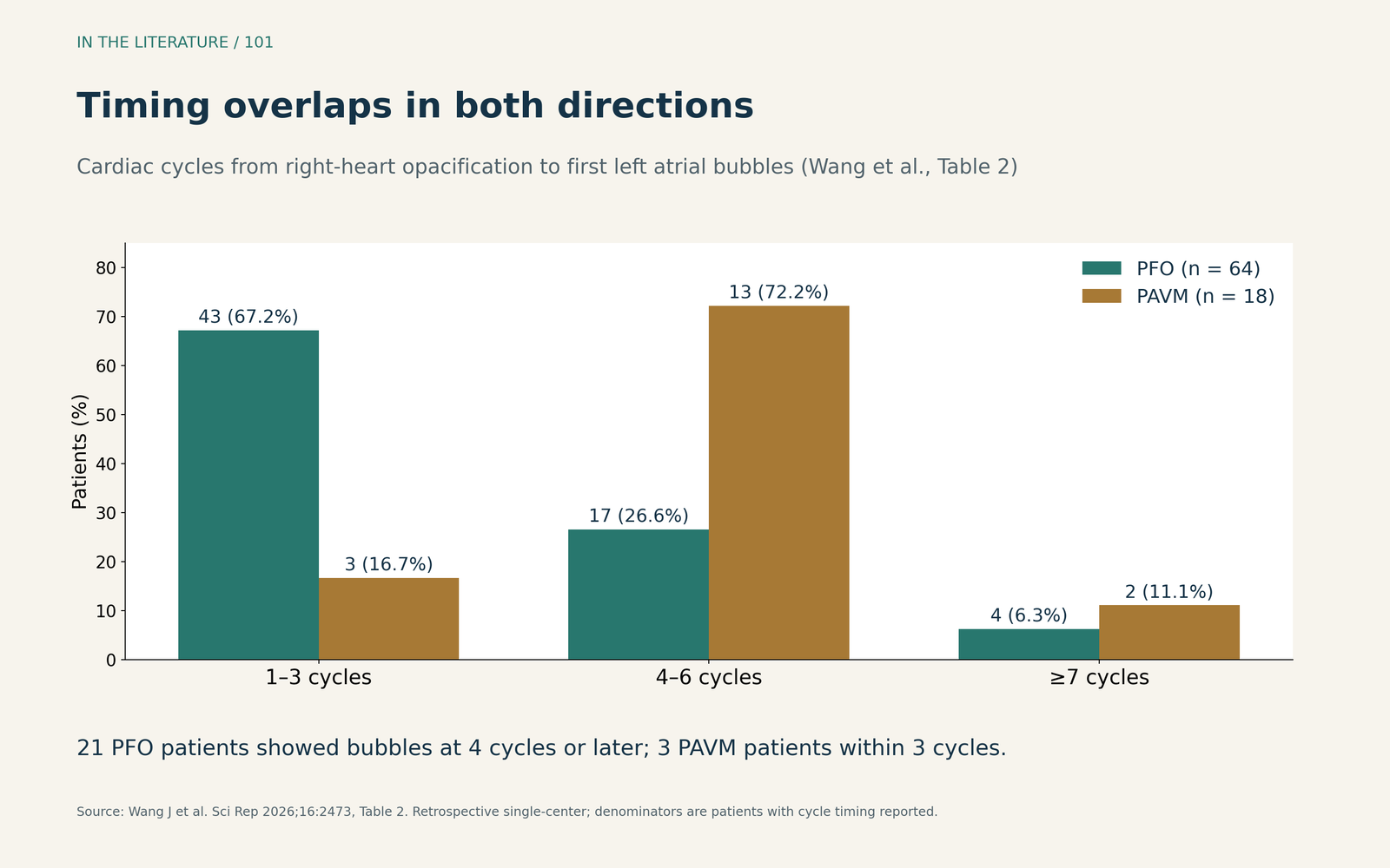
Figure 1. Timing overlaps. Redrawn from Table 2 of Wang et al.; denominators are patients with cycle timing reported (PFO, n = 64; PAVM, n = 18). Retrospective single-center data; TEE and CT/angiography served as reference standards.[3] View larger.

Look for the point of entry and the pattern
Point of entry is the most specific observation. Bubbles seen crossing the interatrial septum strongly support an intracardiac shunt. Bubbles emerging from a pulmonary vein point to intrapulmonary shunting, although this does not exclude a coexisting PFO. Direct observation of septal crossing matters more than the beat count, which varies with hemodynamics.[1]
On TTE, however, entry is often not seen. In the study by Wang and colleagues, bubbles were observed crossing the septum in only 11.8% of PFO patients and emerging from a pulmonary vein in 72.2% of PAVM patients. When the source remains uncertain, the BSE guidance recommends TEE to differentiate flow from the pulmonary veins and flow through the septum.[2], [3]
Pattern over time helps when entry is not visible. A PFO is a flap valve: it opens when right atrial pressure transiently exceeds left atrial pressure, so contrast usually enters the left atrium as a phasic puff. Late contrast from PAVMs or hepatopulmonary syndrome characteristically builds in density over several beats and then decays.[2]
Response to provocation also differs. In the study by Wang and colleagues, a modified Valsalva maneuver increased the left atrial bubble count in PFO patients (median increment 25 bubbles) but not in PAVM patients (median 0). An increment of 10 or more bubbles identified PFO with an area under the curve of 0.94. PAVM shunting was usually large at rest and changed little with Valsalva. The patients had migraine, stroke, hypoxemia or dyspnea; these cutoffs are single-center ROC findings, not validated universal thresholds.[3]
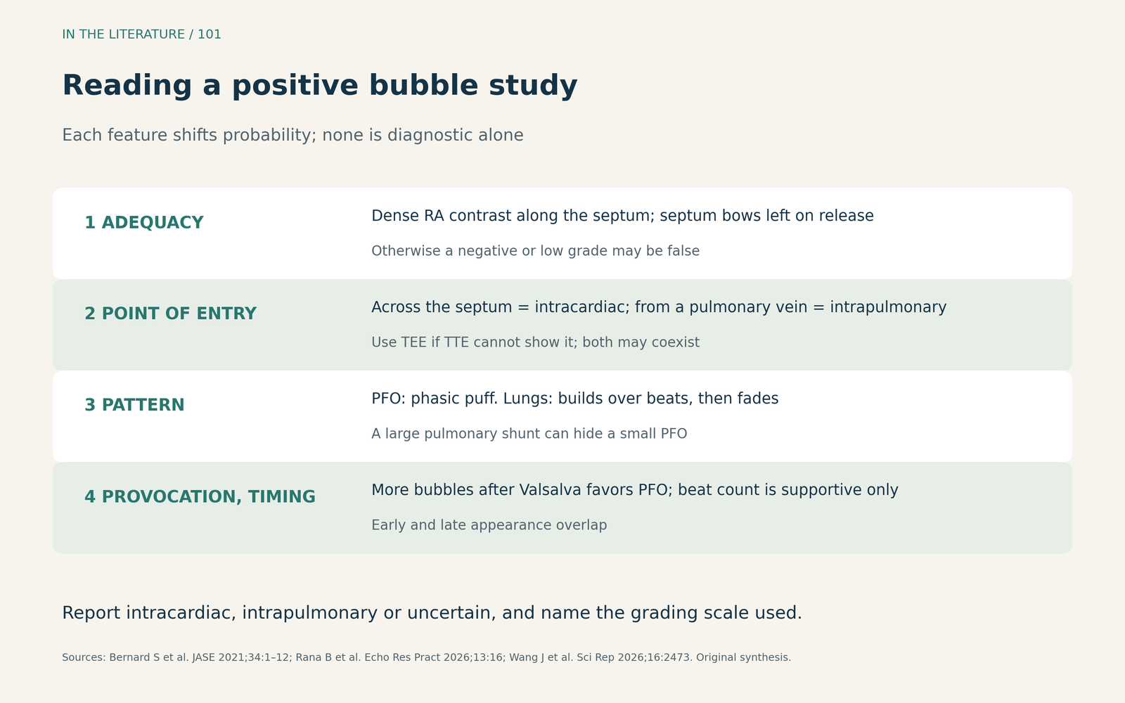
Figure 2. Reading a positive study. A synthesis of the BSE guidance, the Bernard review and the study by Wang and colleagues; each feature shifts probability and none is diagnostic alone.[1]–[3] View larger.

Small pulmonary shunts are common
The differential is not limited to a PFO versus a rare PAVM. Physiological intrapulmonary arteriovenous anastomoses can carry bubbles to the left heart. Chen and colleagues reviewed 407 patients who underwent both contrast TTE and contrast TEE. Patients with pulmonary hypertension, cirrhosis or organic heart disease were excluded. With TEE and Valsalva, a pulmonary right-to-left shunt was seen in 82.2% of patients with PFO and 79.2% without PFO. The corresponding TTE figures were 52.2% and 48.8%. Most pulmonary shunts were low grade; CT angiography in patients with the highest grade identified PAVMs in only four.[4]
Two practical implications follow. A few late bubbles in the left heart do not necessarily indicate a PFO or a treatable malformation. Conversely, a coexisting pulmonary shunt can be mistaken for a larger PFO shunt, which may matter when shunt size contributes to a closure decision. The investigators also observed that the TEE examination and Valsalva maneuver themselves increased pulmonary shunting. The study was retrospective and single-center, and its link to clinical outcomes was not established.[4]
Grading schemes add another source of confusion. The BSE guidance grades intracardiac shunts from 1 to 4 by the maximal bubble count in one frame. Bernard and colleagues proposed a different scheme, and pulmonary shunts are often graded on a separate, higher scale. A report should state which scale was used.[1], [2], [4]
Before calling the study negative
A PFO shunts only when right atrial pressure exceeds left atrial pressure. Inadequate provocation, poor right atrial opacification and elevated left atrial pressure are the main causes of false-negative studies. In one TEE series cited by Bernard and colleagues, PFO was detected in 5% of patients with left-sided heart disease versus 29% without it. A large eustachian valve or brisk inferior vena caval flow can keep contrast away from the septum.[1]
The test is reassuring only when the right atrium is completely opacified along the septum, the septum shifts toward the left atrium during release, and no bubbles appear. In sedated or ventilated patients, maneuvers are less reliable. For an intubated patient, the BSE guidance describes an anesthesiologist-performed Valsalva: increase airway pressure, inject while holding it, then release.[1], [2]
During TEE, a single imaging plane can also undercount. In a prospective series of consecutive patients undergoing TEE for suspected PFO, Xu and colleagues added a live orthogonal plane. During provocation, 19 of 104 examinations graded small in the original plane were graded large in the orthogonal plane. Patients with a concomitant intrapulmonary shunt were excluded.[5]
False positives also occur. Pseudocontrast from pulmonary venous stasis, contrast persisting from a previous injection, a large eustachian valve mistaken for the septum, and over-agitated colloid solutions producing small bubbles can all mislead. Unusual venous anatomy, such as a sinus venosus defect or persistent left superior vena cava, can make contrast appear in the left atrium before or simultaneously with the right. In such cases, the injection site matters; a structured search for the connection is required.[1], [2]
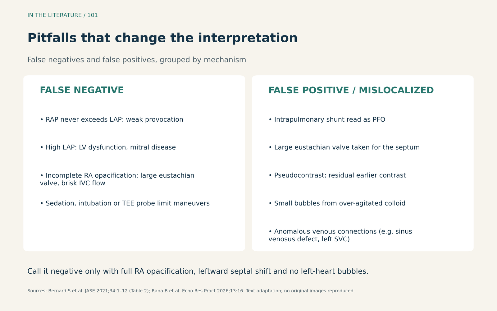
Figure 3. Pitfalls that change the interpretation. Adapted as text from Table 2 of the Bernard review and the BSE guidance; no original images reproduced.[1], [2] View larger.

Safety in the perioperative setting
Agitated saline studies are generally safe. Reported cerebral ischemic events are rare and occur almost exclusively when a shunt is present. Bernard and colleagues advise using less than 1 mL of air and never injecting visible large bubbles. In patients on extracorporeal membrane oxygenation, bubbles may enter the circuit and trigger the bubble detector. Coordinate with the perfusionist before any injection.[1]
The BSE guidance advises against routine agitated saline studies in patients with pulmonary hypertension, atrial septal defects or complex congenital disease, and in the elderly, unless discussed with an expert cardiologist.[2]
Three takeaways
- A positive study proves a shunt, not its location. Combine point of entry, contrast pattern, response to provocation and timing; do not decide on the beat count alone.[1]–[3]
- Low-grade pulmonary shunting is common. Late bubbles, particularly on TEE or after Valsalva, may reflect physiological intrapulmonary pathways rather than a PFO or a treatable malformation.[4]
- Check adequacy before calling a study negative. Confirm septal shift, complete right atrial opacification and an acceptable left atrial pressure; use TEE when the source or the result remains uncertain.[1], [2], [5]
References
- Bernard S, Churchill TW, Namasivayam M, et al. Agitated saline contrast echocardiography in the identification of intra- and extracardiac shunts: connecting the dots. J Am Soc Echocardiogr. 2021;34(1):1–12. doi:10.1016/j.echo.2020.09.013
- Rana B, Robinson S, Ring L, et al. Agitated saline contrast echocardiography in the diagnosis of right to left shunts: guidance and recommendations from the British Society of Echocardiography. Echo Res Pract. 2026;13(1):16. doi:10.1186/s44156-026-00117-3
- Wang J, Zhu Z, Meng H, et al. Differentiation between patent foramen ovale and pulmonary arteriovenous malformations via simultaneous contrast transthoracic echocardiography and transcranial doppler. Sci Rep. 2026;16(1):2473. doi:10.1038/s41598-025-32229-x
- Chen A, Zhu J, Zhu L, et al. Neglected intrapulmonary arteriovenous anastomoses: a comparative study of pulmonary right-to-left shunts in patients with patent foramen ovale. Front Cardiovasc Med. 2023;10:1111818. doi:10.3389/fcvm.2023.1111818
- Xu C, Zhang Y, Peng Y, et al. Live biplane transesophageal echocardiography improves detection of right-to-left shunts through patent foramen ovale. J Am Soc Echocardiogr. 2024;37(6):643–645. doi:10.1016/j.echo.2024.01.012