ITL-ce060bcee0ac
Juan Li · In the Literature · Updated November 16, 2026
Watch the video
Key points
- The two large Sentinel randomized trials did not establish a reduction in their primary endpoint of early clinical stroke with routine use.
- The disabling-stroke signal in PROTECTED TAVR was a secondary finding; the larger BHF PROTECT-TAVI trial did not confirm a significant reduction.
- MRI lesions represent brain injury, but an association with stroke does not prove that reducing those lesions improves patients' clinical outcomes.
- BHF PROTECT-TAVI's cognitive analysis found no benefit on its 6–8-week assessments; it does not settle long-term cognition.
- Selective protection in patients considered at high embolic risk remains an individualized decision with uncertain benefit, rather than a proven exception to the randomized evidence.
Define what “protection” is meant to achieve
Before TAVR, a team may anticipate embolization from a heavily calcified valve or a technically demanding procedure. Adding a filter is intuitively appealing. The clinical question, however, is whether routine use reduces stroke or preserves function, not simply whether a device can intercept material.
The Sentinel system studied in the large trials places filters in the brachiocephalic and left common carotid arteries. It does not directly cover the left vertebral route. Riley and colleagues' JCVA review explains why captured debris, new diffusion-weighted MRI lesions, clinically apparent stroke and cognitive change are different endpoints. Partial coverage is a mechanical limitation; it is not proof that another device with different coverage will improve outcomes. [1]
MRI findings should nevertheless be taken seriously. In Lansky and colleagues' patient-level pooled analysis, 495 of 603 participants from four prospective studies completed postprocedural MRI. Lesion burden was associated with clinical ischemic stroke and disability. That association supports the relevance of brain injury, but the analysis does not show that a treatment-induced reduction in lesions prevents stroke. Incomplete imaging and inconsistent availability of detailed cognitive data also limit what can be inferred. [2]
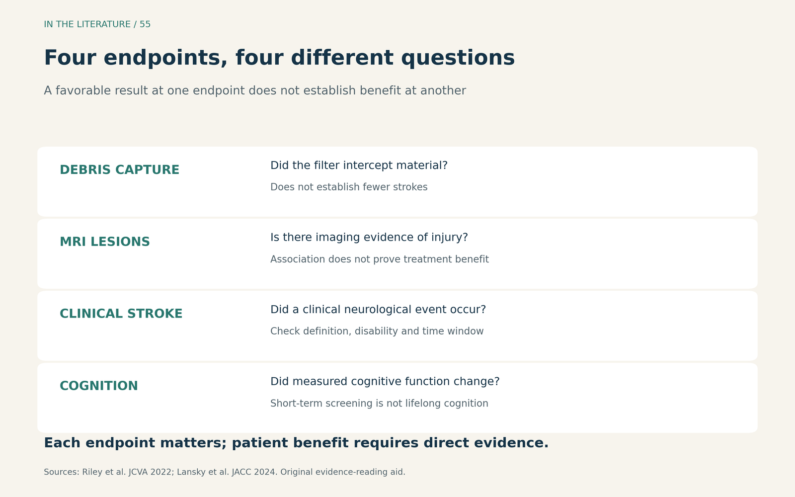
Figure 1. Different observations answer different questions. The diagram is an original evidence-reading aid, not a validated sequence in which success at one level guarantees success at the next. MRI injury is clinically relevant; its relationship with treatment benefit requires direct testing. [1, 2] View figure

Routine Sentinel use has not been shown to reduce early clinical stroke
PROTECTED TAVR randomized 3,000 patients undergoing transfemoral TAVR. Clinical stroke within 72 hours or discharge, whichever came first, occurred in 34/1,501 patients with Sentinel versus 43/1,499 controls: 2.3% versus 2.9%. The absolute difference was −0.6 percentage points (95% CI, −1.7 to 0.5). The trial did not demonstrate superiority; the interval also did not exclude a potentially useful benefit. [3]
BHF PROTECT-TAVI then randomized 7,635 patients. In its primary endpoint population, the corresponding early clinical stroke rates were 81/3,795 versus 82/3,799: 2.1% versus 2.2%. The reported difference was −0.02 percentage points (95% CI, −0.7 to 0.6). This larger pragmatic trial also failed to establish a benefit from routine protection. Neither trial was an equivalence trial. [4]
Deployment matters, but it does not resolve the efficacy question by itself. In BHF PROTECT-TAVI, both filters were correctly positioned throughout the procedure in 3,058/3,768 participants assigned to protection with available deployment data, 81.2%. This helps describe the routine strategy tested. It does not establish that perfect deployment would produce a clinical benefit. [4]
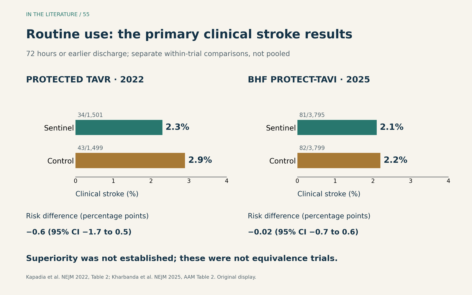
Figure 2. Original display of the two trials' primary clinical stroke results. Each comparison is within its own randomized trial; this is not a pooled estimate. Both endpoints used 72 hours or earlier discharge. Risk differences and confidence intervals are those reported by the studies, not subtraction of the rounded percentages. [3, 4] View figure

The disabling-stroke signal needs its proper weight
In PROTECTED TAVR, disabling stroke occurred in 8/1,501 patients with protection versus 20/1,499 controls: 0.5% versus 1.3%. This was an important secondary signal, but the trial's statistical plan did not correct secondary comparisons for multiplicity. It should not be presented as a definitive treatment effect. Disability was assessed after an early qualifying stroke, at approximately 30 days. [3]
BHF PROTECT-TAVI reported disabling stroke in 47/3,795 versus 53/3,799 patients: 1.2% versus 1.4%, with a difference of −0.2 percentage points (95% CI, −0.7 to 0.4). Here, disability was assessed at 6–8 weeks after TAVI among patients with an early stroke. These are severity assessments of early events, not counts of every new stroke throughout those later follow-up windows. The larger trial did not confirm a significant disabling-stroke benefit. [4]
Cognitive protection is a separate claim
Kennedy and colleagues examined cognition in 3,535 participants from BHF PROTECT-TAVI with baseline and 6–8-week assessments. This is a secondary analysis of the same trial, not another independent randomized population. The adjusted between-group difference in change on the telephone Montreal Cognitive Assessment, or t-MoCA, was −0.07 points (95% CI, −0.22 to 0.09), without a demonstrated benefit from protection. [5]
A decline of at least three points occurred in 154/1,763 participants assigned to protection versus 142/1,772 controls: 8.7% versus 8.0%. The absolute difference was 0.72 percentage points (95% CI, −1.10 to 2.55). Short follow-up, incomplete assessments and use of one screening tool prevent this result from establishing long-term cognitive equivalence or excluding effects in domains the test does not measure. [5]
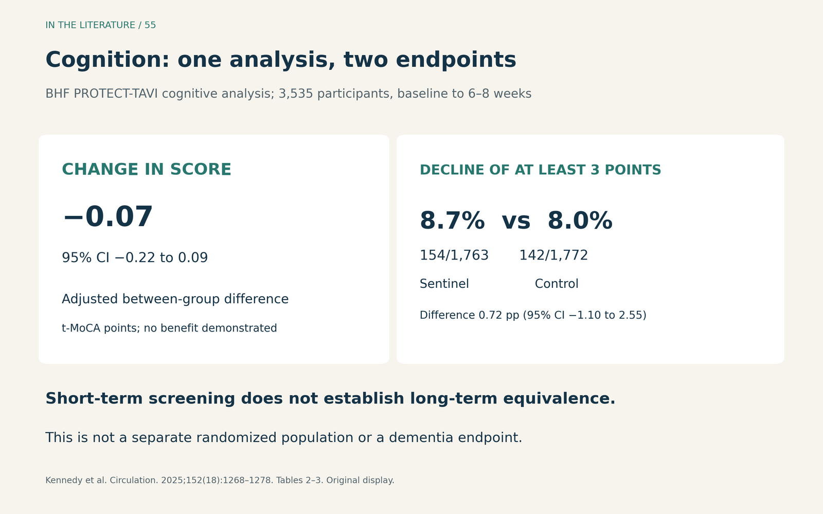
Figure 3. Two different cognitive endpoints from the same analysis. The adjusted mean difference concerns score change; the percentages concern a decline of at least three points. Neither is a dementia diagnosis or a long-term outcome. [5] View figure

High risk does not automatically identify treatment benefit
A recent study by Nakai and colleagues illustrates why selective use remains difficult to interpret. In a retrospective high-risk cohort, patients received no protection when device placement was considered anatomically unsuitable. After propensity matching, in-hospital stroke or TIA occurred in 1/245 protected patients versus 7/245 controls. Stroke alone occurred in 1/245 versus 4/245, without a significant difference. [6]
The combined endpoint must not be relabeled as stroke. More fundamentally, anatomical feasibility influenced who received treatment, and matching cannot eliminate all resulting selection differences. The study generates a hypothesis about selective protection; it does not validate a high-risk rule that guarantees benefit. [6]
For the clinical discussion, specify the anticipated embolic mechanism, whether the particular device can be deployed safely, and the uncertainty about the outcome the patient values. This is an individualized interpretation of the evidence, not a formal recommendation for a particular subgroup. If protection is chosen, its presence should not be understood as eliminating stroke risk. [1, 3, 4]
Three takeaways
- Use patient outcomes to judge routine use. The large Sentinel trials did not establish an early clinical stroke benefit; device deployment or captured debris cannot substitute for that endpoint.
- Keep the outcome and time window explicit. Disabling stroke, MRI injury and short-term cognitive testing are related but distinct; an encouraging secondary signal is not definitive proof.
- Describe selective use honestly. High embolic risk may motivate discussion, but current observational data do not identify a reliably benefiting subgroup or justify a promise of protection.
References
- Riley KJ, Kao LW, Low YH, et al. Neurologic Dysfunction and Neuroprotection in Transcatheter Aortic Valve Implantation. J Cardiothorac Vasc Anesth. 2022;36(8 Pt B):3224–3236. doi:10.1053/j.jvca.2021.11.016.
- Lansky AJ, Grubman D, Dwyer MG 3rd, et al. Clinical Significance of Diffusion-Weighted Brain MRI Lesions After TAVR: Results of a Patient-Level Pooled Analysis. J Am Coll Cardiol. 2024;84(8):712–722. doi:10.1016/j.jacc.2024.05.055.
- Kapadia SR, Makkar R, Leon M, et al. Cerebral Embolic Protection during Transcatheter Aortic-Valve Replacement. N Engl J Med. 2022;387(14):1253–1263. doi:10.1056/NEJMoa2204961.
- Kharbanda RK, Kennedy J, Jamal Z, et al. Routine Cerebral Embolic Protection during Transcatheter Aortic-Valve Implantation. N Engl J Med. 2025;392(24):2403–2412. doi:10.1056/NEJMoa2415120.
- Kennedy J, Blackman DJ, Dodd M, et al. Impact of Cerebral Embolic Protection on Cognitive Function After Transcatheter Aortic Valve Implantation: Data From the BHF PROTECT-TAVI Randomized Trial. Circulation. 2025;152(18):1268–1278. doi:10.1161/CIRCULATIONAHA.125.076761.
- Nakai C, Liu J, Azhagiri N, et al. Outcome of cerebral embolic protection during transcatheter aortic valve replacement in high-risk patient for stroke. JTCVS Struct Endovasc. 2026;9:100094. doi:10.1016/j.xjse.2025.100094.