ITL-fde792a47873
Juan Li, MD · Updated November 18, 2026
Key points
- One pause, two estimates. The expiratory occlusion swing (ΔPocc) converts to muscle pressure and dynamic lung stress, and flagged high effort accurately.[1]
- Good for flags, not for numbers. Neither ΔPocc nor P0.1 predicts exact values, and cutoffs rest on consensus.[2]
- Effort is not the same as drive. High drive does not always mean high effort, and oesophageal pressure remains the reference.[3]
- Technique matters. Average several breaths, beware intrinsic PEEP, and use P0.1 only as a screen for the extremes.[4], [5], [6]
A patient on pressure support breathes calmly at 22 breaths/min with a tidal volume of 7 mL/kg, yet the team suspects hidden strong effort. Can a simple ventilator pause tell?
Where the method comes from
In the derivation study (Bertoni et al., Crit Care 2019), the expiratory occlusion swing (ΔPocc) accurately detected muscle pressure above 10 cm H2O (AUROC 0.92). The authors used conversion factors of −3/4 for muscle pressure and 2/3 for dynamic transpulmonary pressure, so that muscle pressure is about −3/4 × ΔPocc and dynamic lung stress about ΔPaw − 2/3 × ΔPocc. Muscle pressure exceeded 10 cm H2O on 84% of patient-days, but within-subject limits of agreement were relatively wide.[1]
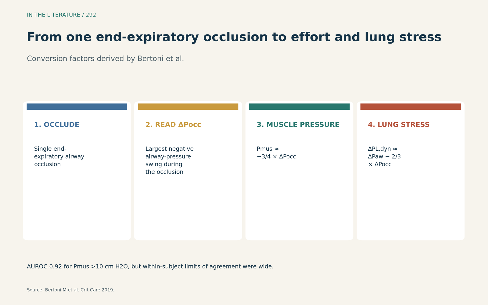
Figure 1. Occlude at end expiration, read ΔPocc, and convert to muscle pressure and dynamic lung stress. Data from Bertoni et al.[1] View larger.

How well it works
Against a double-balloon reference (de Vries et al., Anesthesiology 2023), neither P0.1 nor Pocc should be used to predict exact values of effort or lung stress; Pocc is more accurate than P0.1 in detecting high diaphragm effort, and current cutoffs are based only on consensus.[2]
ΔPocc is the maximum airway pressure deflection during a whole-breath occlusion, while the gold standard for global effort is the oesophageal pressure–time product; elevated respiratory drive does not always result in elevated effort (Goligher et al., Intensive Care Med 2020).[3]
How to do it, and its limits
P0.1 is the airway pressure drop during the first 100 ms of an occluded breath and is more useful as a screening tool for the extremes of effort. A single random airway occlusion does not significantly alter the breathing pattern, but ΔPocc may underestimate effort when intrinsic PEEP is not equilibrated at occlusion. Clinicians should average at least 3–5 breaths and repeat measurements, and if precise quantification is needed, oesophageal manometry remains the gold standard (Tonelli et al., Crit Care 2025).[4]
Absolute ΔPocc values of 6–13 cm H2O have been proposed as a safe range, but there are no definite safe cutoffs for ΔPocc (Chiumello et al., Ann Intensive Care 2026).[5] Ventilator-measured P0.1 correlated poorly with work of breathing, and the authors do not recommend ventilator P0.1 as a reliable measure of effort (Smits et al., Crit Care Med 2025).[6]
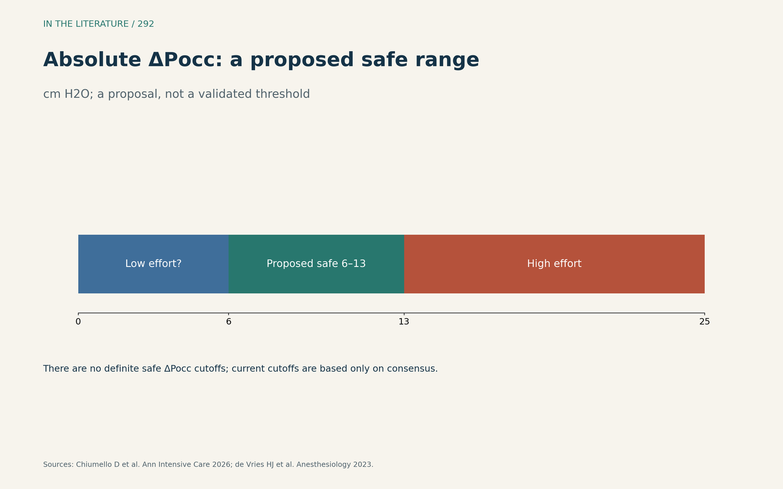
Figure 2. A 6–13 cm H2O range has been proposed, but no cutoff is validated. Data from Chiumello et al. and de Vries et al.[2], [5] View larger.

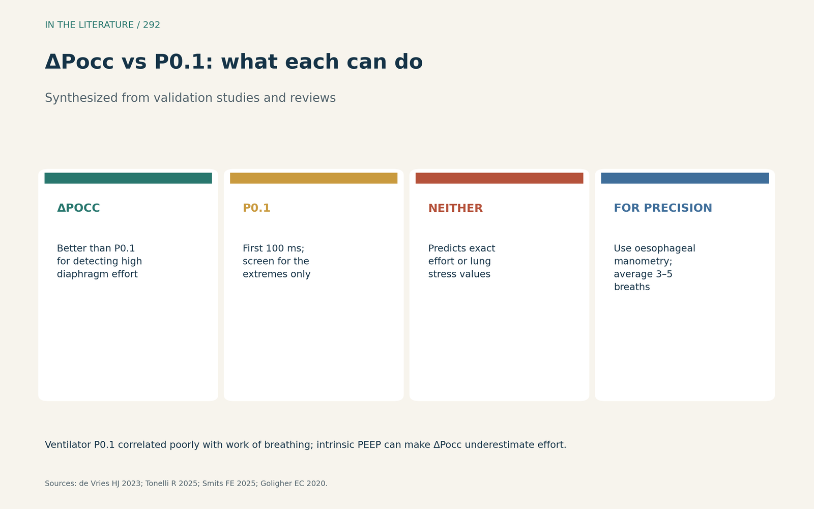
Figure 3. ΔPocc is the better flag for high effort; P0.1 is a rough screen; neither gives exact values. Synthesized from de Vries et al., Tonelli et al., Smits et al. and Goligher et al.[2], [3], [4], [6] View larger.

Three takeaways
- Use an end-expiratory occlusion to flag excessive effort and lung stress during assisted ventilation.[1], [2]
- Treat converted values as estimates; average 3–5 breaths and watch for intrinsic PEEP.[4], [5]
- Do not rely on ventilator P0.1 alone, and use oesophageal manometry when precise values matter.[3], [6]
References
- Bertoni M, Telias I, Urner M, et al. A novel non-invasive method to detect excessively high respiratory effort and dynamic transpulmonary driving pressure during mechanical ventilation. Crit Care. 2019;23:346. doi:10.1186/s13054-019-2617-0
- de Vries HJ, Tuinman PR, Jonkman AH, et al. Performance of noninvasive airway occlusion maneuvers to assess lung stress and diaphragm effort in mechanically ventilated critically ill patients. Anesthesiology. 2023;138(3):274–288. doi:10.1097/ALN.0000000000004467
- Goligher EC, Jonkman AH, Dianti J, et al. Clinical strategies for implementing lung and diaphragm-protective ventilation: avoiding insufficient and excessive effort. Intensive Care Med. 2020;46(12):2314–2326. doi:10.1007/s00134-020-06288-9
- Tonelli R, Protti A, Spinelli E, et al. Assessing inspiratory drive and effort in critically ill patients at the bedside. Crit Care. 2025;29:339. doi:10.1186/s13054-025-05526-0
- Chiumello D, Muscas A, Mancusi S, Fioccola A. The invasive and non-invasive monitoring of inspiratory effort and drive during assisted mechanical ventilation. Ann Intensive Care. 2026;16:100003. doi:10.1016/j.aicoj.2025.100003
- Smits FE, Rietveld PJ, Snoep JWM, et al. P0.1 is an unreliable measure of effort in support mechanical ventilation in comparison with esophageal-derived measures of effort: a comparison study. Crit Care Med. 2025;53(8):e1650–e1658. doi:10.1097/CCM.0000000000006745