
ITL-e9572f47a7d0
Watch the video

Key points
- Suspected device thrombosis requires prompt assessment of perfusion and hospital evaluation with the LVAD team; do not wait for circulatory collapse.[1]
- Displayed pump flow is an estimate. Increased power can coexist with reduced true flow, and a reassuring display does not exclude dysfunction.[1], [2]
- Modest LDH elevation does not rule out outflow graft obstruction: in one small mixed-device series, most affected patients had LDH below twice the laboratory upper limit.[3]
- HeartMate 3 has changed the thrombosis landscape, but external graft compression and other mechanical failures remain important alternatives.[1], [4], [5]
- Echocardiography evaluates unloading and competing causes; CT angiography helps localize graft disease, but a negative CT does not reliably exclude an intrapump clot.[1], [2]
A patient with a durable LVAD develops dark urine, worsening breathlessness or a low-flow alarm. “Pump thrombosis” belongs on the differential, but these findings do not identify where the problem lies. A clot within the pump, an obstructed outflow graft and inadequate ventricular filling can produce overlapping signs. The useful question is whether the evidence points to blood trauma within the device, a mechanical flow restriction, or another cause of inadequate support.[1], [2]
Start with the patient, then compare the device with its baseline
Assess perfusion while contacting the implanting or responsible LVAD team. Altered consciousness, hypotension, worsening organ function or new neurologic findings make the evaluation urgent. ISHLT recommends admission for expedited investigation when device thrombosis is suspected, with serial reassessment rather than a single reassuring set of observations.[1]
Record the pump model, programmed speed, power, displayed flow and alarm history, and compare them with the patient's usual values. Manufacturer log analysis can show changes that a snapshot misses. Rotor-related thrombosis may increase power consumption while effective flow falls; because the controller estimates flow, the displayed value can become misleadingly high. Obstruction outside the pump may instead reduce flow without the same power rise. These are clues, not a device-independent diagnostic rule.[1], [2]
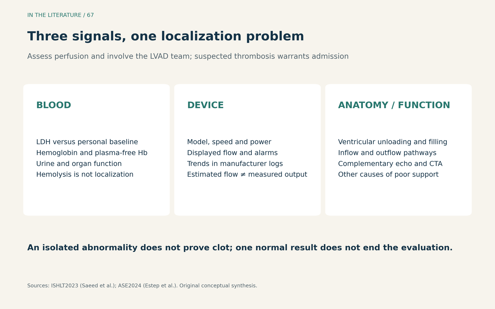
Figure 1. Bring the evidence together. Trends and anatomy determine what an abnormal number means. Original synthesis of ISHLT and ASE recommendations.[1], [2] View larger.

Interpret hemolysis as a pattern, not a universal cutoff
The initial laboratory assessment includes LDH, hemoglobin, plasma-free hemoglobin and urinalysis, alongside tests for end-organ dysfunction. Review the anticoagulation history and current INR with the LVAD team. A rising hemolysis signal strengthens concern, but its interpretation depends on the device, the patient's baseline and the accompanying clinical findings. Older-pump thresholds should not be imported into HeartMate 3 practice as universal diagnostic cutoffs.[1]
The opposite mistake is to regard a modest LDH as reassurance against all device obstruction. Peters and colleagues identified 11 outflow graft obstructions in a retrospective mixed-device series: 10 patients presented with low-flow alarms, and eight had LDH below twice their laboratory upper limit. This supports looking beyond intrapump thrombosis when the flow is abnormal but hemolysis is limited. It does not establish the sensitivity or negative predictive value of LDH for pump thrombosis.[3]
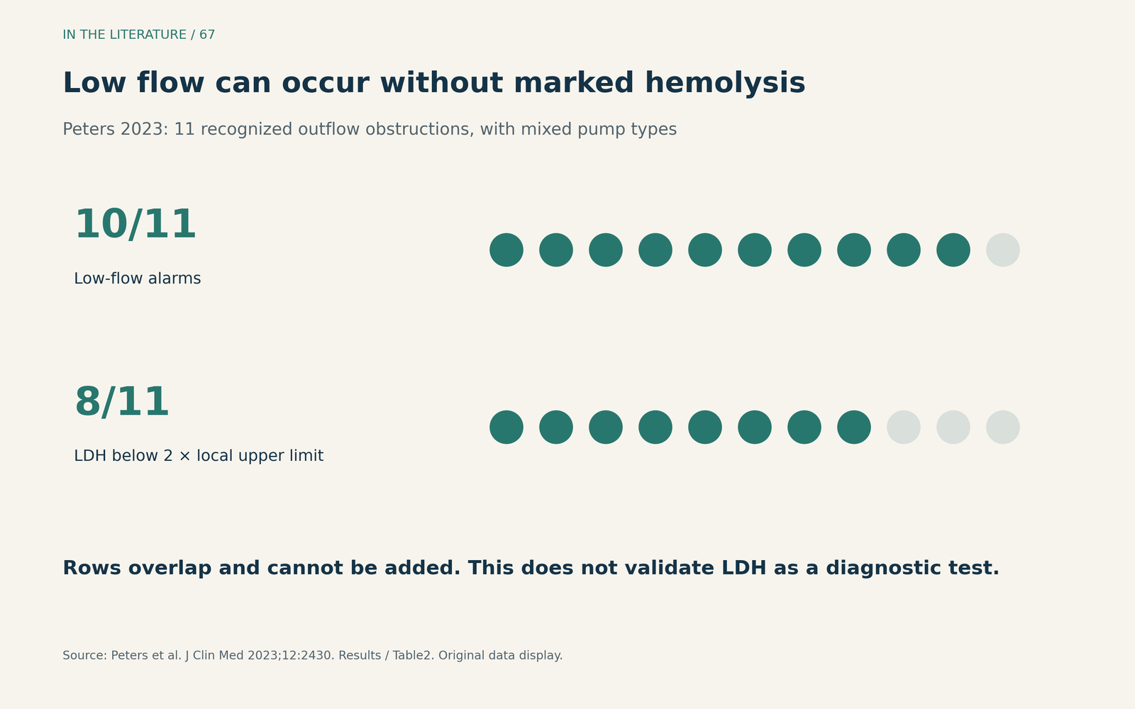
Figure 2. Outflow obstruction can have little biochemical evidence of hemolysis. The rows describe overlapping findings in the same 11 patients, not separate groups. Original display of Peters et al., Results and Table 2.[3] View larger.

Use imaging to locate the problem
Echocardiography asks whether the LV is being unloaded and whether another explanation is present: RV dysfunction, inadequate filling, tamponade, inflow cannula malposition or aortic regurgitation. Comparison with earlier studies is often more informative than an isolated image. Specialist-guided speed adjustment or a ramp study may help assess the response to support; an abnormal response is not, by itself, proof of an intrapump clot. HeartMate 3's artificial-pulse cycle can also vary cannula velocities and should not be mistaken for intermittent obstruction.[1], [2]
When outflow obstruction remains a concern, CT angiography helps evaluate the graft for kinking, twisting, external compression or intraluminal disease. In the Peters series, CTA localized the obstruction in eight of the nine patients who underwent scanning. That selected case series is not a diagnostic-accuracy study, and the fraction should not be presented as a universal sensitivity.[3]
CT is also much less reliable for excluding thrombus inside the pump. A normal-looking graft does not settle a persistent combination of hemolysis, altered power and inadequate unloading. Conversely, a narrowed graft does not automatically mean that thrombolysis will address the cause. Hemodynamic assessment and, in selected cases, catheter-based evaluation may be needed when the clinical, echocardiographic and CT findings do not agree.[1], [2]
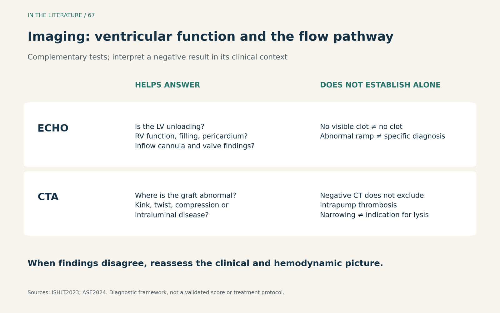
Figure 3. Choose imaging for the question it can answer. This is a diagnostic framework, not a validated decision score or a treatment protocol.[1], [2] View larger.

HeartMate 3 lowers one concern without removing the others
Jahangiri and colleagues studied 125 HeartMate 3 recipients with assessable CTA. Using a study definition of at least 25% graft-diameter reduction, they found external outflow stenosis in 23 patients. Five of the 12 moderate-to-severe cases were detected during routine follow-up scanning. The cohort shows that external compression can exist without a typical alarm presentation; it does not establish a universal CT surveillance interval or an automatic intervention threshold.[4]
A separate 2026 single-center report of 215 primary HeartMate 3 recipients recorded no intrinsic pump thrombosis, while seven patients experienced other device failures, including outflow obstruction or mechanical problems. Individual support duration varied; the median was 603 days. Zero observed intrapump events in that cohort does not make thrombosis impossible in an individual patient. It does make careful separation of “pump thrombosis” from broader “device failure” essential.[5]
The diagnosis changes the treatment discussion
Significant hemolysis or convincing device dysfunction belongs in an inpatient specialist pathway. The 2024 ISHLT consensus describes escalation of parenteral anticoagulation with unfractionated heparin or a direct thrombin inhibitor in stable patients with significant hemolysis, with close monitoring for response and bleeding. Failure to respond or hemodynamic instability prompts consideration of device exchange, accounting for operative risk and transplant options.[6]
Thrombolysis is not a default response to a low-flow alarm. Specialist teams may consider it in selected patients after anticoagulation failure when neither device exchange nor transplantation is suitable. Its reported experience is largely from heterogeneous, often older-device case series, and bleeding or embolic complications can be substantial. A kinked or externally compressed graft requires a different discussion from an intrapump clot. After a thrombotic event, the team should also investigate medication adherence, infection and device position rather than assuming that a higher long-term anticoagulation target is the answer.[1], [6]
Three takeaways
- Escalate on clinical suspicion and perfusion, not on the alarm alone. Suspected thrombosis warrants admission and coordinated assessment.[1]
- Localize before labeling. Combine hemolysis trends, device logs and ventricular unloading; limited hemolysis still leaves outflow obstruction on the table.[1], [2], [3]
- Match the next step to the mechanism. CTA can clarify graft disease, but negative CT does not exclude intrapump clot, and unselected thrombolysis is not a substitute for diagnosis.[1], [2], [6]
References
- Saeed D, Feldman D, Banayosy AE, et al. The 2023 International Society for Heart and Lung Transplantation Guidelines for Mechanical Circulatory Support: A 10-Year Update. J Heart Lung Transplant. 2023;42(7):e1-e222. doi:10.1016/j.healun.2022.12.004.
- Estep JD, Nicoara A, Cavalcante J, et al. Recommendations for Multimodality Imaging of Patients With Left Ventricular Assist Devices and Temporary Mechanical Support: Updated Recommendations from the American Society of Echocardiography. J Am Soc Echocardiogr. 2024;37(9):820-871. doi:10.1016/j.echo.2024.06.005.
- Peters CJ, Zhang RS, Vidula MK, et al. Durable Left Ventricular Assist Device Outflow Graft Obstructions: Clinical Characteristics and Outcomes. J Clin Med. 2023;12(6):2430. doi:10.3390/jcm12062430.
- Jahangiri P, Sjatskig J, Sener YZ, et al. External Outflow Graft Stenosis in Patients With Heartmate 3 Left Ventricular Assist Devices. ASAIO J. 2026;72(4):284-290. doi:10.1097/MAT.0000000000002452.
- Karaca S, Kahraman U, Kapkin AY, et al. A Decade of Durable Mechanical Circulatory Support with HeartMate 3 Across Age Groups in a Single-Center Experience. Life (Basel). 2026;16(10):1640. doi:10.3390/life16101640.
- Hollis IB, Jennings DL, Krim S, et al. An ISHLT consensus statement on strategies to prevent and manage hemocompatibility related adverse events in patients with a durable, continuous-flow ventricular assist device. J Heart Lung Transplant. 2024;43(8):1199-1234. doi:10.1016/j.healun.2024.04.065.