ITL-a72313212933
Juan Li, MD · Updated November 17, 2026
Watch the video
Key points
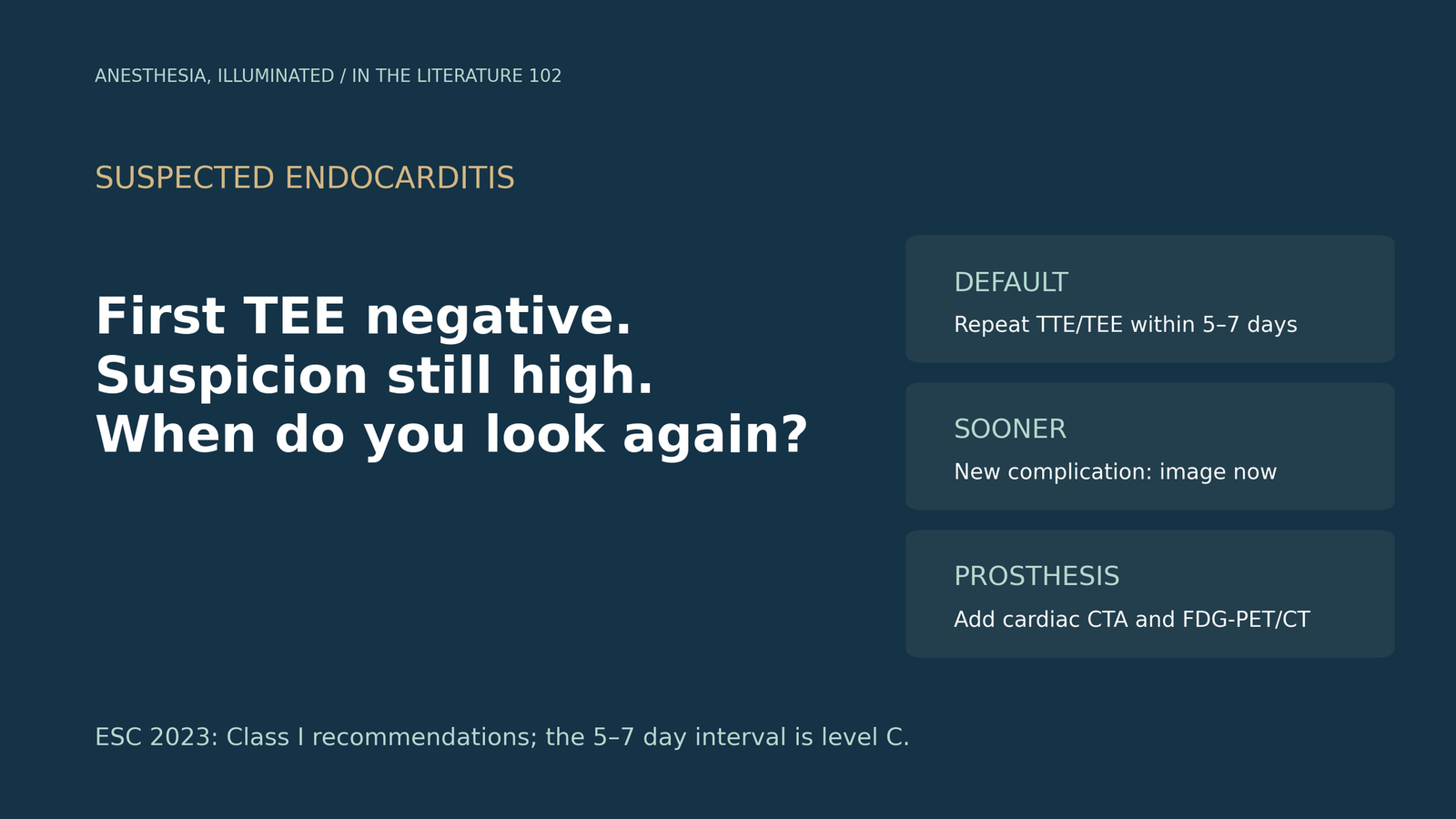
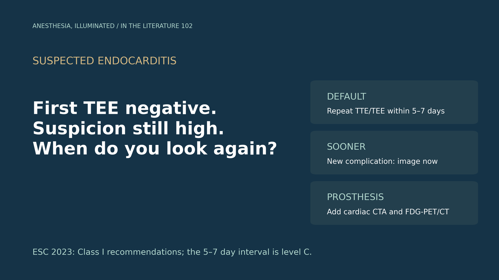
- A negative transesophageal echocardiogram (TEE) does not exclude infective endocarditis (IE) when clinical suspicion remains high. The 2023 European Society of Cardiology (ESC) guideline recommends repeating TTE and/or TEE within 5–7 days (Class I, level of evidence C).[1]
- That interval is expert consensus, not trial-derived. Older European guidance used 7–10 days, and the yield of a planned second TEE after a modern first study appears low.[1], [2], [3]
- Repeat sooner when the clinical picture changes: new murmur, embolism, persistent fever or bacteremia, heart failure, suspected abscess or atrioventricular block.[1]
- Prosthetic valves and cardiac devices are where echocardiography most often fails. In possible prosthetic valve IE, the ESC recommends cardiac CT angiography and [18F]FDG-PET/CT rather than relying on echocardiography alone.[1]
- Repeat TEE still changes management in selected patients, whereas a second transthoracic study rarely changes the diagnosis once multimodality imaging is available.[4], [5]
Consider a common situation: blood cultures grow Staphylococcus aureus in a patient with a prosthetic aortic valve, and the first TEE shows no vegetation. The team asks whether endocarditis can now be set aside, or whether the patient should return for another study, and when. For anesthesiologists, the same question arises from the other direction: the patient may come to the operating room for a different procedure, or for valve surgery, with a negative or equivocal preoperative TEE. Which patients need repeat imaging after a negative TEE, how soon, and with which modality?
Why a negative TEE can be wrong
TEE is the most sensitive echocardiographic test, but it is not perfect. The 2010 European Association of Echocardiography recommendations estimated that echocardiography is negative in about 15% of patients with IE. The usual reasons are very small vegetations, early disease before a vegetation forms, preexisting valve lesions and prosthetic material. They cited one series in which 3 of 65 patients with an initially negative TEE later developed visible vegetations.[2]
Abscesses pose a similar problem. Very early in the disease, immediately after valve surgery, or when a prosthesis or annular calcification shadows the field, a perivalvular abscess may not yet be visible. The 2023 ESC guideline states that echocardiography can be normal or inconclusive in up to 30% of patients with prosthetic material despite the presence of IE.[1], [2]
The 5–7 day interval: what it rests on
The ESC 2023 guideline recommends repeating TTE and/or TOE within 5–7 days after an initially negative or inconclusive examination when clinical suspicion remains high. It also advises repeat imaging in patients with diagnosed IE at high risk of complications, such as aggressive microorganisms or prosthetic valves. The recommendation is Class I but level of evidence C, meaning expert consensus. The 2010 European echocardiography recommendations had suggested 7–10 days, and earlier when the presentation justified it, for example in suspected early postoperative staphylococcal prosthetic valve endocarditis.[1], [2]
The interval reflects biology rather than a measured optimum: small vegetations may grow and abscesses may cavitate. It is a default for a stable patient, not a waiting period to be observed when the patient deteriorates. The ESC recommends repeating TTE and/or TOE as soon as a new complication is suspected, including a new murmur, embolism, persistent fever and bacteremia, heart failure, abscess or atrioventricular block (Class I, level B).[1]
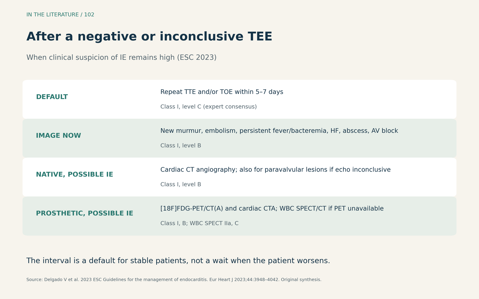
Figure 1. A practical pathway after a negative first TEE. Based on the ESC 2023 recommendations and diagnostic algorithms; classes and levels are those stated in the guideline. Original synthesis, not a validated algorithm.[1] View larger.

How often does a second TEE change the answer?
Direct evidence is limited and retrospective. Montané and colleagues reviewed 242 patients with confirmed IE who underwent at least two TEEs within 6 months at one tertiary center, comparing 2011 with 2019. The first TEE detected IE in 85.7% in 2011 and 95.3% in 2019, driven by better detection of prosthetic valve endocarditis (70.8% versus 93.7%). A second TEE newly showed IE after a negative first study in 8 (11.4%) patients in 2011 and 6 (3.5%) in 2019. Most second TEEs in 2019 were intraoperative, and the study included only patients with confirmed IE, so it cannot estimate how often a repeat study finds IE in all patients with a negative first TEE.[3]
In a population-based Mayo Clinic cohort, Shafiyi and colleagues examined non-routine repeat TEEs (excluding intraoperative and end-of-therapy studies). Of 46 such repeat TEEs in 39 patients, 8 (17.4%) were useful for the diagnosis of IE, 8 (17.4%) affected antimicrobial therapy and 11 (23.9%) influenced surgical decisions. The median interval from the first TEE to the first repeat was 7.5 days. Patients who underwent repeat TEE more often had a prosthetic valve or annuloplasty ring. The study was small and unadjusted.[4]
Together, these studies suggest that modern TEE misses few cases on the first examination. However, repeat TEE still matters in selected patients, particularly those with prosthetic material, persistent bacteremia or new complications.[3], [4]
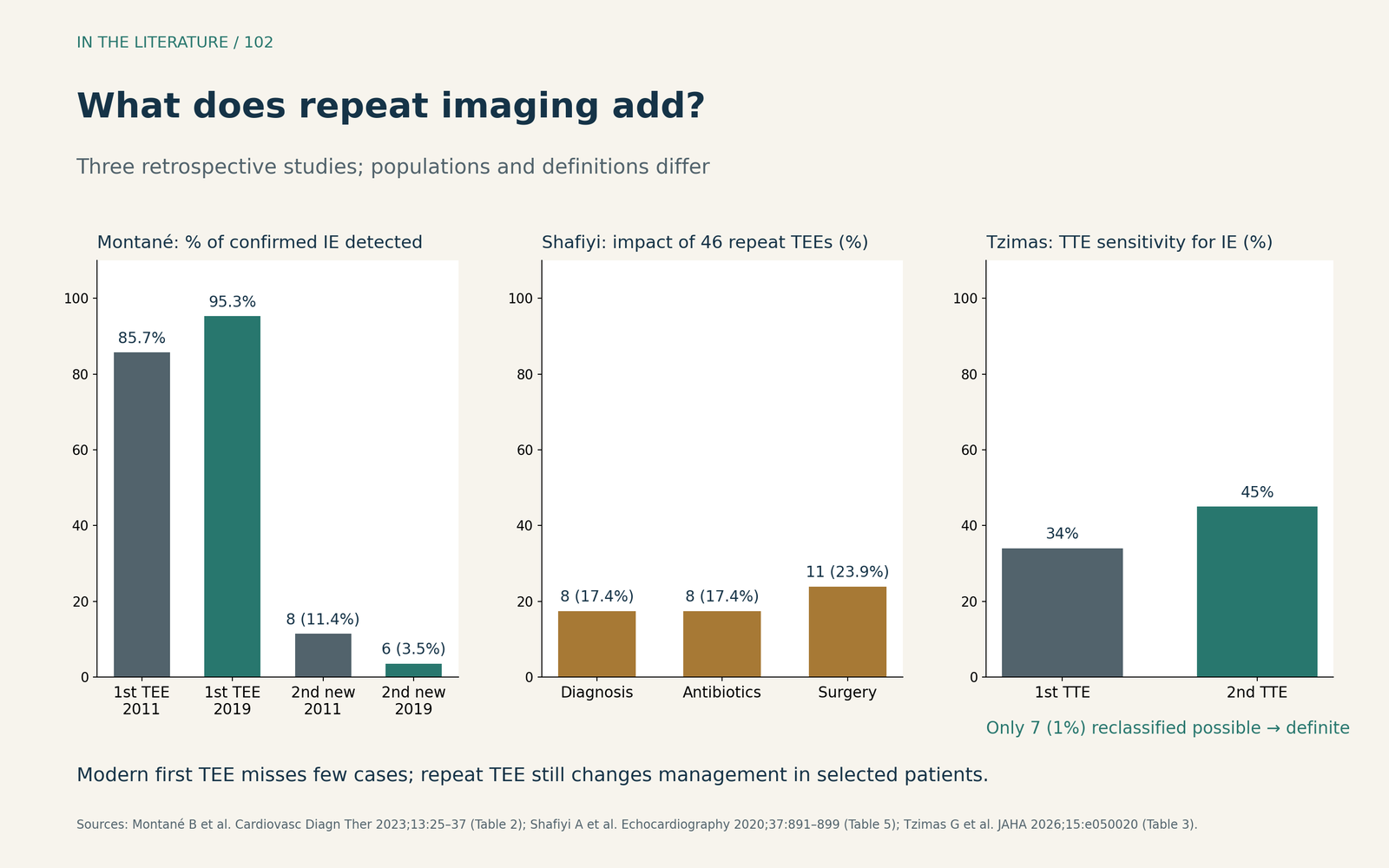
Figure 2. Repeat imaging yield, redrawn from the original tables. Montané: patients with confirmed IE and at least two TEEs (2011, n = 70; 2019, n = 172). Shafiyi: non-routine repeat TEEs (n = 46). Tzimas: possible-to-definite reclassification after a second TTE (n = 503 episodes). Populations and definitions differ; the bars are not directly comparable.[3]–[5] View larger.

Repeat TEE, CT or PET: choosing the next test
The ESC recommendation allows either TTE or TEE. Recent data favor the more sensitive test. Tzimas and colleagues analyzed 503 episodes of suspected IE in which a second TTE was performed at a median of 8 days after the first. Within a pathway that already used TEE, CT and PET, the second TTE reclassified only 7 (1%) episodes from possible to definite IE. Its sensitivity for IE rose from 34% to 45%; in patients with prosthetic valves or TAVI it rose from 17% to 33%. The authors concluded that TEE, cardiac CT or PET should be preferred when suspicion persists. The study was retrospective and single-center.[5]
The ESC guideline now gives CT and nuclear imaging a defined role:
- Cardiac CT angiography is recommended in possible native valve IE and to detect paravalvular or periprosthetic complications when echocardiography is inconclusive (Class I, level B). The guideline notes that CT is more accurate than TOE for perivalvular complications such as abscesses and pseudoaneurysms, whereas echocardiography remains better for small vegetations and leaflet perforation.[1]
- [18F]FDG-PET/CT(A) and cardiac CTA are recommended in possible prosthetic valve IE (Class I, level B). In native valve IE, PET sensitivity is low (about 31%), so a negative scan does not exclude infection.[1]
Sifaoui and colleagues compared TEE and cardiac CT with surgical findings in 68 patients with definite left-sided IE. TEE performed better for valvular lesions (area under the curve 0.881 versus 0.720) and vegetations (0.863 versus 0.693). The two were similar for paravalvular lesions (0.830 versus 0.816); CT detected pseudoaneurysms with higher sensitivity (100% versus 66.7%). Because every patient had definite IE, the study does not show how well CT excludes the diagnosis.[6]
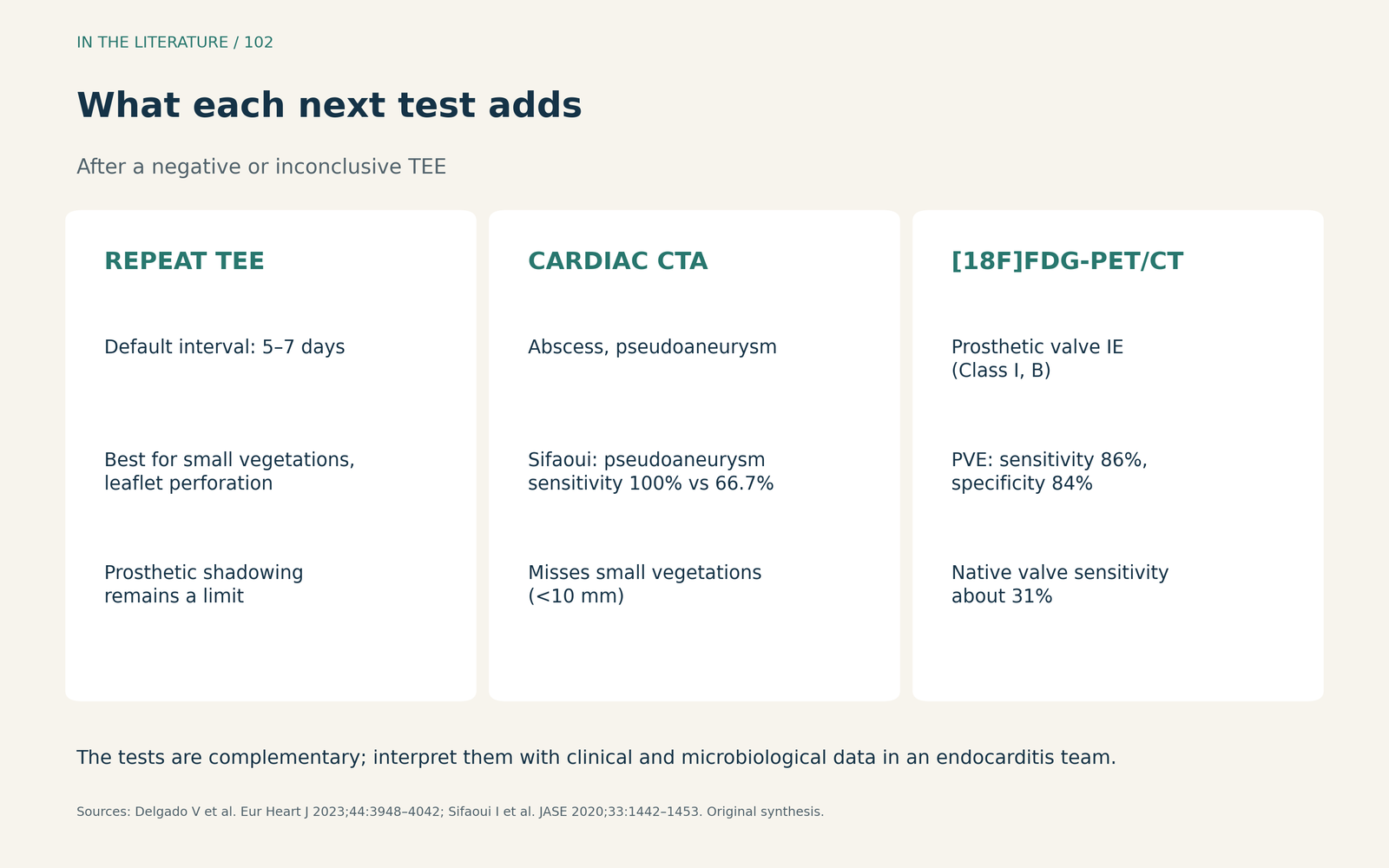
Figure 3. Complementary roles of repeat TEE, cardiac CT and PET/CT. Summarized from the ESC 2023 guideline and the surgical comparison by Sifaoui and colleagues.[1], [6] View larger.

In the operating room
The ESC guideline recommends intraoperative echocardiography in all patients with IE who require surgery (Class I, level C). It describes the intraoperative TOE as reassessing the extent of infection, the stability of known vegetations, previously uninvolved valves and biventricular function. A post-repair study establishes the immediate result and the baseline for follow-up.[1]
A negative or equivocal preoperative TEE therefore does not shorten the intraoperative examination. Perivalvular extension, a second valve or a new lesion may appear between studies. In the 2019 Montané cohort, most second TEEs were intraoperative.[1], [3]
Three takeaways
- A negative TEE does not close the question when suspicion remains high. Repeat imaging within 5–7 days is the ESC default (Class I, level C), and earlier imaging is indicated when a new complication appears.[1]
- Prosthetic material changes the pathway. Echocardiography can be normal in up to 30% of patients with prosthetic material; use cardiac CTA and [18F]FDG-PET/CT in possible prosthetic valve IE.[1]
- Choose the more informative repeat test. A second TTE rarely changes the diagnosis in a multimodality pathway; repeat TEE, CT or PET are more likely to answer the question.[3]–[6]
References
- Delgado V, Ajmone Marsan N, de Waha S, et al. 2023 ESC Guidelines for the management of endocarditis. Eur Heart J. 2023;44(39):3948–4042. doi:10.1093/eurheartj/ehad193
- Habib G, Badano L, Tribouilloy C, et al. Recommendations for the practice of echocardiography in infective endocarditis. Eur J Echocardiogr. 2010;11(2):202–219. doi:10.1093/ejechocard/jeq004
- Montané B, Chahine J, Fiore A, et al. Diagnostic performance of contemporary transesophageal echocardiography with modern imaging for infective endocarditis. Cardiovasc Diagn Ther. 2023;13(1):25–37. doi:10.21037/cdt-22-431
- Shafiyi A, Anavekar NS, Virk A, et al. Repeat transesophageal echocardiography in infective endocarditis: an analysis of contemporary utilization. Echocardiography. 2020;37(6):891–899. doi:10.1111/echo.14676
- Tzimas G, Monney P, Antiochos P, et al. Real-world diagnostic performance of repeat transthoracic echocardiography within a multimodality imaging approach in patients with suspected infective endocarditis. J Am Heart Assoc. 2026;15(16):e050020. doi:10.1161/JAHA.126.050020
- Sifaoui I, Oliver L, Tacher V, et al. Diagnostic performance of transesophageal echocardiography and cardiac computed tomography in infective endocarditis. J Am Soc Echocardiogr. 2020;33(12):1442–1453. doi:10.1016/j.echo.2020.07.017