ITL-2998bcbeb351
Juan Li, MD · Updated November 17, 2026
Watch the video
Key points
- Perivalvular extension occurs in 10% to 30% of native valve infective endocarditis (IE) and more often with prosthetic valves. It is most common in aortic valve IE, especially in the mitral-aortic intervalvular fibrosa.[1]
- Persistent fever, new atrioventricular block, chest pain, a new murmur, recurrent embolism or heart failure should prompt a deliberate search for it.[1]
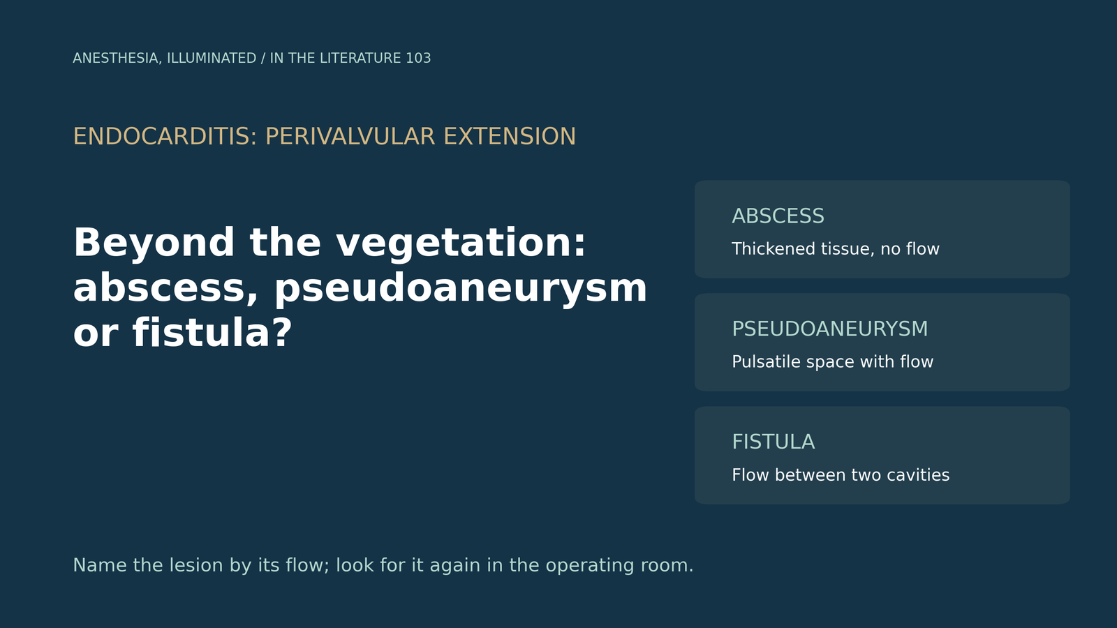
- The echo signatures differ: an abscess is thickened, non-homogeneous perivalvular tissue without flow; a pseudoaneurysm is a pulsatile echo-free space with color flow; a fistula is a color Doppler communication between two cavities.[2], [3]
- Transesophageal echocardiography (TEE) and cardiac CT are complementary. CT helps when TEE is inconclusive, particularly for prosthetic valves and pseudoaneurysms; TEE remains strong for fistulae and valvular lesions.[1], [3], [4]
- About one quarter of aortic root abscesses in one surgical series were found only at operation, so the intraoperative TEE should search for them actively.[5]
Consider a common situation: a patient with aortic valve endocarditis remains febrile despite appropriate antibiotics and develops a new conduction delay. The preoperative TEE showed a vegetation and a slightly thickened posterior aortic root, but no definite cavity. Is the infection still confined to the valve, or has it spread into the annulus and surrounding tissue? The answer changes the urgency of surgery, the operation itself and what the anesthesiologist should look for on the intraoperative TEE.[1], [2]
Where perivalvular extension starts, and who is at risk
The 2023 European Society of Cardiology (ESC) guideline describes perivalvular extension as a sign of locally uncontrolled infection. It occurs in 10% to 30% of native valve IE and is more frequent with prosthetic valve endocarditis. It is more common in aortic than mitral IE. In aortic valve IE, extension most often involves the mitral-aortic intervalvular fibrosa; in mitral IE, abscesses are usually posterior or lateral.[1]
The clinical clues are not specific, but they are important: persistent fever and infection, new atrioventricular block, chest pain, a new heart murmur, recurrent embolism and heart failure. The guideline recommends confirming the diagnosis with TEE, which is more sensitive and specific than transthoracic echocardiography. It also notes that mitral annular calcification can hide small regions of posterior mitral extension.[1]
Three lesions, three echocardiographic signatures
The European Association of Echocardiography recommendations define the lesions both anatomically and by echocardiography:[2]
- Abscess: a perivalvular cavity with necrosis and purulent material that does not communicate with the cardiovascular lumen. On echo it appears as a thickened, non-homogeneous perivalvular area, either echodense or echolucent.
- Pseudoaneurysm: a perivalvular cavity that does communicate with the lumen. On echo it is a pulsatile, echo-free perivalvular space with color Doppler flow.
- Fistula: a communication between two neighboring cavities through a perforation, shown by color Doppler flow between them.
The distinction is clinically meaningful. Color flow into the cavity separates a pseudoaneurysm from a closed abscess; flow between two chambers or between the aortic root and a chamber defines a fistula. Abscess formation in aortic IE is also dynamic. The recommendations describe it as beginning with aortic root wall thickening and advise repeat echocardiography when slight root thickening is seen. Pulsed, continuous-wave and color Doppler localize fistulae and quantify the resulting intracardiac shunt.[2]
On CT, Sordelli and colleagues describe an abscess as a low-attenuation necrotic center with a peripherally enhancing rim, whereas early abscess formation may appear only as soft-tissue thickening. A pseudoaneurysm is a contrast-filled perivalvular cavity connected to the root or a chamber. A fistula appears as a contrast-filled tract between two cavities, and their review considers TEE more effective than CT for depicting intracardiac fistulae.[3]
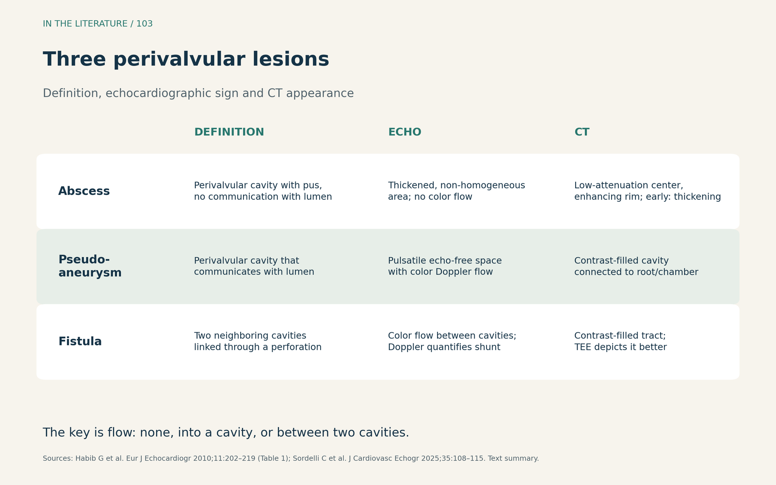
Figure 1. Recognizing the three perivalvular lesions. Definitions and echocardiographic signs from the European Association of Echocardiography recommendations; CT appearances from the review by Sordelli and colleagues. Text summary; no source images reproduced.[2], [3] View larger.

TEE and CT: complementary, not interchangeable
The ESC guideline recommends cardiac CT angiography to detect paravalvular or periprosthetic complications when echocardiography is inconclusive (Class I, level B). Cardiac CT is described as an accurate alternative for evaluating perivalvular extension, and PET/CT may be particularly helpful in prosthetic valve endocarditis.[1]
Sifaoui and colleagues compared TEE and ECG-gated cardiac CT with surgical inspection in 68 patients with definite left-sided IE. For paravalvular lesions overall, the two performed similarly (area under the curve 0.830 for TEE versus 0.816 for CT). For surgically confirmed abscess, sensitivity was 72.7% with TEE and 77.3% with CT, and both were less sensitive for mitral than aortic IE. CT detected pseudoaneurysms with higher sensitivity (100% versus 66.7%), whereas TEE was better for valvular lesions and vegetations. Both modalities performed less well in prosthetic valve IE. Because every patient had definite IE and underwent surgery, the study describes lesion detection, not how well a negative scan excludes extension.[4]
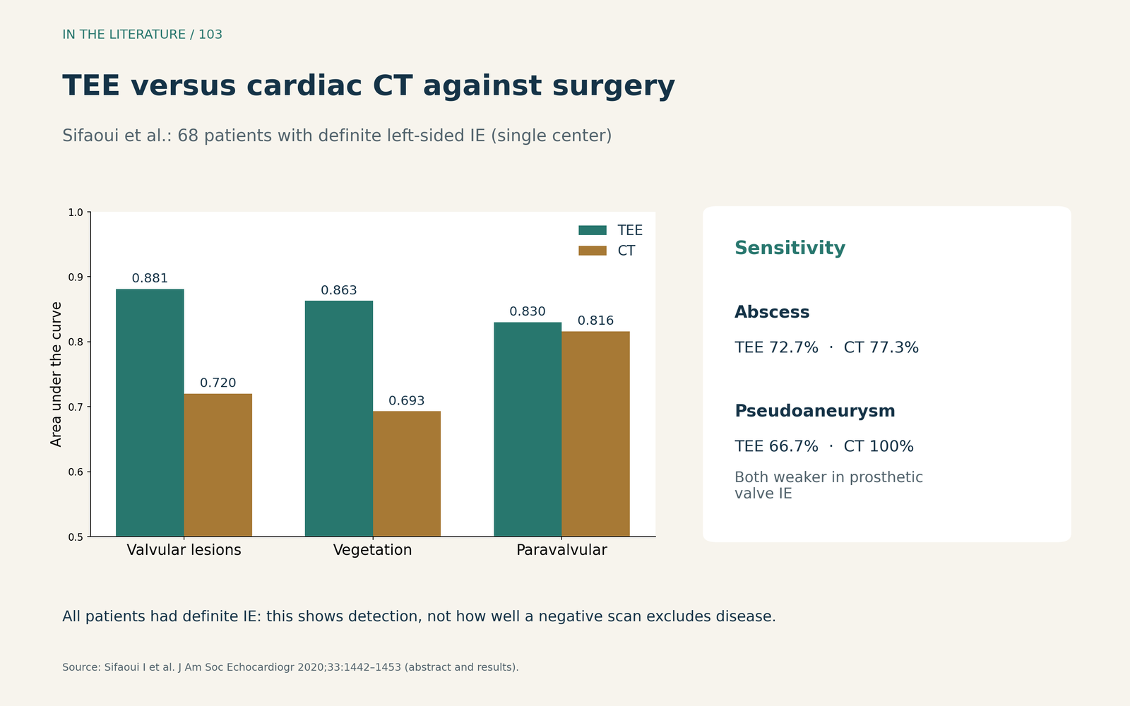
Figure 2. TEE versus cardiac CT against surgical inspection. Redrawn from the abstract and results of Sifaoui et al. (n = 68, single center, all definite IE undergoing surgery).[4] View larger.

Why the anesthesiologist should keep looking
Degife and colleagues reviewed 218 patients operated on for active aortic valve endocarditis at one center between 2011 and 2022; 64 had a perivalvular abscess. Patients with abscess more often had previous cardiac surgery, prosthetic valve endocarditis (46.9% versus 18.2%), aortocavitary fistula (25.0% versus 3.25%) and heart block (21.9% versus 7.79%). In 15 of 64 (23.4%), the abscess had not been detected by preoperative echocardiography or CT angiography and was found only at surgery.[5]
Abscess was associated with operative mortality (odds ratio 5.12), and the association remained after adjustment (adjusted odds ratio 3.51). These are retrospective, single-center associations rather than proof that earlier detection changes survival. They do show that a negative preoperative study does not exclude perivalvular extension.[5]
The ESC guideline recommends intraoperative echocardiography in all patients with IE who undergo surgery. It describes reassessment of the extent of infection as part of the intraoperative TEE.[1] In practice, this means examining the posterior aortic root, the mitral-aortic intervalvular fibrosa and the annulus with color Doppler, rather than looking only at the known vegetation. This is a clinical inference from the guidance and the surgical data, not a validated checklist.
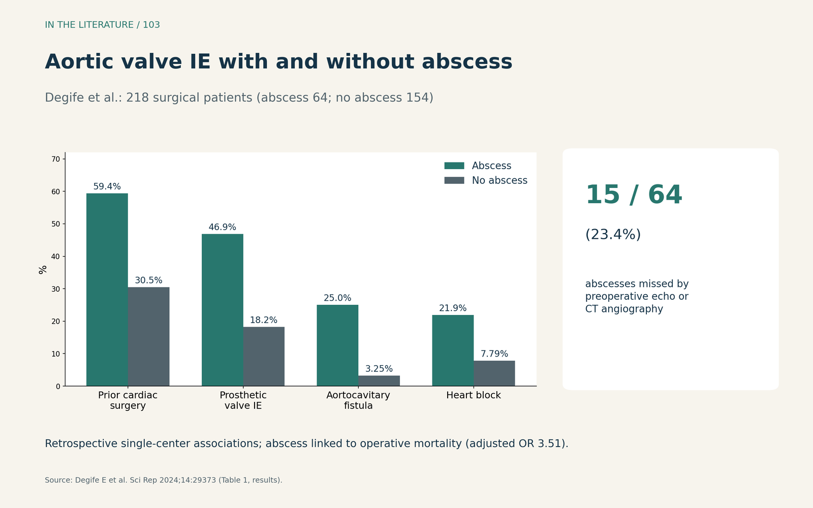
Figure 3. What accompanied aortic root abscess in a surgical series. Redrawn from Table 1 of Degife et al. (n = 218; abscess, n = 64). Retrospective single-center data.[5] View larger.

Three takeaways
- Suspect extension when infection is not behaving. Persistent fever, new atrioventricular block or a new murmur in aortic or prosthetic valve IE should prompt targeted TEE of the annulus and intervalvular fibrosa.[1]
- Name the lesion by its flow. No flow suggests abscess; flow into a pulsatile perivalvular space defines pseudoaneurysm; flow between two cavities defines fistula.[2], [3]
- A negative preoperative study does not exclude it. Use CT when TEE is inconclusive, and search actively on the intraoperative TEE; a quarter of abscesses in one series were found only at surgery.[1], [4], [5]
References
- Delgado V, Ajmone Marsan N, de Waha S, et al. 2023 ESC Guidelines for the management of endocarditis. Eur Heart J. 2023;44(39):3948–4042. doi:10.1093/eurheartj/ehad193
- Habib G, Badano L, Tribouilloy C, et al. Recommendations for the practice of echocardiography in infective endocarditis. Eur J Echocardiogr. 2010;11(2):202–219. doi:10.1093/ejechocard/jeq004
- Sordelli C, Liguori C, Fele N, et al. Perivalvular complication in infective endocarditis: an integrated imaging approach in the diagnostic workup. J Cardiovasc Echogr. 2025;35(2):108–115. doi:10.4103/jcecho.jcecho_92_24
- Sifaoui I, Oliver L, Tacher V, et al. Diagnostic performance of transesophageal echocardiography and cardiac computed tomography in infective endocarditis. J Am Soc Echocardiogr. 2020;33(12):1442–1453. doi:10.1016/j.echo.2020.07.017
- Degife E, Morrison A, Mahmood SBU, et al. Surgical treatment and outcomes in aortic valve endocarditis and perivalvular abscesses. Sci Rep. 2024;14(1):29373. doi:10.1038/s41598-024-79256-8