ITL-eb84e200d1bb
Juan Li, MD · Updated November 18, 2026
Key points
- Very common. Asynchrony occurs in up to 80% of patients at some point.[1]
- The eye misses a lot. Visual inspection of flow and pressure waveforms is not very sensitive.[2]
- Some are hidden. Experts judged auto-triggering and delayed cycling unlikely to be detectable from waveforms alone.[3]
- Read by phase. Ineffective triggering shows a pressure dip with flow rise; flow starvation gives a scooped-out pressure curve.[4]
- It matters. An asynchrony index above 10% was associated with longer mechanical ventilation.[5]
A sedated patient on pressure support has a respiratory rate on the ventilator of 18, but you count 26 chest movements per minute. What is the ventilator missing, and how can you see it on the screen?[2], [4]
Trigger: missed and extra breaths
A quarter to 38% of ventilated patients have ineffective triggering, seen as a fall in airway pressure with a rise in expiratory flow, and trigger asynchronies go undetected in about 20% without invasive monitoring (De Oliveira et al., J Clin Med 2021).[4] When auto-PEEP causes ineffective efforts, Costa et al. (J Clin Med 2025) set external PEEP below 75 to 85% of measured auto-PEEP,[1] and Oto et al. (Anaesth Intensive Care 2021) suggest about 75%.[6] In the SYNAPsE Delphi (Molenaar et al., Intensive Care Med 2026), double triggering was unanimously considered identifiable from waveforms and ranked first by 82%.[3] Breath stacking is common during low tidal volume ventilation in ARDS.[4]
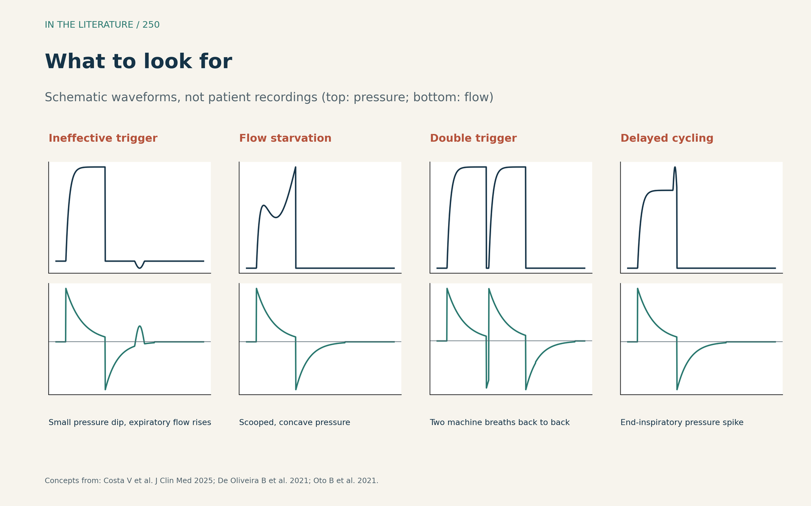
Figure 1. What to look for on pressure and flow (schematic). Concept from Costa et al., De Oliveira et al. and Oto et al.[1], [4], [6] View larger.

Flow and cycling
Flow starvation gives the pressure-time curve a scooped-out look;[4] with flow starvation, the pressure curve flattens and then becomes upwardly concave,[6] and in pressure-assisted modes it gives a rounded inspiratory flow curve.[1] Delayed cycling shows a positive pressure inflection at the end of inspiration.[1]
When the screen is not enough
Experts judged auto-triggering and delayed cycling unlikely to be detectable from waveforms alone, and clinicians' ability to recognize asynchronies is generally low.[3] For reverse triggering, which appears during the transition from deep sedation, agreement rose from kappa 0.5 to 0.84 when diaphragm electrical activity was added (Mellado Artigas et al., Anesthesiology 2021).[2] An automated pressure-and-flow tool detected reverse triggering with 95.5% accuracy, 83.1% sensitivity and 99.4% specificity, and found it in 24% of breaths (Pham et al., Crit Care 2021).[7] In a simulation trial, adding an estimated muscle-pressure curve raised detection sensitivity (Silva et al., Crit Care 2023).[8] In one study cited by a clinical focus review, automated detection was twice as sensitive as clinicians (83.2% vs 41.1%) (Bailey, Anesthesiology 2021).[9] Another review proposes a stepwise reading of expiratory flow, inspiratory flow, pressure and trigger waveforms (Flynn et al., Anesthesiology 2022),[10] and during NIV, diaphragm ultrasound detected auto-triggering and delayed cycling more accurately than waveform observation alone (Vivier et al., Anesthesiology 2020).[11]
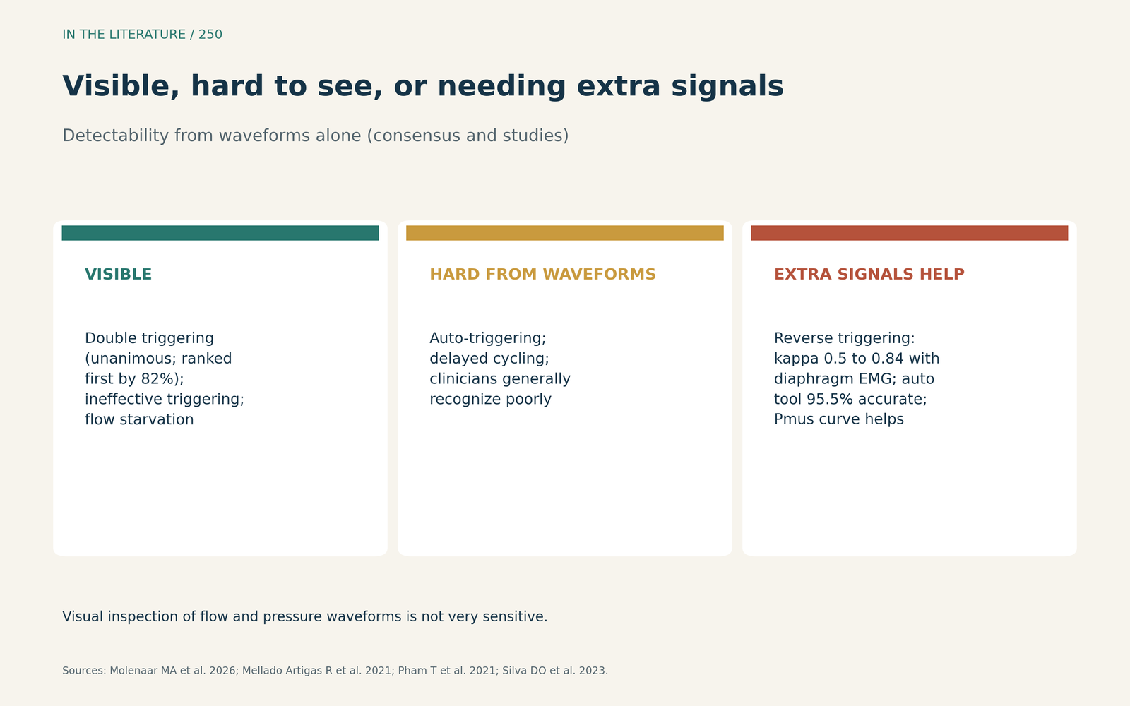
Figure 2. Visible, hard to see, or needing extra signals. Data from Molenaar et al., Mellado Artigas et al., Pham et al. and Silva et al.[3], [2], [7], [8] View larger.

Why it matters
An asynchrony index above 10% defines a high incidence.[1] A systematic review of 19 studies with 2672 patients (de Bie et al., Crit Care Med 2025) found that an index above 10% was associated with longer mechanical ventilation, but there is no widely accepted gold standard for detection.[5]
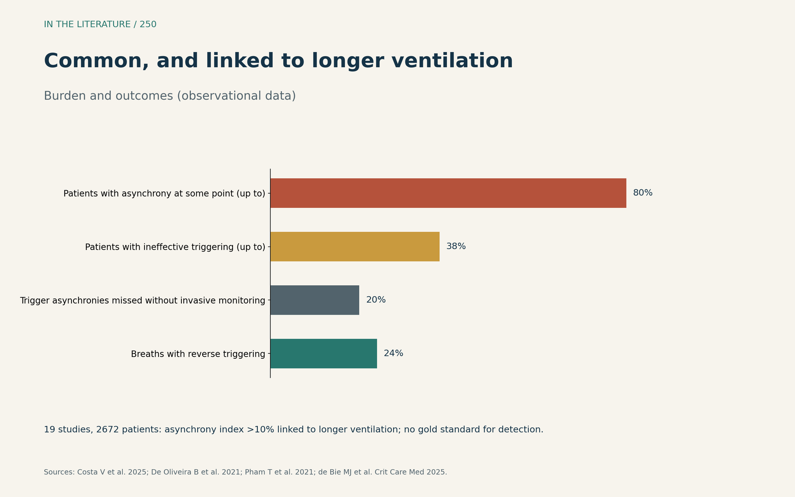
Figure 3. Common, and linked to longer ventilation. Data from Costa et al., De Oliveira et al. and de Bie et al.[1], [4], [5] View larger.

Three takeaways
- Read the waveform by phase: trigger, flow, then cycling.[1], [4]
- Look for pressure dips with flow rise (missed triggers), scooped pressure (flow starvation) and end-inspiratory pressure spikes (late cycling).[4], [1]
- Remember that some asynchronies are invisible on the screen; use oesophageal or diaphragm signals where available.[3], [2]
References
- Costa V, Cidade JP, Medeiros I, Povoa P. Optimizing mechanical ventilation: a clinical and practical bedside method for the identification and management of patient-ventilator asynchronies in critical care. J Clin Med. 2025;14(1):214. doi:10.3390/jcm14010214
- Mellado Artigas R, Damiani LF, Piraino T, et al. Reverse triggering dyssynchrony 24 h after initiation of mechanical ventilation. Anesthesiology. 2021;134(5):760–769. doi:10.1097/ALN.0000000000003726
- Molenaar MA, Nasa P, Damiani LF, et al. Consensus on identifying and ranking ventilator asynchronies in invasively ventilated ICU patients: a modified Delphi study (SYNAPsE). Intensive Care Med. 2026;52(3):423–433. doi:10.1007/s00134-026-08328-2
- De Oliveira B, Aljaberi N, Taha A, et al. Patient-ventilator dyssynchrony in critically ill patients. J Clin Med. 2021;10(19):4550. doi:10.3390/jcm10194550
- de Bie MJ, Rietveld PJ, van der Velde-Quist F, et al. The association between patient-ventilator asynchrony and clinical outcomes in mechanically ventilated patients: a systematic review. Crit Care Med. 2025;53(11):e2261–e2270. doi:10.1097/CCM.0000000000006816
- Oto B, Annesi J, Foley RJ. Patient-ventilator dyssynchrony in the intensive care unit: a practical approach to diagnosis and management. Anaesth Intensive Care. 2021;49(2):86–97. doi:10.1177/0310057X20978981
- Pham T, Montanya J, Telias I, et al. Automated detection and quantification of reverse triggering effort under mechanical ventilation. Crit Care. 2021;25(1):60. doi:10.1186/s13054-020-03387-3
- Silva DO, de Souza PN, de Araujo Sousa ML, et al. Impact on the ability of healthcare professionals to correctly identify patient-ventilator asynchronies of the simultaneous visualization of estimated muscle pressure curves on the ventilator display: a randomized study (Pmus study). Crit Care. 2023;27(1):128. doi:10.1186/s13054-023-04414-9
- Bailey JM. Management of patient-ventilator asynchrony. Anesthesiology. 2021;134(4):629–636. doi:10.1097/ALN.0000000000003704
- Flynn BC, Miranda HG, Mittel AM, Moitra VK. Stepwise ventilator waveform assessment to diagnose pulmonary pathophysiology. Anesthesiology. 2022;137(1):85–92. doi:10.1097/ALN.0000000000004220
- Vivier E, Haudebourg AF, Le Corvoisier P, et al. Diagnostic accuracy of diaphragm ultrasound in detecting and characterizing patient-ventilator asynchronies during noninvasive ventilation. Anesthesiology. 2020;132(6):1494–1502. doi:10.1097/ALN.0000000000003239