
ITL-eb84e200d1bb
Juan Li, MD · 更新于2026年11月18日
核心要点
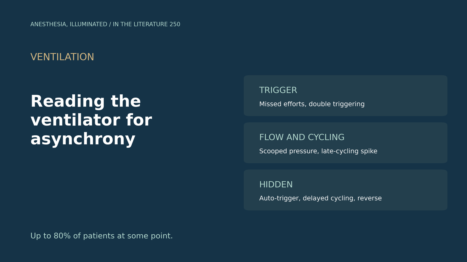
- 非常常见。多达80%的患者在某个时间点出现人机不同步。[1]
- 肉眼容易漏看。目测流量和压力波形的敏感性不高。[2]
- 有些看不见。专家认为仅凭波形难以识别自动触发和延迟切换。[3]
- 按时相阅读。无效触发表现为压力下降伴流量上升;流量不足使压力曲线呈“挖空”样。[4]
- 有临床意义。不同步指数超过10%与机械通气时间延长相关。[5]
一位镇静的压力支持通气患者,呼吸机显示呼吸频率18次/分,但你数到胸廓起伏每分钟26次。呼吸机漏掉了什么?屏幕上怎样看出来?[2], [4]
触发:漏掉的呼吸和多出的呼吸
四分之一至38%的机械通气患者存在无效触发,表现为气道压下降伴呼气流量上升;没有有创监测时,约20%的触发不同步会被漏诊(De Oliveira等,J Clin Med 2021)。[4] 当内源性PEEP导致无效努力时,Costa等(J Clin Med 2025)将外源性PEEP设在实测内源性PEEP的75%–85%以下,[1] Oto等(Anaesth Intensive Care 2021)建议约75%。[6] 在SYNAPsE德尔菲研究中(Molenaar等,Intensive Care Med 2026),专家一致认为双触发可从波形识别,82%将其列为最重要。[3] ARDS低潮气量通气时常见呼吸叠加。[4]
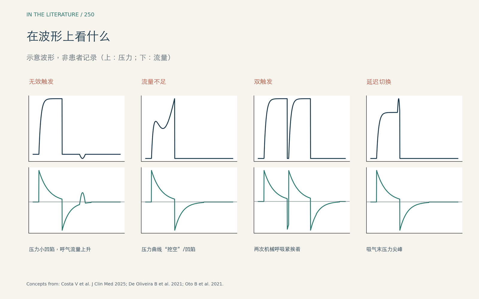
图1. 在压力和流量波形上看什么(示意)。概念来自Costa等、De Oliveira等与Oto等。[1], [4], [6] 查看大图。

流量与切换
流量不足使压力-时间曲线呈“挖空”样;[4] 流量不足时,压力曲线先变平,然后向上凹陷,[6] 在压力辅助模式下吸气流量曲线变为圆钝形。[1] 延迟切换表现为吸气末压力出现正向拐点。[1]
只看屏幕不够时
专家认为仅凭波形难以识别自动触发和延迟切换,临床医生识别不同步的能力普遍较低。[3] 反向触发常出现在从深镇静转出的阶段;加入膈肌电活动后,识别的一致性从kappa 0.5升至0.84(Mellado Artigas等,Anesthesiology 2021)。[2] 一款基于压力和流量的自动工具识别反向触发的准确率为95.5%,敏感性83.1%,特异性99.4%,并发现24%的呼吸存在反向触发(Pham等,Crit Care 2021)。[7] 在一项模拟试验中,加入估算的呼吸肌压力曲线提高了识别敏感性(Silva等,Crit Care 2023)。[8] 一篇临床聚焦综述引用的研究显示,自动识别的敏感度是临床医生的两倍(83.2% 对 41.1%)(Bailey,Anesthesiology 2021)。[9] 另一篇综述建议按呼气流量、吸气流量、压力和触发波形的顺序逐步判读(Flynn 等,Anesthesiology 2022);[10] 无创通气时,膈肌超声识别自动触发和延迟切换比单看波形更准确(Vivier 等,Anesthesiology 2020)。[11]
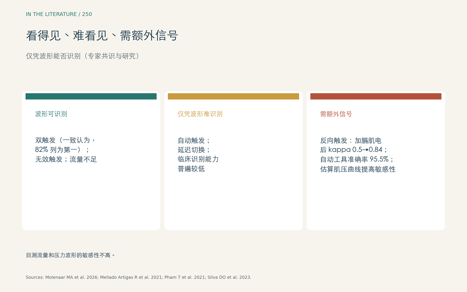
图2. 看得见、难看见,还是需要额外信号。数据来自Molenaar等、Mellado Artigas等、Pham等与Silva等。[3], [2], [7], [8] 查看大图。

为什么重要
不同步指数超过10%被定义为高发生率。[1] 一项纳入19项研究、2672例患者的系统综述(de Bie等,Crit Care Med 2025)发现,指数超过10%与机械通气时间延长相关,但目前没有公认的识别金标准。[5]
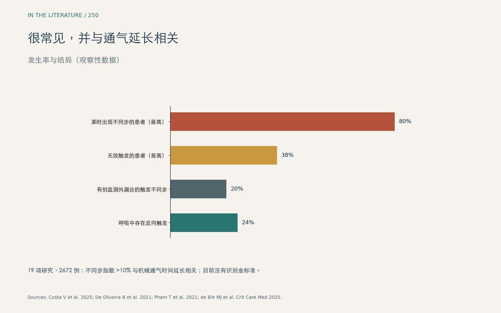
图3. 很常见,并与通气时间延长相关。数据来自Costa等、De Oliveira等与de Bie等。[1], [4], [5] 查看大图。

临床带走的三点
- 按时相阅读波形:触发、流量,然后是切换。[1], [4]
- 寻找压力下降伴流量上升(漏触发)、压力“挖空”(流量不足)和吸气末压力尖峰(延迟切换)。[4], [1]
- 记住有些不同步在屏幕上看不出来;条件允许时使用食管压或膈肌电信号。[3], [2]
参考文献
- Costa V, Cidade JP, Medeiros I, Povoa P. Optimizing mechanical ventilation: a clinical and practical bedside method for the identification and management of patient-ventilator asynchronies in critical care. J Clin Med. 2025;14(1):214. doi:10.3390/jcm14010214
- Mellado Artigas R, Damiani LF, Piraino T, et al. Reverse triggering dyssynchrony 24 h after initiation of mechanical ventilation. Anesthesiology. 2021;134(5):760–769. doi:10.1097/ALN.0000000000003726
- Molenaar MA, Nasa P, Damiani LF, et al. Consensus on identifying and ranking ventilator asynchronies in invasively ventilated ICU patients: a modified Delphi study (SYNAPsE). Intensive Care Med. 2026;52(3):423–433. doi:10.1007/s00134-026-08328-2
- De Oliveira B, Aljaberi N, Taha A, et al. Patient-ventilator dyssynchrony in critically ill patients. J Clin Med. 2021;10(19):4550. doi:10.3390/jcm10194550
- de Bie MJ, Rietveld PJ, van der Velde-Quist F, et al. The association between patient-ventilator asynchrony and clinical outcomes in mechanically ventilated patients: a systematic review. Crit Care Med. 2025;53(11):e2261–e2270. doi:10.1097/CCM.0000000000006816
- Oto B, Annesi J, Foley RJ. Patient-ventilator dyssynchrony in the intensive care unit: a practical approach to diagnosis and management. Anaesth Intensive Care. 2021;49(2):86–97. doi:10.1177/0310057X20978981
- Pham T, Montanya J, Telias I, et al. Automated detection and quantification of reverse triggering effort under mechanical ventilation. Crit Care. 2021;25(1):60. doi:10.1186/s13054-020-03387-3
- Silva DO, de Souza PN, de Araujo Sousa ML, et al. Impact on the ability of healthcare professionals to correctly identify patient-ventilator asynchronies of the simultaneous visualization of estimated muscle pressure curves on the ventilator display: a randomized study (Pmus study). Crit Care. 2023;27(1):128. doi:10.1186/s13054-023-04414-9
- Bailey JM. Management of patient-ventilator asynchrony. Anesthesiology. 2021;134(4):629–636. doi:10.1097/ALN.0000000000003704
- Flynn BC, Miranda HG, Mittel AM, Moitra VK. Stepwise ventilator waveform assessment to diagnose pulmonary pathophysiology. Anesthesiology. 2022;137(1):85–92. doi:10.1097/ALN.0000000000004220
- Vivier E, Haudebourg AF, Le Corvoisier P, et al. Diagnostic accuracy of diaphragm ultrasound in detecting and characterizing patient-ventilator asynchronies during noninvasive ventilation. Anesthesiology. 2020;132(6):1494–1502. doi:10.1097/ALN.0000000000003239