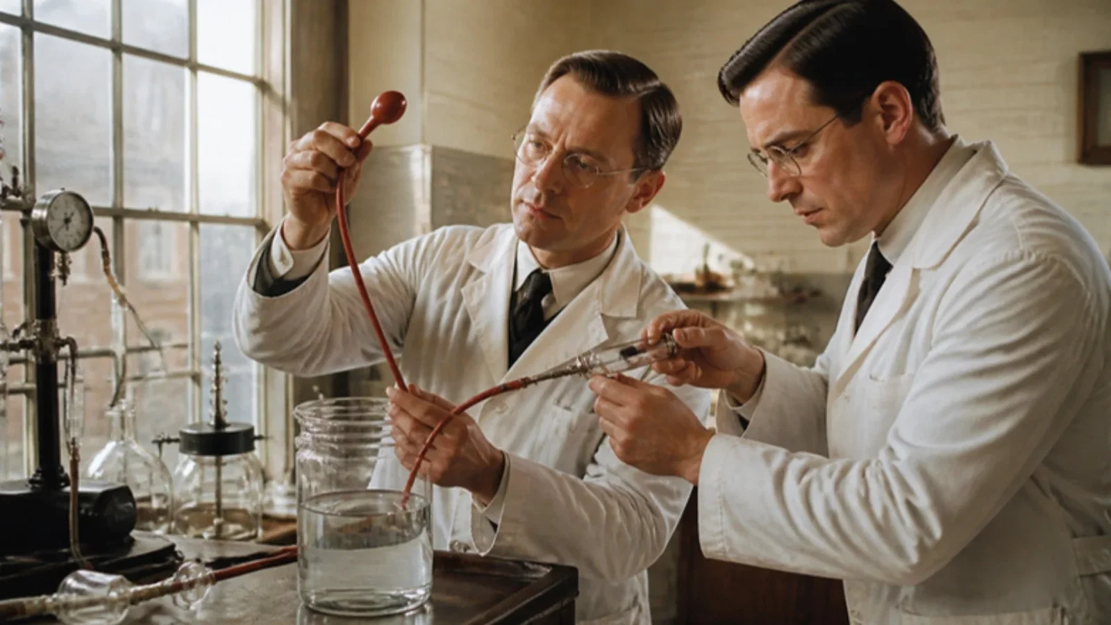
In December 1931 Arthur E. Guedel and Ralph M. Waters described in the Annals of Otology, Rhinology and Laryngology the method they had used since 1928: a cuff made of two pieces of Penrose tubing on a 26 to 30 French catheter, placed under direct laryngoscopy just below the cords, inflated with 4 to 10 c.c. of air from a 10 c.c. syringe, and connected to a closed circuit with a soda-lime canister, to keep debris from the lungs in mouth and nose surgery.
Reading Library · Anesthesia Epochs · 中文版
Watch the education video (for clinicians)
1931: Guedel and Waters' Cuffed Tube · Anesthesia Epochs Education Series
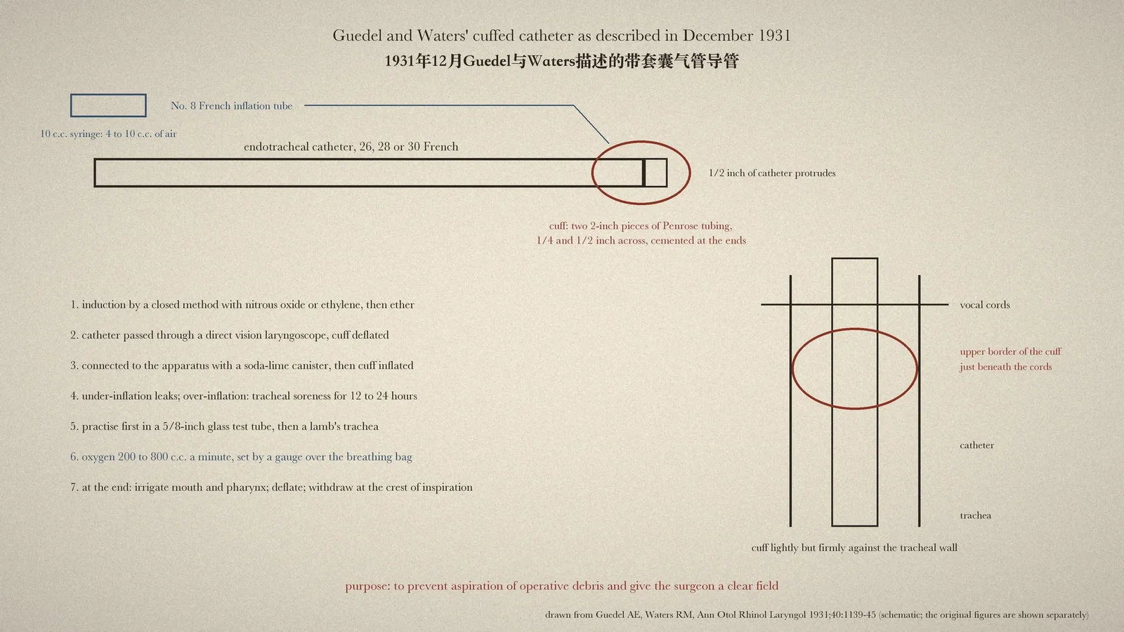
The cuff on an endotracheal tube does two jobs that every anaesthetist relies on: it lets gas be delivered without a leak, and it keeps blood, debris and stomach contents out of the lungs. The History of Anaesthesia Society (HAS) timeline records simply that in 1931 "cuffed endotracheal tubes are introduced into clinical practice". This article reads the paper in which Arthur E. Guedel of Beverly Hills and Ralph M. Waters of Madison, Wisconsin, set out their cuff and its use, "Endotracheal anesthesia: a new technic", in the Annals of Otology, Rhinology and Laryngology for December 1931. [1][2]
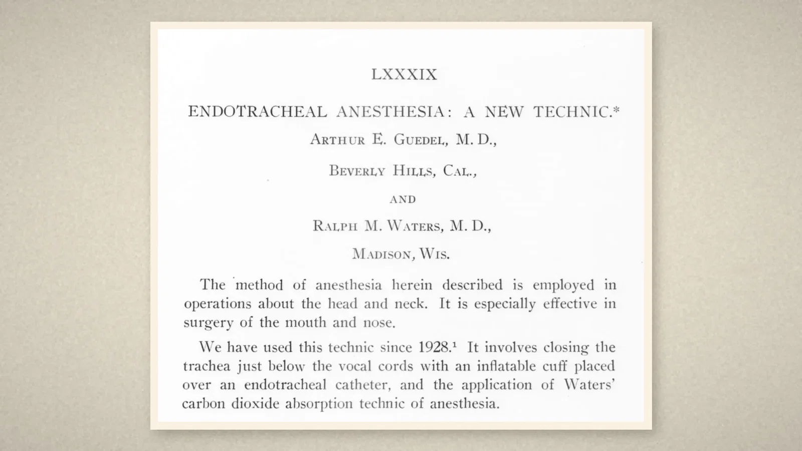
A cuff from two rubber tubes
The method was "employed in operations about the head and neck" and was "especially effective in surgery of the mouth and nose". The authors wrote: "We have used this technic since 1928. It involves closing the trachea just below the vocal cords with an inflatable cuff placed over an endotracheal catheter, and the application of Waters' carbon dioxide absorption technic of anesthesia." Their first note, "A new endotracheal catheter", had appeared in Anesthesia and Analgesia in 1928. [1]
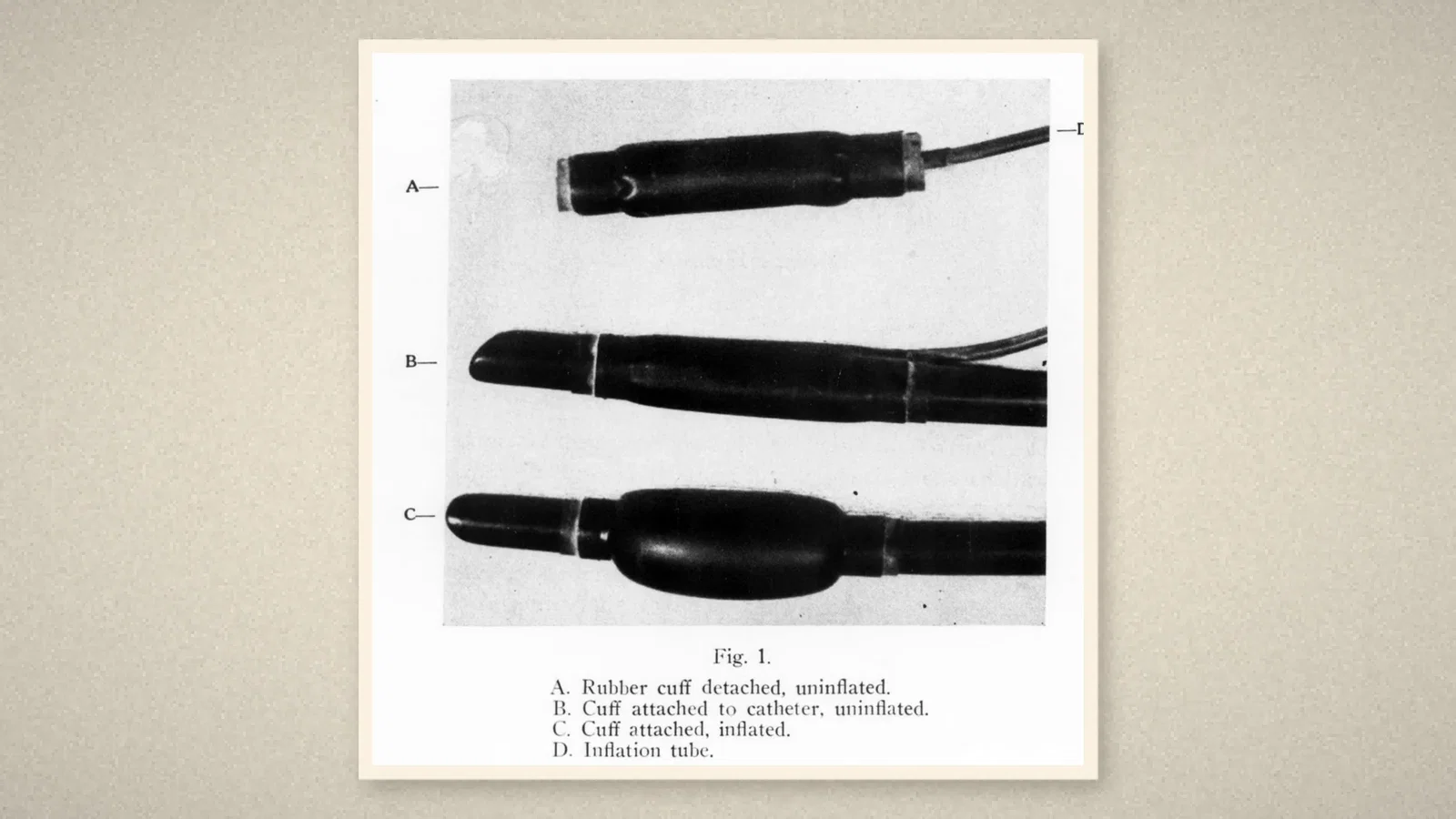
The catheters were 26, 28 and 30 French, and the technique had not been used in any patient under nine. The cuff was home-made: two pieces of Penrose rubber tubing, a quarter and a half inch in diameter and both two inches long, "placed one inside of the other and their ends cemented together". A No. 8 French soft rubber catheter cemented into the end served as the inflation tube. The cuff was slipped over the catheter so that half an inch of catheter protruded beyond it. [1]
Placing and inflating it
Anaesthesia began with nitrous oxide or ethylene "by an entirely closed method", then ether was added for six to ten minutes. At that point the authors added carbon dioxide, about 10 per cent, which changed breathing from hyperpnoea to near arrest; during the twenty to forty seconds of apnoea the vocal cords lay widely separated (their Fig. 2 compares the cords). The catheter, cuff deflated, was "introduced through a direct vision laryngoscope, so that the upper border of the cuff lies just beneath the vocal cords". The anaesthetic apparatus, "now with a soda-lime cannister for carbon dioxide absorption in place", was connected, and the cuff inflated. [1]
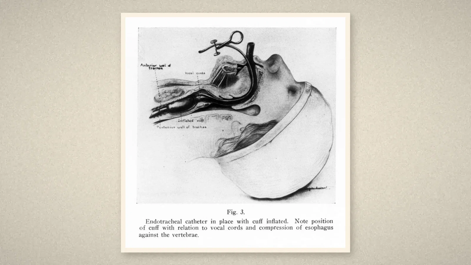
Inflation was a matter of touch. Air was injected from a ten cubic centimetre ground glass syringe, and "the degree of inflation is determined by the sense of pressure within the syringe", until the cuff "lightly but firmly contacts the wall of the trachea". "Under-inflation will permit leakage between the cuff and the tracheal wall. Over-inflation will produce tracheal soreness lasting from twelve to twenty-four hours." Four to ten cubic centimetres were needed, more with advancing age. The authors advised practising first in a five-eighths inch glass test tube, where the inflation could be seen, then in a freshly removed lamb's trachea. [1]
Maintenance, emergence and the point of it
Maintenance followed Waters' absorption technique. Nitrous oxide and ethylene are not destroyed in the body, so once anaesthesia is established it is maintained "without the addition of more gases, so long as the escape of those gases present in the body be prevented"; oxygen is added to meet metabolism, 200 to 800 cubic centimetres a minute, and soda-lime granules take up the expired carbon dioxide. The inspired gases were warm and moist, 100 to 104 °F after twenty minutes. A stationary gauge over the breathing bag regulated the oxygen: too much and the bag rose toward the gauge, too little and it fell away, before any clinical sign appeared. [1]
At the end the mouth and pharynx were irrigated, the apparatus disconnected, and the patient allowed to emerge until the swallowing reflex returned; then "the cuff is deflated and the catheter withdrawn at the crest of inspiration." Although many patients retched with the cuff inflated, "but two or three cases have succeeded in forcing vomitus into the pharynx", each less than an ounce. The authors attributed this to the cuff compressing the oesophagus against the vertebrae (Fig. 3). "The purposes of this technic are to prevent aspiration of operative débris into the lungs and to provide a clear field of work for the surgeon." [1]
What the record shows
Almost every element of modern cuff management is in these seven pages: a cuff placed just below the cords, inflated only enough to seal, the trade-off between leak and mucosal injury, a pilot line for inflation, practice on a model, and extubation after reflexes return. The 1931 cuff was judged by feel; later practice measures cuff pressure because the tracheal soreness the authors describe is a pressure injury. Their apnoeic induction with added carbon dioxide has not survived.
Sources and reading limits
Read: Guedel AE, Waters RM, Ann Otol Rhinol Laryngol 1931;40:1139-45, in full; the HAS timeline entry for 1931.
Not covered: the 1928 paper in Anesthesia and Analgesia (not openly available; cited here only as the authors' own reference); Waters' papers on carbon dioxide filtration; the "dunked dog" demonstration, known from later accounts and not described in these pages; earlier cuffed tubes by others. The authors' "since 1928" and the HAS's 1931 are both given; the article is dated to the 1931 publication. The carbon dioxide induction is historical practice, not a recommendation; the observations are the authors'.
Three illustrations are crops of the December 1931 issue, including Figs. 1 and 3; one is drawn from the text.
- Guedel AE, Waters RM. Endotracheal anesthesia: a new technic. Ann Otol Rhinol Laryngol. 1931;40:1139-45. Internet Archive
- History of Anaesthesia Society. Timeline. HAS
Continue exploring
Related reading connects shared questions and contexts; it does not by itself establish historical influence.
Anesthesia Epochs · The Source Room · About & Editorial Method
Published November 2, 2026. Sources and reading limits are stated above. Illustrations are labelled as original source images or AI-made. Historical accounts are educational; they are not current treatment instructions.