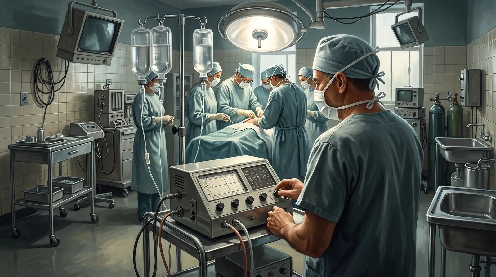
Surgery, Gynecology and Obstetrics for December 1963 (vol. 117) carried 'Homotransplantation of the liver in humans' by Starzl, Marchioro, von Kaulla and colleagues of the University of Colorado and the Denver Veterans Administration Hospital: three orthotopic liver transplantations, the first failing on the operating table from haemorrhage, the other two establishing that the operation is feasible in humans although both patients died of pulmonary emboli, one after twenty-two days and one after seven and a half. Coagulation was the central perioperative problem. Profound clotting defects appeared in all three at the time of transplantation, and the authors argued that these came not from an acute loss of the clotting factors the liver makes but from an explosive rise in plasma fibrinolytic activity within minutes, both while the liver was being mobilised and after it was removed; as a working hypothesis they suggested that the liver normally elaborates a substance restraining the activation of plasminogen. During the operation the venous return from the splanchnic and inferior caval systems was temporarily occluded, and the authors concluded that a single external bypass from the inferior to the superior vena cava suffices. Afterwards the picture reversed into hypercoagulability, which, perhaps together with the bypass itself, contributed to the fatal emboli. The whole course was followed with thrombelastograms and serial euglobulin lysis times.
Reading Library · Anesthesia Epochs · 中文版
Watch the education video (for clinicians)
1963: Starzl's First Liver Transplants — The Anhepatic Phase · Anesthesia Epochs Education Series
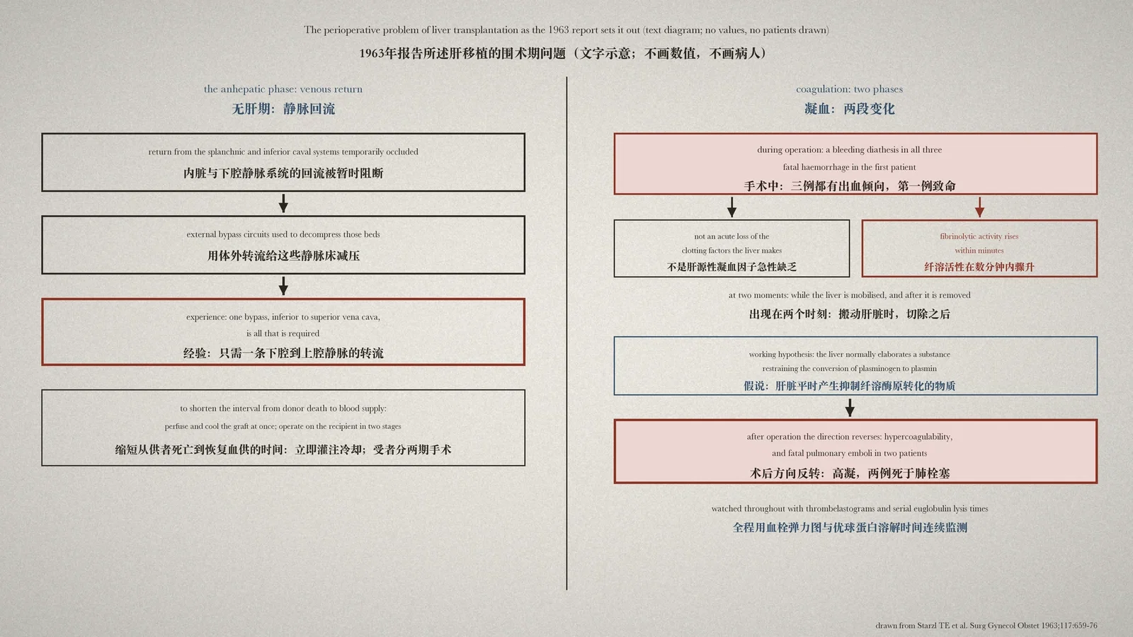
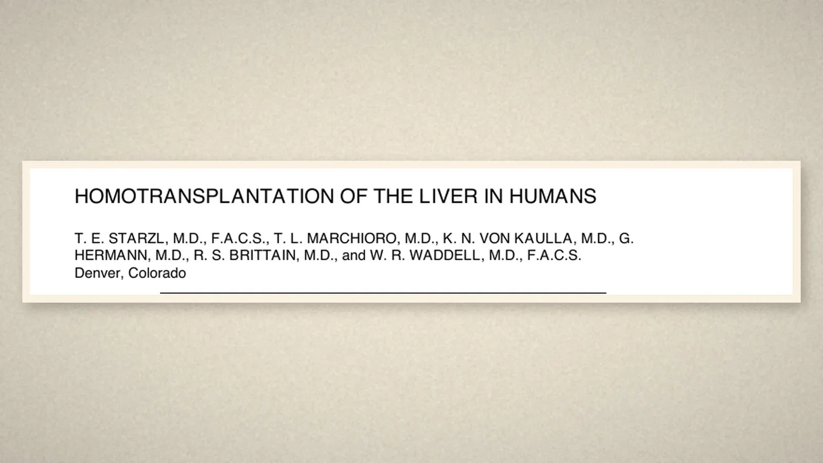
Moving a liver from a cadaver into a patient is only half a technical problem. The other half sits on the far side of the drapes: during the time the patient has no liver, how does blood get back to the heart, and why will the bleeding not stop? In December 1963 Surgery, Gynecology and Obstetrics, vol. 117, carried "Homotransplantation of the liver in humans" by Starzl, Marchioro, von Kaulla, Hermann, Brittain and Waddell of the University of Colorado School of Medicine and the Denver Veterans Administration Hospital. This article reads that paper, with the weight on its perioperative half. [1, p. 659]
Three operations
The article opens by saying that for several kinds of liver disease an ideal treatment would be removal of the diseased organ and orthotopic replacement with a hepatic homograft, and that applying such therapy depends first on a satisfactory operative procedure and second on suitable measures against immunological rejection. Both, the authors judged, had reached at least partly satisfactory solutions: the technical requirements for canine hepatic transplantation had been defined, and a regimen developed in recipients of kidney grafts was giving consistently prolonged survival of foreign tissue. [1, p. 659]
The paper reports those advances applied to three patients. The first attempt failed on the operating table. The other two established the feasibility of such an operation in humans, although both patients died of pulmonary emboli, one twenty-two days and one seven and a half days after transplantation. [1, p. 659]
The anhepatic phase: where does the blood go?
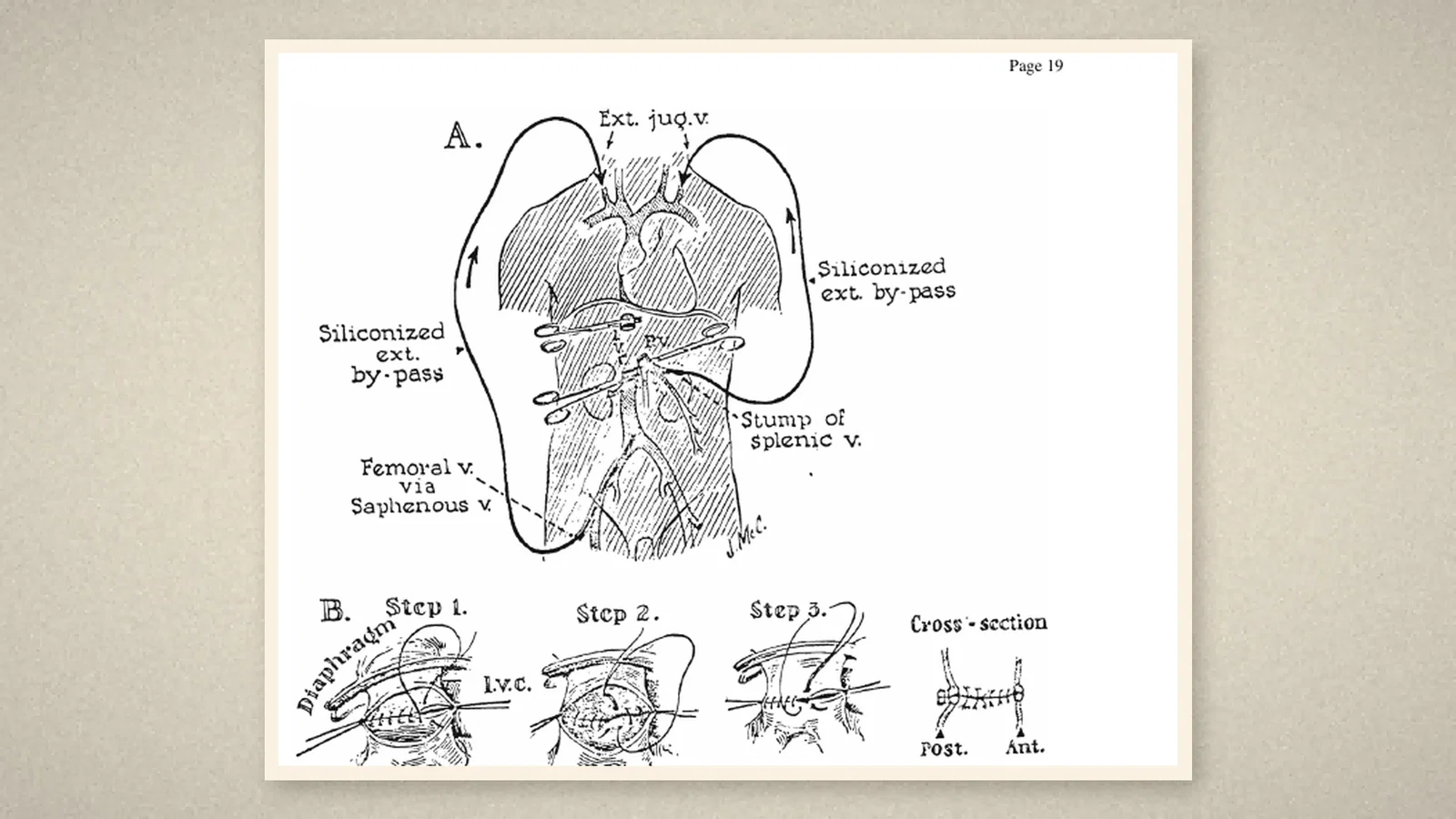
The operation itself sets the circulation a problem. While the transplantation is performed, the venous return from the splanchnic and inferior vena caval systems is temporarily occluded. To decompress those beds the authors used external bypass circuits, drawn in the paper's Figure 4: siliconised external bypasses, one taken from the femoral vein by way of the saphenous and led to the external jugular, another from the splenic vein to decompress the portal system. [1, p. 675]
Experience then pared the arrangement down. In two of the three patients no external bypass was used, and in the third flow ceased after a few minutes, from which the authors concluded that a single effective bypass from the inferior vena cava to the superior vena cava is all that is required for adequate venous return to the heart from both the caval and the portal systems. [1, p. 672]
To shorten the interval between the donor's death and the restoration of a blood supply to the graft, two measures were used: an extracorporeal circuit that perfused and cooled the liver immediately after death, and operating on the recipient in two stages, the preliminary one skeletonising the structures above and below the liver so that the hepatectomy and the several anastomoses could be done at the definitive procedure. [1, p. 675]
Bleeding: not missing factors but fibrinolysis
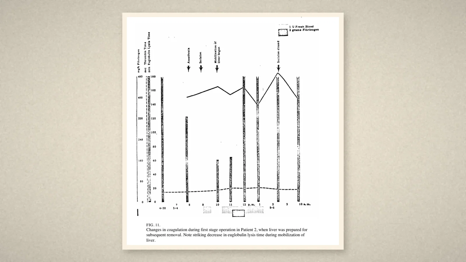
Experience with these three patients, the authors wrote, stresses the need for close control of the coagulation processes during and after transplantation. Profound clotting defects were demonstrated in all three at the time of transplantation, and in the first they resulted in fatal haemorrhage. [1, p. 672]
What matters is how the cause was read. The haemorrhagic tendency did not result from an acute deficiency of the clotting factors synthesised in the liver: during the anhepatic phase in the second and third patients, plasma fibrinogen showed no clinically significant fall, in accord with experimental work showing that a substantial decrease in clotting factors after total hepatectomy takes several hours to develop. The important finding instead was an explosive increase in plasma fibrinolytic activity which developed within minutes, both during manipulation of the liver and after its removal. [1, p. 672]
As a working hypothesis the authors suggested that the liver normally elaborates a substance which inhibits the conversion of plasminogen to plasmin, presumably by acting on a plasminogen activator; absence of the liver, or severe injury to it during operative manipulation, could remove that restraint and let the activator system run unchecked. [1, p. 672]
These changes were visible because they were watched for. The overall clotting process was monitored by serial thrombelastograms, by von Kaulla's method, giving a continuous record of the onset and progress of fibrin formation and fibrinolysis, supplemented by determinations of the prothrombin complex, plasma fibrinogen and the thrombin time. Fibrinolytic activity was measured serially by the euglobulin lysis time, a method chosen for speed and simplicity and because its results are affected neither by heparin nor by the antifibrinolytic drug used. [1, p. 663]
Afterwards, the direction reverses
Bleeding during the operation gave way to its opposite. During operation, the summary says, a bleeding diathesis is regularly detectable by laboratory examination; postoperatively, a state of hypercoagulability developed, which probably contributed to the lethal multiple pulmonary embolisation in two patients, and the authors add that the external bypass itself may have contributed to the formation of the emboli. [1, p. 675]
As for the grafts, hepatic function was immediately deranged after operation, probably from injury incurred during transplantation, with progressive improvement thereafter. No biochemical evidence of rejection was later observed, and at autopsy in the last two patients the graft structure was preserved surprisingly well, which the authors attributed to the anti-rejection therapy forestalling the process. [1, p. 675]
What the record shows
The paper gives the perioperative problem of liver transplantation its first concrete shape: the blood returning from below needs somewhere to go while the liver is out; bleeding has to be sorted into factors lost and clot dissolved too fast; and the risk after the operation may run opposite to the risk during it. The authors did not stop at "severe bleeding": continuous monitoring let them put the change at the moment the liver was handled and the moment it was removed, and from that they proposed a hypothesis that could be tested further.
Sources and reading limits
Read: Starzl TE, Marchioro TL, von Kaulla KN, Hermann G, Brittain RS, Waddell WR, "Homotransplantation of the liver in humans", Surgery, Gynecology and Obstetrics, December 1963, vol. 117, pp. 659-76 (NIH author manuscript, PMC2634660), in full, with page images of the title page, Figure 4 (the bypasses and anastomoses) and Figure 11 (the coagulation record of a first-stage operation) checked.
Not covered: the canine work on the technical requirements, and the two papers on the anti-rejection regimen, all cited in this article.
Limits: no doses and no laboratory values are reproduced here, and the individual patients are not recounted; the paper's photographs of patients are not used as illustrations. Anti-rejection drugs are mentioned only by category. The exact dates of the three operations are in the original; this article gives month and year.
Three illustrations are taken from the 1963 paper (title page, Figure 4 and Figure 11). One is a diagram drawn from the text, with no people drawn.
- Starzl TE, Marchioro TL, von Kaulla KN, Hermann G, Brittain RS, Waddell WR. Homotransplantation of the liver in humans. Surg Gynecol Obstet. 1963;117:659-76. PMC2634660
Continue exploring
Related reading connects shared questions and contexts; it does not by itself establish historical influence.
Anesthesia Epochs · The Source Room · About & Editorial Method
Published November 4, 2026. Sources and reading limits are stated above. Illustrations are labelled as original source images or AI-made. Historical accounts are educational; they are not current treatment instructions.