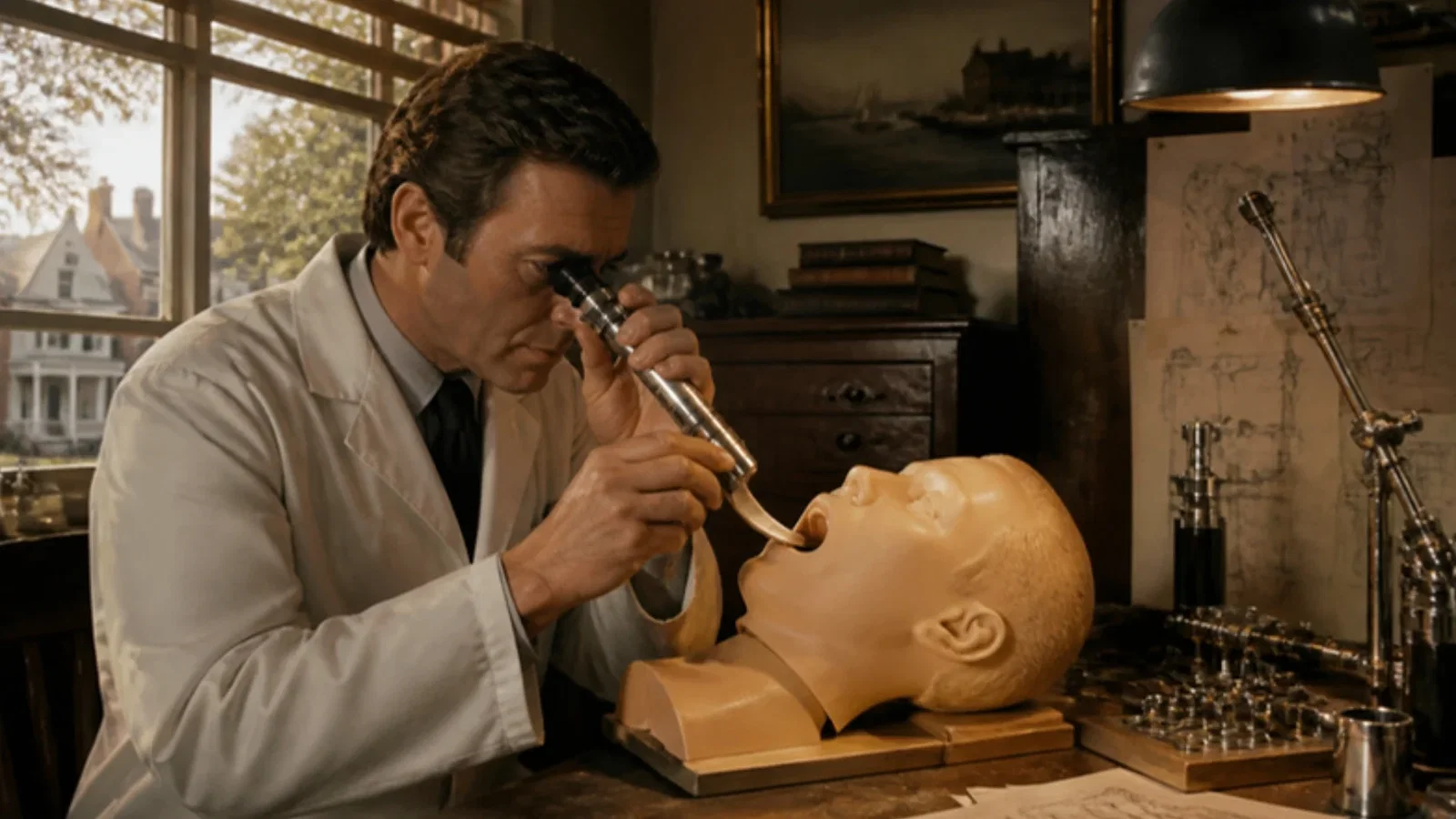
On 9 July 1976 James R. Bullard of Charleston, South Carolina, filed the application that became US Patent 4,086,919, 'Laryngoscope', granted on 2 May 1978: a thin rigid blade curved to the oral and pharyngeal passage, carrying a fibre-optic bundle to an eyepiece, so that the glottis could be seen indirectly without disturbing the normal position of a supine patient's head. Direct Miller and Macintosh blades, the patent says, demanded forced head tilt, neck extension and a wide mouth, could injure patients with spinal, neck or facial injuries, and often levered on the upper teeth. A later Bullard patent from Augusta added a working channel. The ASA timeline dates production of the Bullard laryngoscope by Circon to 1989, and credits the first video laryngoscopy that year to the Augsburg ENT surgeon Peter Bumm.
Reading Library · Anesthesia Epochs · 中文版
Watch the education video (for clinicians)
1976: The Bullard Laryngoscope — Seeing Around the Tongue · Anesthesia Epochs Education Series
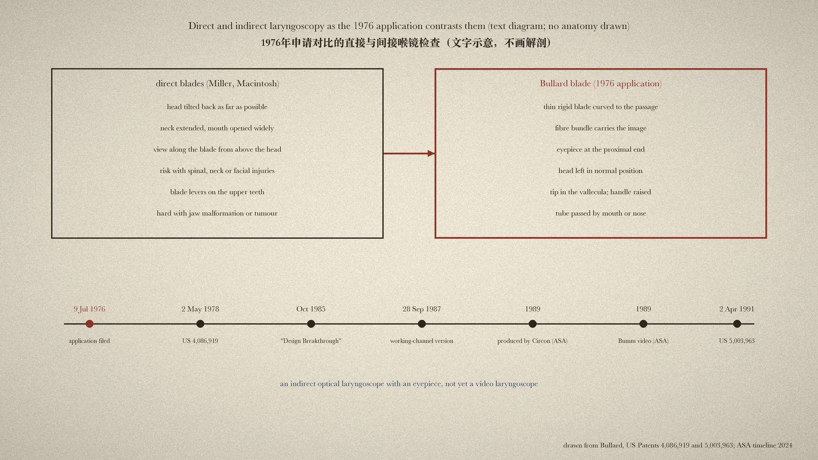
Every laryngoscope that shows the glottis on a screen rests on one idea: the person intubating does not have to see the larynx in a straight line. Long before video, that idea was written into a patent application by an anaesthetist in Charleston, South Carolina. The blade it described carried its own eye. [1]
The problem with looking straight
James R. Bullard filed the application for "Laryngoscope" on 9 July 1976; it became US Patent 4,086,919 on 2 May 1978. The patent describes how intubation was usually done with a straight Miller or slightly curved Macintosh blade: "place the patient in supine position, tilt the head backwards as far as possible, and distend the lower jaw to widely open the mouth", then view the larynx "from an observation position just above and beyond the head of the patient". [1, col. 1]
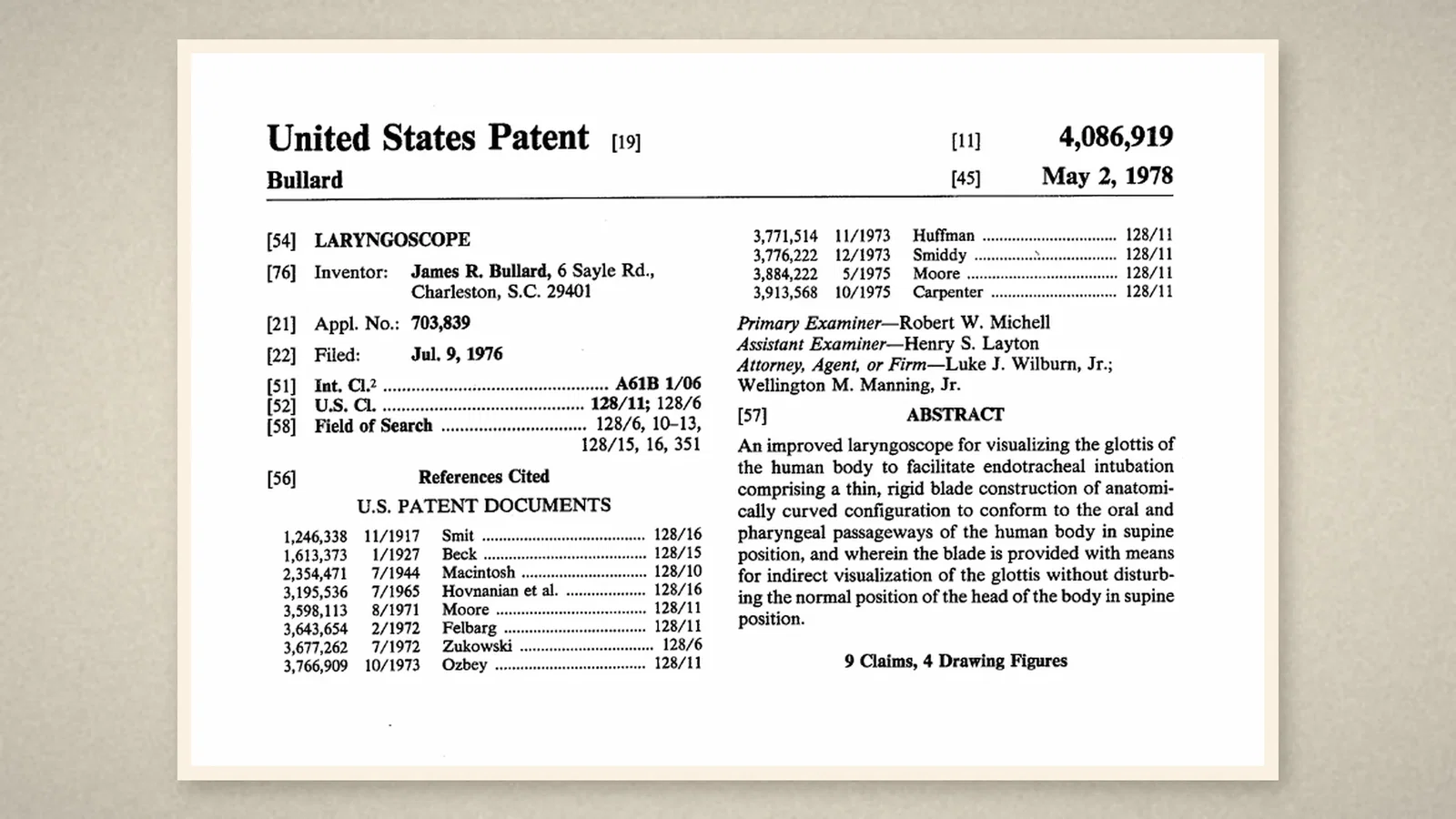
That method had costs. Forcing the head back and the neck into extension could "further complicate and severely injure patients having spinal, neck or facial injuries, such as fractures"; the blades often pivoted on the upper teeth, and "breakage or injury of the teeth often results"; in patients with malformed jaws or throat tumours direct vision could be impossible. Fibre-optic probes already existed, but they needed large tubes and could not lift the tongue and epiglottis, and a straight rigid fibre-optic instrument still required "hyperextension of the head and neck". [1, cols. 1-2]
A blade with an eyepiece
Bullard's answer was "a thin, rigid blade construction of anatomically curved configuration" that followed the passage from the mouth to the epiglottis, with "means for indirect visualization of the glottis without disturbing the normal position of the head". Inside the blade a bundle of optical fibres carried the image from an opening at the tip to an eyepiece at the proximal end; another bundle carried light down to the tip. [1, abstract; cols. 2-3]
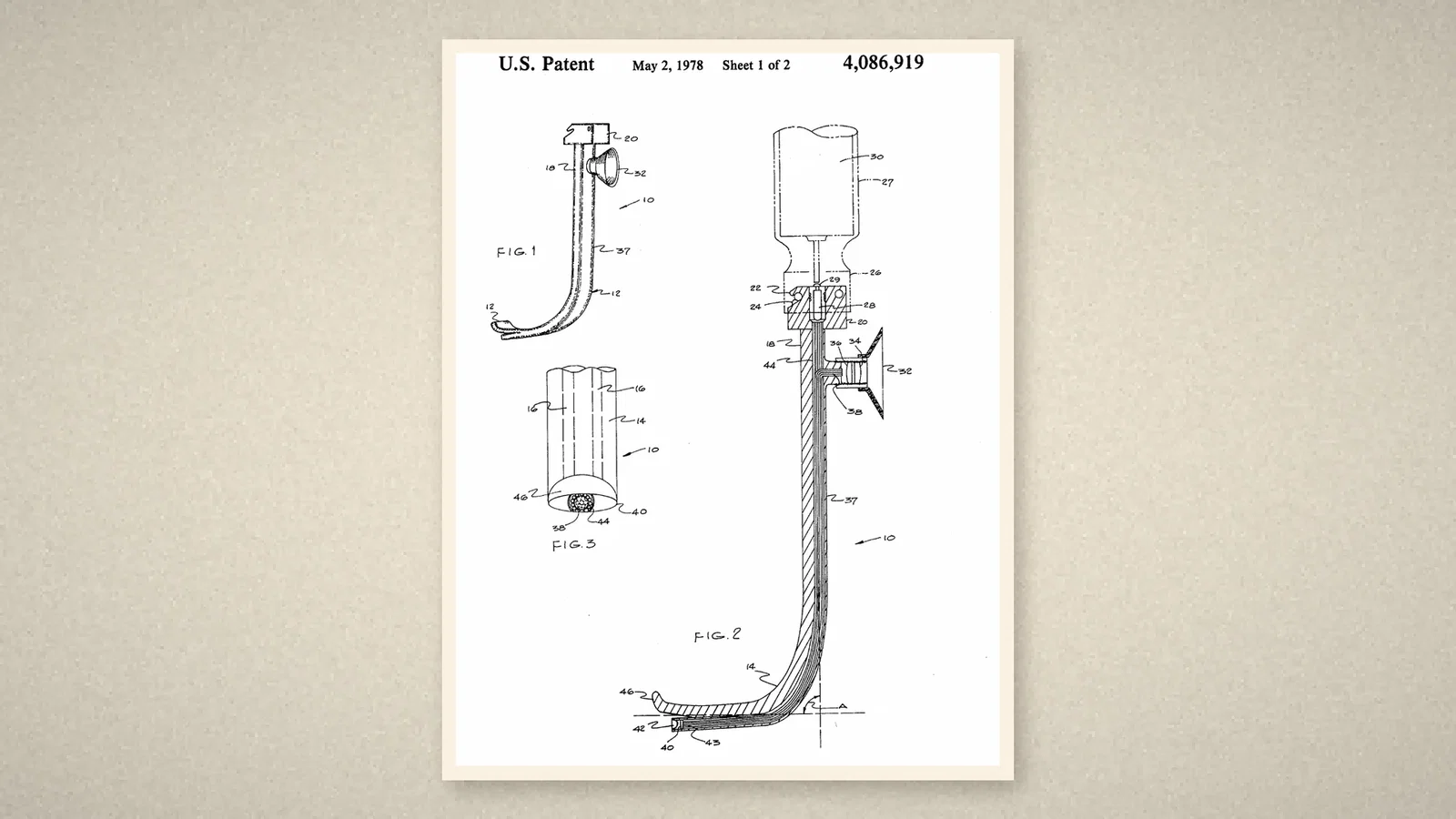
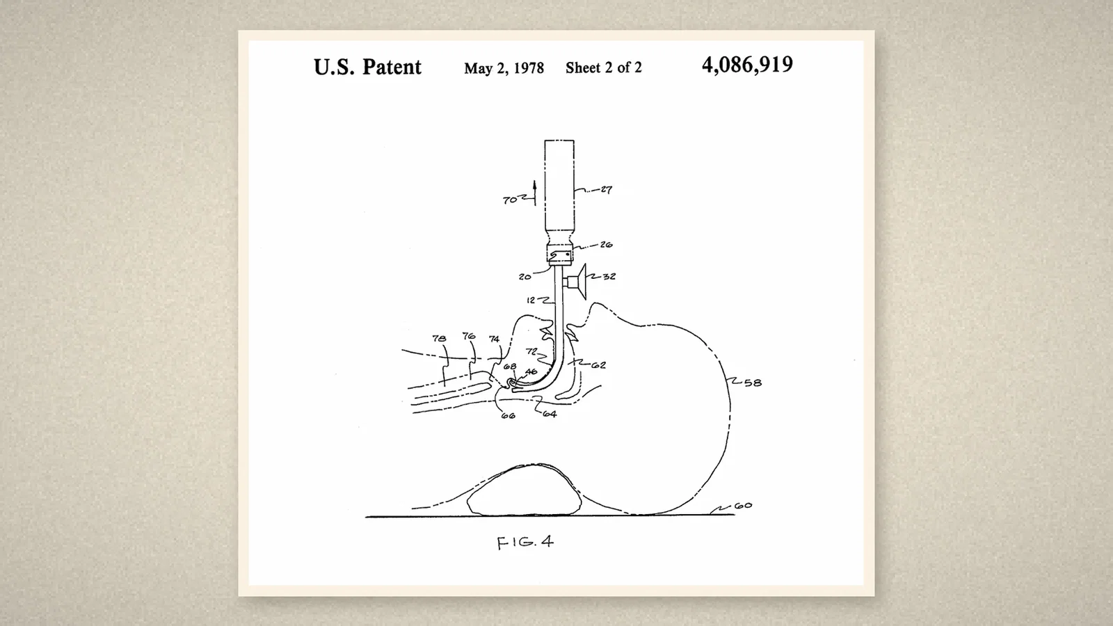
The patent's fourth figure shows it in use. With the patient supine and the head resting normally on the table, the anaesthetist at the head end slips the thin blade between the teeth, places the upturned tip in the vallecula and, "by merely raising laryngoscope handle 27 a short distance in the vertical direction", lifts tongue and epiglottis to see "the glottis and larynx through the eyepiece". The tube can then be passed by mouth or nose. [1, col. 4]
From patent to product
Bullard kept working on the design. A later patent, 5,003,963, granted on 2 April 1991 to Bullard, Richard P. Muller, Benedetto Grossi and John Cristine and assigned to Bullard in Augusta, Georgia, goes back to an application of 28 September 1987; it describes a laryngoscope with a working channel and a device for moving objects in the larynx, and lists among its references Bullard's own "Design Breakthrough in Laryngoscopy" of October 1985. [2][3]
The Australian Society of Anaesthetists (ASA) timeline lists the "Bullard laryngoscope blade produced" under 1989: designed by Dr James Roger Bullard, director of obstetrical anesthesia at the Medical College of Georgia in Augusta, for difficult intubation in children and adults, with a right-angled rigid blade, a fibre-optic light attachment, an external eyepiece and two ports, manufactured by Circon Corporation. Under the same year it records the first video laryngoscopy, by the Augsburg ENT surgeon Peter Bumm with a rigid endoscope. [4, p. 81]
What the record shows
The step from seeing the larynx to seeing it around a corner. The 1976 application reasoned from injuries, teeth and awkward anatomy to a blade that leaves the head where it lies and brings the view back up through glass. The camera came later; the geometry was already here.
Sources and reading limits
Read: US Patent 4,086,919 in full (patent PDF with text layer, page images of the front page and both drawing sheets); US Patent 5,003,963, front page, related applications, references and the description of the working channel (text layer and front page image, with the Google Patents record); the ASA timeline 2024, p. 81.
Not covered: Bullard's "Design Breakthrough in Laryngoscopy" (1985); clinical reports on the Bullard laryngoscope; the manufacturer's literature; Bumm's video laryngoscopy, named here only from the ASA.
Limits: the patents describe a design, not its use or its spread. The 1989 production date and the manufacturer are the ASA's. This article does not call the Bullard a video laryngoscope; it is an optical laryngoscope with an eyepiece. Dimensions and angles in the claims are not repeated. Quotations are from the English originals.
Three illustrations are crops of US Patent 4,086,919; one is drawn from the patents.
- Bullard JR. Laryngoscope. US Patent 4,086,919. 2 May 1978 (filed 9 Jul 1976). Google Patents
- Bullard JR, Muller RP, Grossi B, Cristine J. Laryngoscope. US Patent 5,003,963. 2 Apr 1991. Google Patents
- Google Patents. US5003963A: cited and citing documents. Google Patents
- Australian Society of Anaesthetists. Anaesthesia history timeline. 2024:81. ASA
Continue exploring
Related reading connects shared questions and contexts; it does not by itself establish historical influence.
Anesthesia Epochs · The Source Room · About & Editorial Method
Published November 3, 2026. Sources and reading limits are stated above. Illustrations are labelled as original source images or AI-made. Historical accounts are educational; they are not current treatment instructions.