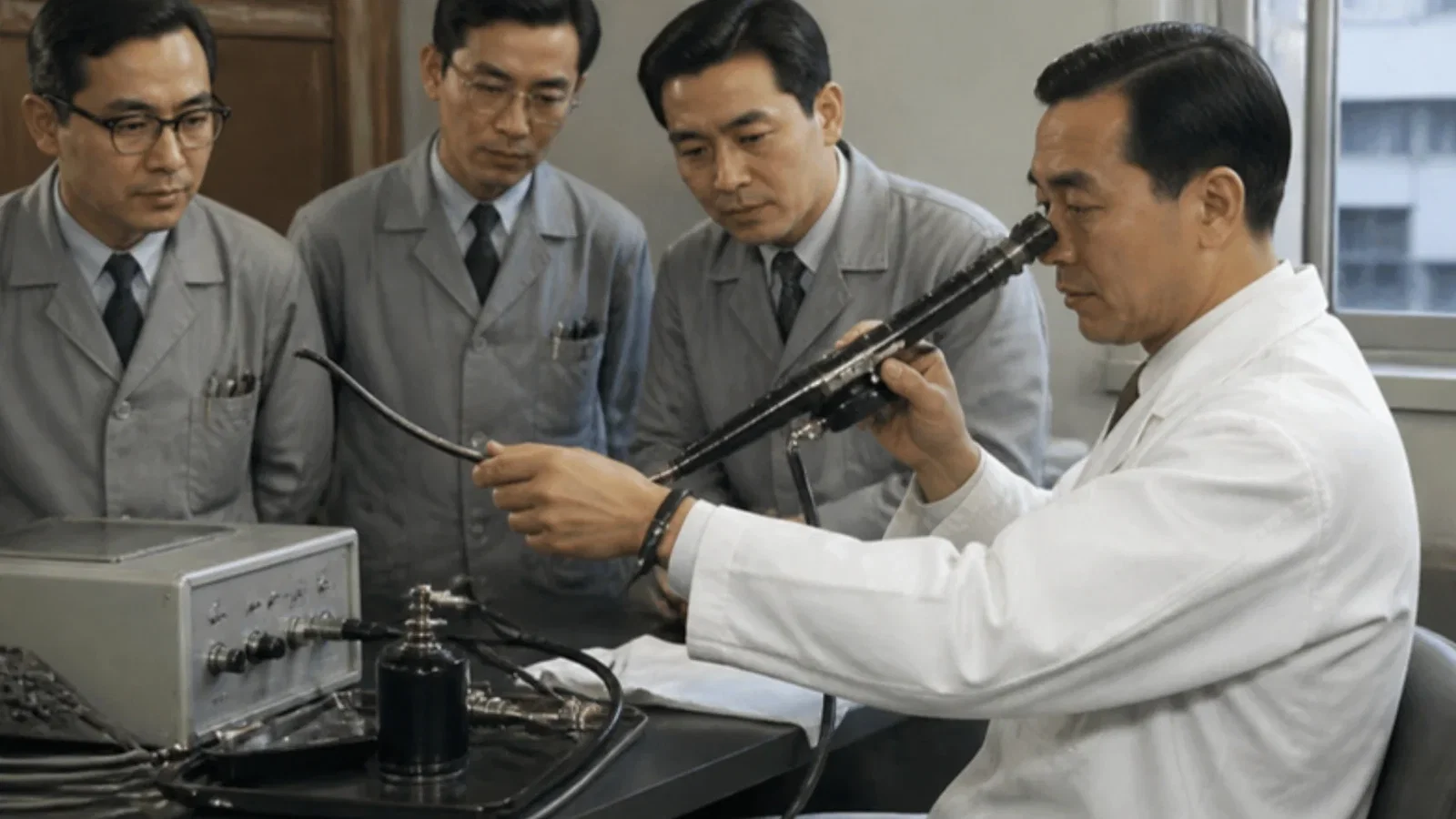
In March 1968 the Keio Journal of Medicine published 'Flexible bronchofiberscope' by Shigeto Ikeda, Noboru Yanai and Shichiro Ishikawa of the National Cancer Center Hospital, Tokyo: because the rigid bronchoscope could not see many segmental bronchi of the upper lobes, they gave a design outline to Machida Endoscope Co. and, at the end of 1965, to Olympus Optical Co.; the first test instrument was completed in July 1966 and five more followed by July 1967, with growing tip flexion, a channel for local anaesthetic and biopsy forceps. The flexible scope could be passed through a tracheal tube, so the patient need not take an unnatural position. The ASA timeline dates the flexible bronchoscope to 1966 and credits the first fibreoptic nasal intubation, in 1965, to the London anaesthetist Peter Murphy.
Reading Library · Anesthesia Epochs · 中文版
Watch the education video (for clinicians)
1966: Ikeda's Flexible Bronchofiberscope — Tokyo · Anesthesia Epochs Education Series
Every anaesthetist who has threaded a fibreoptic scope through a tracheal tube is using an instrument that began as a cancer surgeon's tool. In Tokyo in the middle of the decade, a bronchoscopist at the National Cancer Center Hospital wanted to see into parts of the lungs that a straight steel tube could not reach, and asked two optical companies to build something that would bend. The account of how it was done appeared in the Keio Journal of Medicine in March 1968. [1]
The problem
"Flexible bronchofiberscope", by Shigeto Ikeda, Noboru Yanai and Shichiro Ishikawa of the National Cancer Center Hospital and the Department of Surgery, Keio University, was received on 6 February 1968. Pulmonary cancer, the authors wrote, had markedly increased, and since it usually arose from a bronchus, bronchoscopy mattered more than ever. But "it is only natural that limitations are found with a straight instrument such as a bronchoscope"; even with a glass-fibre light guide, the segmental bronchi of both upper lobes were "still not visible". Their own results with the rigid telescope in lung cancer from November 1962 to the end of 1966, and the finding that many small early cancers lay in the upper lobes, set the task. [1, pp. 1-4]
Six instruments in a year
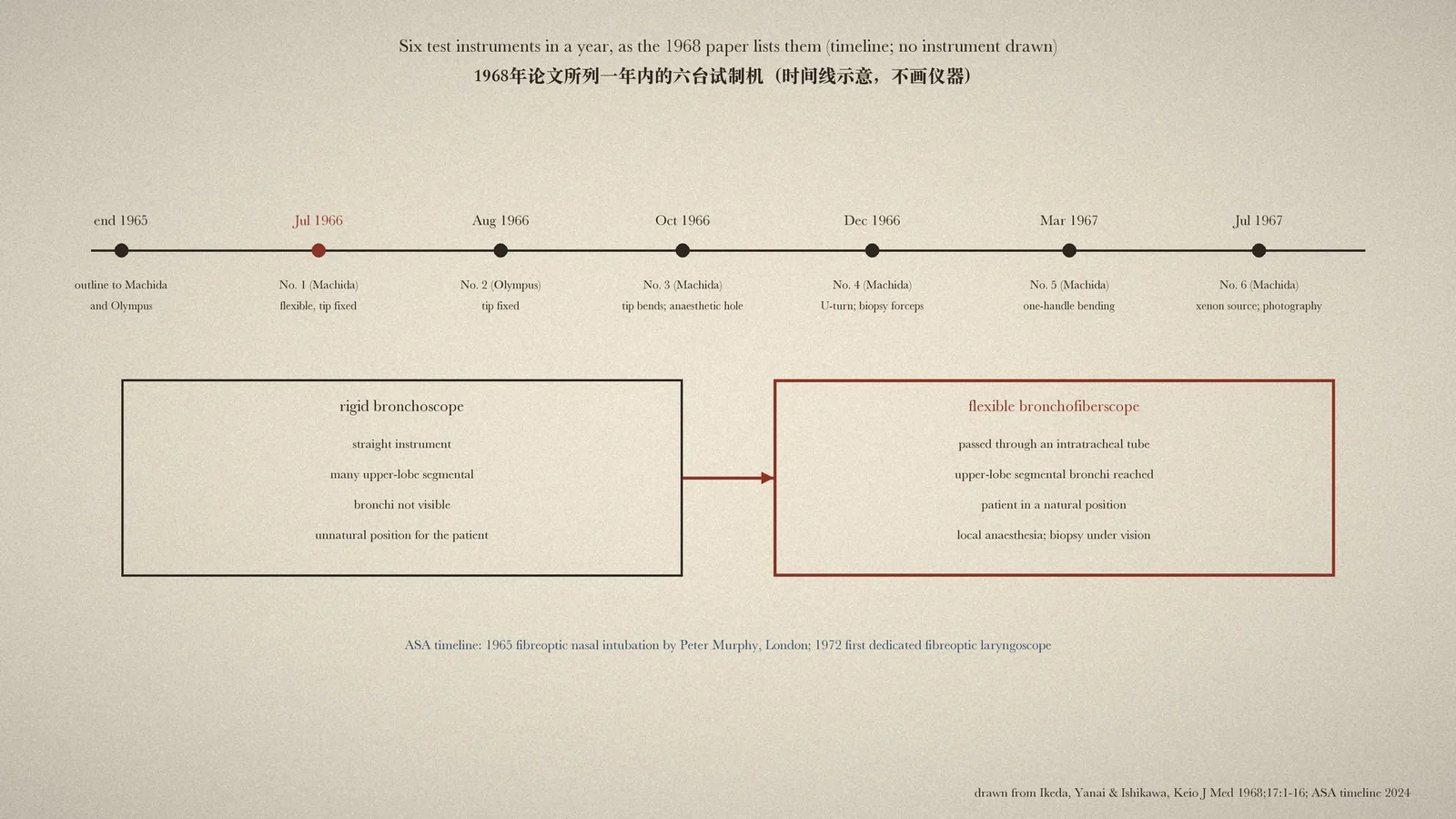
The authors drew up a nine-point outline, covering diameter, the number of image and light fibres, a flexible tip, direct vision and length. "The production of this instrument was entrusted to Machida Endoscope Co., Ltd. under the outlined scheme. Towards the end of 1965, a similar outline was shown to Olympus Optical Co., Ltd." In July 1966 Machida finished the first instrument for test use: very soft, "giving a very distinct picture", but its tip could not be bent. [1, p. 5]
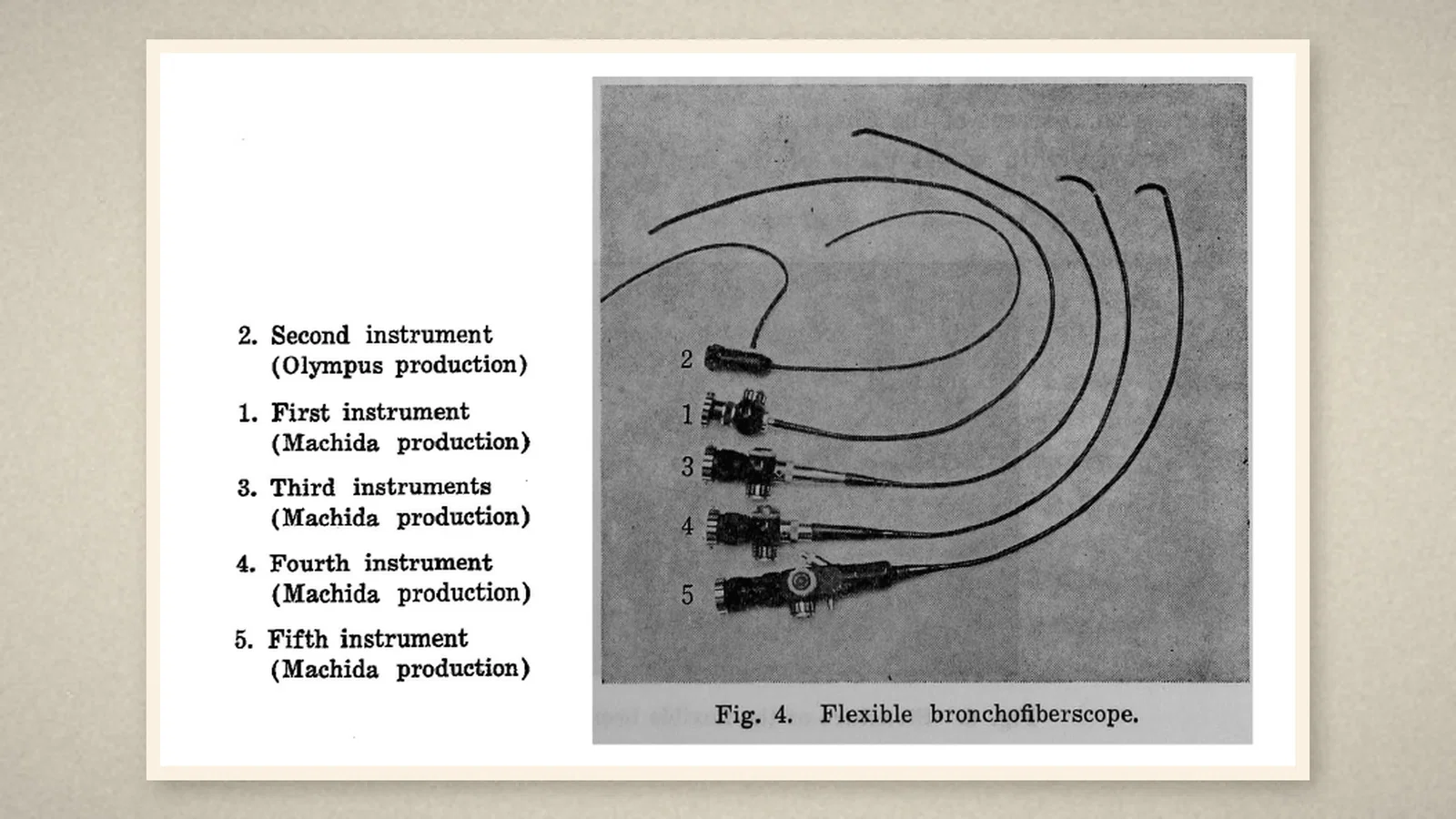
Even that first model did something the rigid scope could not. Because it was flexible, it could be used to look inside the bronchus "provided there was a prior intratracheal anesthesia tube"; the patient could be examined "in a natural position". Olympus delivered a second model in August 1966. The authors called these two "the first practical bronchoscope in the world" and reported them to the IX International Congress on Diseases of the Chest. [1, pp. 5-6]
Use showed the defects, and each new model answered one. The third, from Machida in October 1966, had a tip that could flex and a small hole for instilling anaesthetic. The fourth, in December, could bend into a U and carried biopsy forceps, and for the first time reached the segmental bronchi of both upper lobes. The fifth (March 1967) bent with a single handle; the sixth (July 1967) was more durable, with an external xenon light source that made photography and cinematography possible. [1, pp. 7-9]
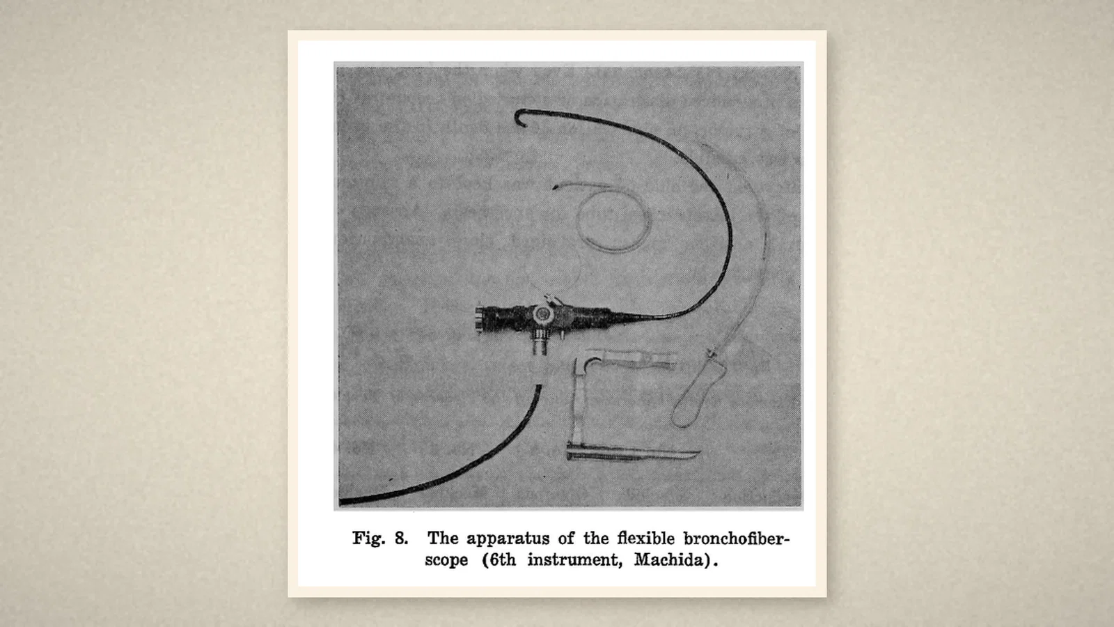
The tube
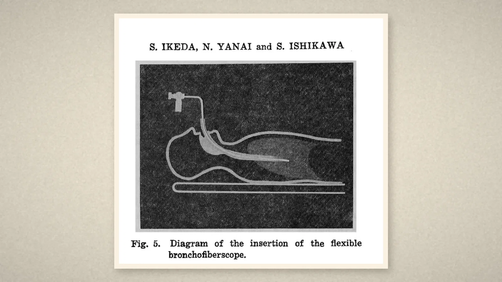
The airway came into the design. To insert the scope, "the rigid bronchoscope or intratracheal anesthesia tube should be inserted into the trachea and the flexible bronchofiberscope inserted through it", to keep the airway open, to suction sputum and to clean the tip. An ordinary anaesthesia tube "markedly alleviated" the patient's discomfort, but it deflected the tip upward so that only the front wall of the trachea could be seen, so the team built a soft intratracheal tube using Rüsch's armoured tube as the outer sheath. Since January 1967 they had examined 184 cases with the new scopes. [1, pp. 11-13]
The timeline's account
The Australian Society of Anaesthetists (ASA) timeline lists "Flexible bronchoscope invented" under 1966, by Dr Shigeto Ikeda "working in concert with Machida Endoscope Company (later Pentax) & Olympus Optical Company". Under 1965 it records the first flexible fibreoptic laryngoscopy for tracheal intubation, performed nasally by Dr Peter Murphy, a senior registrar in anaesthesia at the National Hospital for Nervous Diseases in London, after reading Shore and Lippman's Lancet article on a flexible choledochoscope; and under 1972 the first dedicated flexible fibreoptic laryngoscope, from the American Optical Company. [2, pp. 70, 73]
What the record shows
The year in which the bronchoscope learned to bend. The 1968 paper is a record of iteration, an outline handed to two firms and six machines in twelve months, each fixing what the last could not do. It also shows the airway and the scope becoming partners: from the first model, the flexible instrument went in through a tracheal tube, the arrangement anaesthetists would later turn around to put the tube in over the scope.
Sources and reading limits
Read: Ikeda, Yanai and Ishikawa, "Flexible bronchofiberscope", Keio Journal of Medicine 1968;17(1), in full (J-STAGE PDF with text layer, page images of pp. 1-12); the ASA timeline 2024, pp. 70 and 73.
Not covered: Murphy's report, "A fibre-optic endoscope used for nasal intubation", Anaesthesia 1967;22:489-91 (only the volume index is on the Internet Archive); Shore and Lippman's Lancet article; the congress report; the American Optical laryngoscope and the later fibreoptic intubation literature.
Limits: "the first practical bronchoscope in the world" is the authors' claim, not this article's. The 1965 Murphy intubation and the 1972 laryngoscope are the ASA's accounts and their sources were not read. Fibre counts, dimensions and case percentages are not repeated. Quotations are from the English original.
Three illustrations are crops of the Keio Journal of Medicine (pp. 5, 6 and 9); one is drawn from the paper.
- Ikeda S, Yanai N, Ishikawa S. Flexible bronchofiberscope. Keio J Med. 1968;17(1):1-16. J-STAGE
- Australian Society of Anaesthetists. Anaesthesia history timeline. 2024:70, 73. ASA
Continue exploring
Related reading connects shared questions and contexts; it does not by itself establish historical influence.
Anesthesia Epochs · The Source Room · About & Editorial Method
Published November 3, 2026. Sources and reading limits are stated above. Illustrations are labelled as original source images or AI-made. Historical accounts are educational; they are not current treatment instructions.