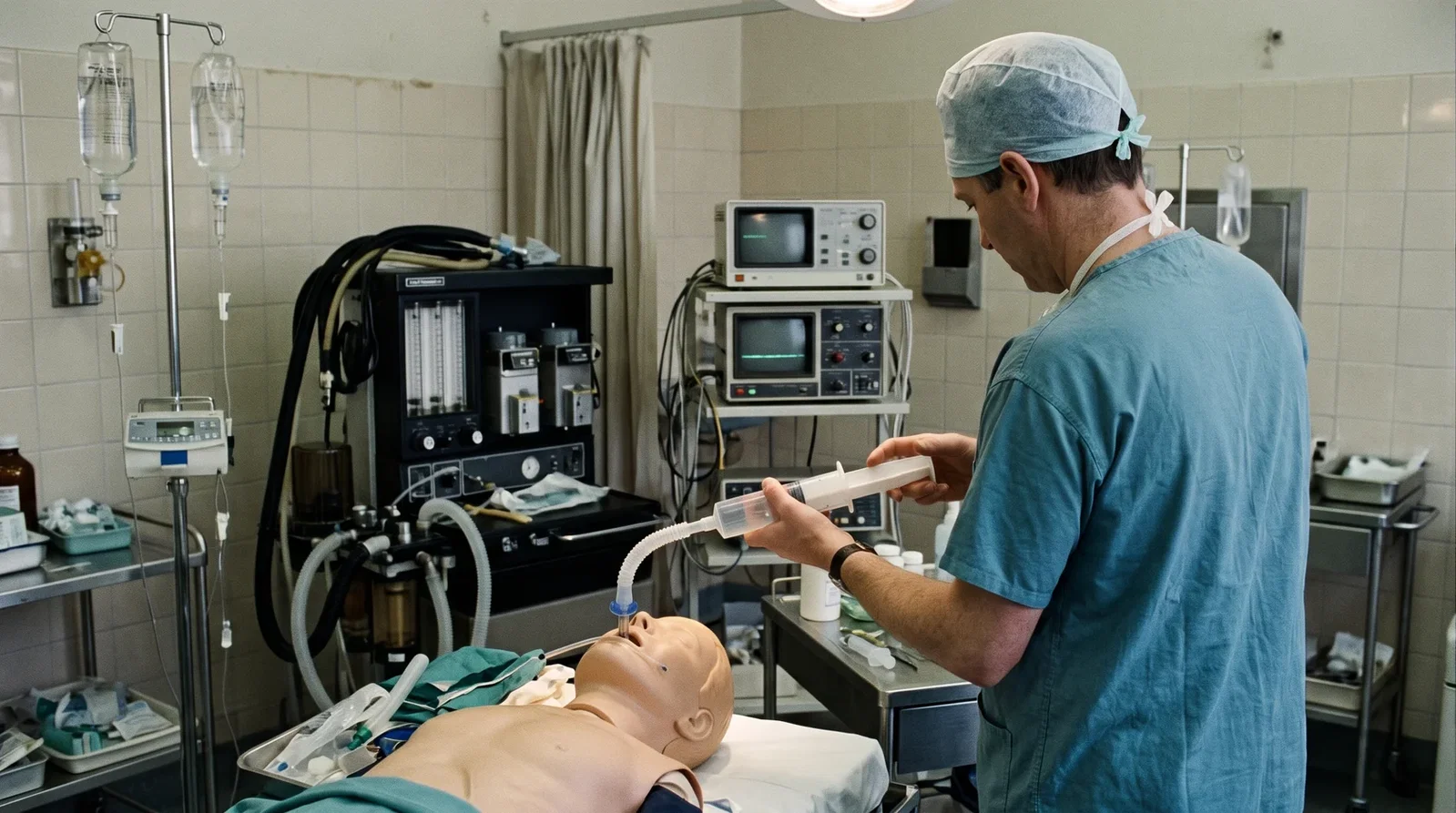
Anaesthesia 1988, vol. 43, no. 1, pp. 27-9 carried 'The oesophageal detector device: assessment of a new method to distinguish oesophageal from tracheal intubation' by M. Y. K. Wee of the Department of Anaesthetics, Ninewells Teaching Hospital, Dundee. The paper opens with the confidential enquiry into maternal deaths in England and Wales for 1979-1981: of twenty two deaths attributable to anaesthesia, eight were directly related to difficulty with tracheal intubation, and in four the tube was definitely in the oesophagus without this being recognised. The only test then considered totally reliable was the absence of carbon dioxide in exhaled gas, but the cost of analysers made them far from universal. The device rests on an anatomical difference: the oesophagus is a fibromuscular tube with no intrinsic structure to keep it open, while the trachea is held patent by its C-shaped cartilages. A large catheter-tip syringe is fitted through a standard catheter mount to the tracheal tube and the plunger withdrawn: from the trachea, gas is aspirated freely; from the oesophagus, the walls are drawn together around the tube, occluding the lumen and producing resistance or negative pressure. In a randomised, single-blind trial in 100 healthy adults, observers of differing experience made 99 correct identifications on the first test, 51 oesophageal and 48 tracheal; the one inconclusive test led to the finding of a tracheal tube almost completely blocked by secretions. The author stresses that the device must be checked for airtightness before use, and notes that patients with oesophageal or tracheal disease were not studied.
Reading Library · Anesthesia Epochs · 中文版
Watch the education video (for clinicians)
1988: The Oesophageal Detector Device — Pull Back and Feel · Anesthesia Epochs Education Series
Once the tube is in, how do you know it is in the trachea and not the oesophagus? By 1988 the question still killed people. In 1988 Anaesthesia, vol. 43, no. 1, pp. 27-9, published "The oesophageal detector device: assessment of a new method to distinguish oesophageal from tracheal intubation" by M. Y. K. Wee of the Department of Anaesthetics, Ninewells Teaching Hospital, Dundee. This article reads that paper. [1, p. 27]
Why a cheap method was wanted
The paper opens with the report on confidential enquiries into maternal deaths in England and Wales for 1979-1981: of twenty two deaths attributable to anaesthesia, eight were directly related to difficulty in tracheal intubation, and in four patients the tube was definitely placed in the oesophagus without this being recognised, with dire consequences. The author also cites anaesthetic accidents reported to the Medical Defence Union, where inadvertent misplacement of the tube in the oesophagus produced thirty seven deaths and thirteen cases of cerebral damage. [1, p. 27]
Many tests had been suggested, but the only one found to be totally reliable was the absence of carbon dioxide in exhaled gas, and that required analysers in operating theatres, intensive care, accident and emergency and resuscitation units, at a cost that was clearly prohibitive. Most anaesthetists relied on a high index of suspicion and clinical observation; faced with hypoxia after intubation many followed the adage "when in doubt, take it out", which causes its own problems if a correctly placed tube is removed and the hypoxia has another cause. Clearly, the paper concludes, there was still a need for an effective, simple and rapid method. [1, p. 27]
One anatomical difference
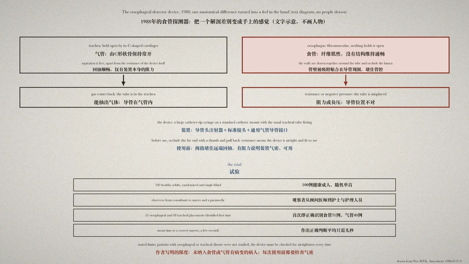
The device rests on the difference between the two tubes. The oesophagus is a fibromuscular tube about twenty five centimetres long in adults, running from the cricopharyngeal sphincter to the cardia of the stomach, with no intrinsic structure to maintain its patency. The trachea runs from the cricoid cartilage to the bifurcation and remains constantly patent because of its C-shaped cartilages, joined vertically by fibro-elastic tissue and closed behind by the trachealis muscle. [1, p. 27]
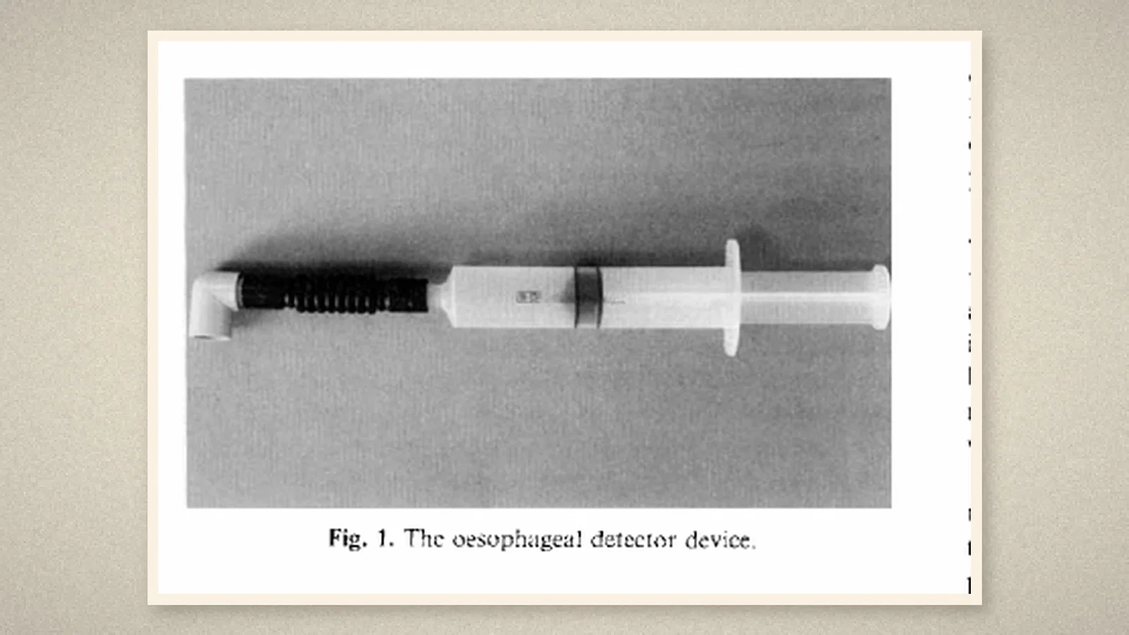
The apparatus is barely an invention: a large catheter-tip syringe fitted to one end of a standard catheter mount whose distal end carries the international 15 mm tracheal tube fitting. The author named it the oesophageal detector device. It must be airtight to work, which is tested by occluding the distal end with a thumb and pulling back the plunger; if resistance or negative pressure is felt, it is fit for use. [1, p. 27]
Using it takes seconds. Attach it to the tracheal tube, ensure an airtight seal, and withdraw the plunger. If the tube lies in the trachea, gas is aspirated from the lungs without resistance beyond that inherent in the device. If it lies in the unsupported oesophagus, withdrawal draws the walls of the oesophagus into apposition around the tube, occluding the lumen and causing negative pressure or resistance, which immediately alerts the clinician that the tube is misplaced. [1, p. 27]
A randomised, single-blind trial
One hundred healthy adults took part. So that the test could be judged blind, the oesophagus was intubated as well, under direct vision to minimise trauma. The lungs were ventilated through the tracheal tube with oxygen and a deepened volatile anaesthetic, the oesophageal tube was then inflated with two tidal volume compressions of the reservoir bag to simulate the usual procedure after intubation, and the breathing system was disconnected from both tubes. A second observer, who had not witnessed the anaesthesia, was then called in to judge one of the two tubes using only the device. [1, p. 28]
The observers ranged widely in experience: eleven consultants, three senior registrars, eleven registrars and six senior house officers, together with three surgeons, five anaesthetic nurses and an RAF paramedic. Tracheal tubes from seven makers were used, in internal diameters from 7.5 to 9.5 mm. [1, p. 28]
There were ninety nine first-time correct identifications, 51 oesophageal and 48 tracheal. The mean time taken to diagnose placement correctly was 6.9 seconds, with a range of 5 to 16 seconds. [1, p. 28]
On one occasion an observer was unsure about a tube in the trachea: gas aspirated freely at first, then met some resistance, then lost it again. That patient had been intubated into the right bronchus before the test and the tube repositioned; airway pressure stayed high while the chest was clear on auscultation. After the inconclusive result the tube was replaced and found almost totally blocked by thick purulent secretions; repeated in the same patient, the test indicated correct placement unequivocally. [1, p. 28]
The author's cautions
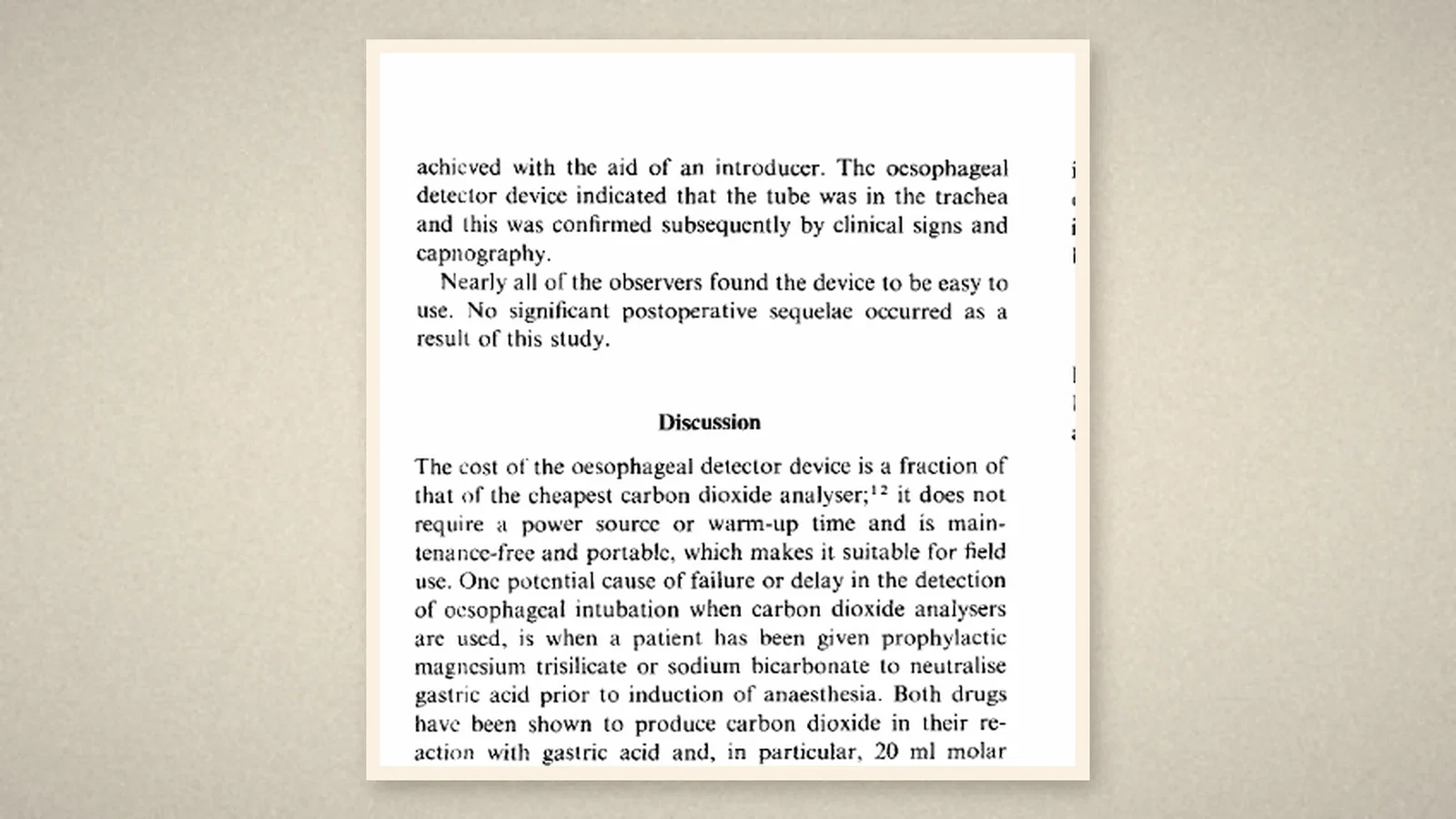
The discussion begins with cost: the device costs a fraction of the cheapest carbon dioxide analyser, needs no power source or warm-up time, is maintenance-free and portable, and so suits field use. The author also notes one way a carbon dioxide analyser can mislead or delay, when drugs given before induction to neutralise gastric acid produce carbon dioxide in reacting with it, though no such case had yet been reported. [1, p. 29]
The limits are set out plainly. The study did not include patients with oesophageal or tracheal disease, where the normal anatomy may be distorted or altered. It cannot be overemphasised, the paper says, that the device must always be checked for airtightness before use and that the fittings between catheter mount, syringe and tracheal connector must be airtight. A confirmatory test is mentioned, injecting a bolus of air through the tube while listening over the epigastrium for bubbling, but it was not found necessary in this study. [1, p. 29]
What the record shows
The reasoning is plain: if the two tubes differ in structure, use something that turns that difference into a feel in the hand. A syringe and a connector gave, in a few seconds at the bedside, an answer that otherwise needed costly apparatus, and the author set out in the same paper where it does not apply and what must be checked before it is trusted.
Sources and reading limits
Read: Wee MYK, "The oesophageal detector device: assessment of a new method to distinguish oesophageal from tracheal intubation", Anaesthesia 1988, vol. 43, no. 1, pp. 27-9, in full, including the summary, the anatomical rationale, the apparatus and technique, the results, the discussion and the references.
Not covered: the confidential enquiry report and the case reports and reviews cited in the paper; later evaluations and variants of the device.
Limits: no doses or injected volumes are reproduced here, so figures such as the cuff inflation volume in the original are omitted. The deliberate oesophageal intubation used to blind the trial is reported as the paper states it. The make-up of the observers and the tube sizes are given as listed. The paper's own summary gives "51 oesophageal and 49 tracheal intubations", counting the blocked tracheal tube; the results section gives ninety nine first-time correct identifications, 51 oesophageal and 48 tracheal, and this article follows the results. Figure 2 of the paper is a photograph of the device in use on a patient and is not used as an illustration here, because this site does not reproduce patient photographs.
Three illustrations are taken from the 1988 paper (p. 27, Figure 1, and p. 29). One is a diagram drawn from the text, with no people drawn.
- Wee MYK. The oesophageal detector device: assessment of a new method to distinguish oesophageal from tracheal intubation. Anaesthesia. 1988;43(1):27-9. doi:10.1111/j.1365-2044.1988.tb05419.x
Continue exploring
Related reading connects shared questions and contexts; it does not by itself establish historical influence.
Anesthesia Epochs · The Source Room · About & Editorial Method
Published November 5, 2026. Sources and reading limits are stated above. Illustrations are labelled as original source images or AI-made. Historical accounts are educational; they are not current treatment instructions.