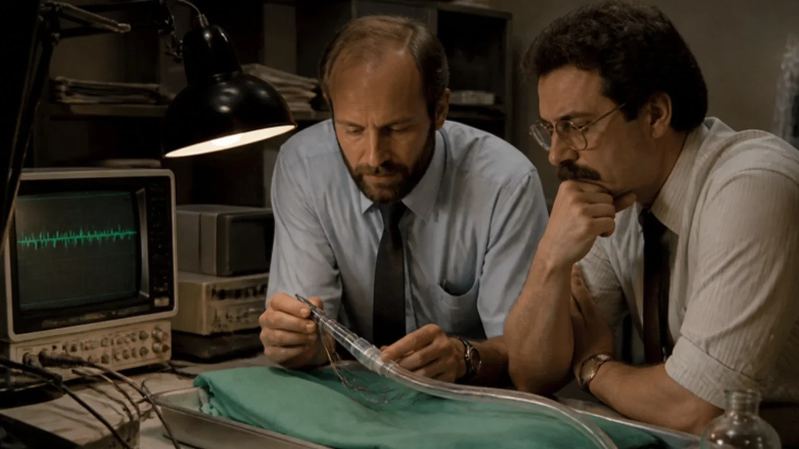
On 29 November 1989 Andrew C. Goldstone and Raymond L. Schettino of Baltimore filed the application that became US Patent 5,024,228, 'Electrode endotracheal tube', granted on 18 June 1991: insulated electrode wires run parallel to the axis of an ordinary ventilating tube and are exposed only where they lie against the vocal cords once the tube is in place, so that laryngeal muscle EMG can warn a surgeon of contact with a recurrent laryngeal nerve. Nerve damage, the patent says, is a common complication of neck surgery, causing paralysis, loss of speech and disturbed breathing; pressure cuffs, needle electrodes and oesophageal electrodes all had drawbacks, while anaesthesiologists already knew how to place tracheal tubes. The ASA timeline dates the EMG electrode endotracheal tube to 1990.
Reading Library · Anesthesia Epochs · 中文版
Watch the education video (for clinicians)
1989: The EMG Endotracheal Tube — The Tube That Listens · Anesthesia Epochs Education Series
During thyroid and other neck operations, the tube the anaesthetist places already runs between the two vocal cords. In 1989 two inventors in Baltimore saw in that position an opportunity: if the tube itself carried electrodes, it could listen to the laryngeal muscles while it ventilated the lungs, and tell the surgeon when an instrument touched the nerve that moves them. [1]
The nerve at risk
Andrew C. Goldstone and Raymond L. Schettino filed "Electrode endotracheal tube" on 29 November 1989; it was granted as US Patent 5,024,228 on 18 June 1991, and a divisional patent, 5,125,406, followed on 30 June 1992. The background is a surgeon's problem. The two recurrent laryngeal nerves run through the neck to the muscles of the larynx, each controlling a vocal cord, and "damage to laryngeal nerves is a common complication of neck surgery". Tumour, inflammation or trauma can make it nearly impossible to dissect the nerve without harm, and paralysis of the laryngeal muscles "results in loss of speech and may also disrupt breathing". [1][2]
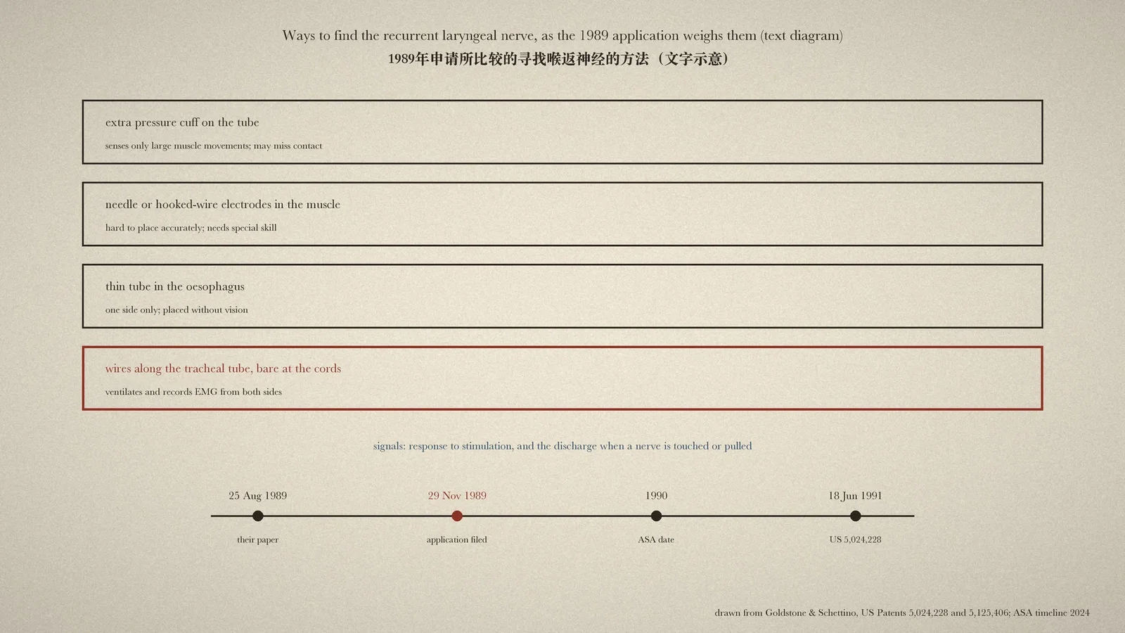
The patent then weighs what existed. A tube with an extra cuff, which registered pressure changes when the laryngeal muscles contracted after the nerve was stimulated, could "only detect relatively large movements" and might miss the smaller discharge when an instrument touched the nerve. Needles, needle pairs and hooked wires placed in the muscle could record that discharge, but were "difficult to accurately emplace" and demanded special skill. A thin tube with ring electrodes in the oesophagus could watch only one side and had to be placed blind. [1, cols. 1-2]
Wires along the tube
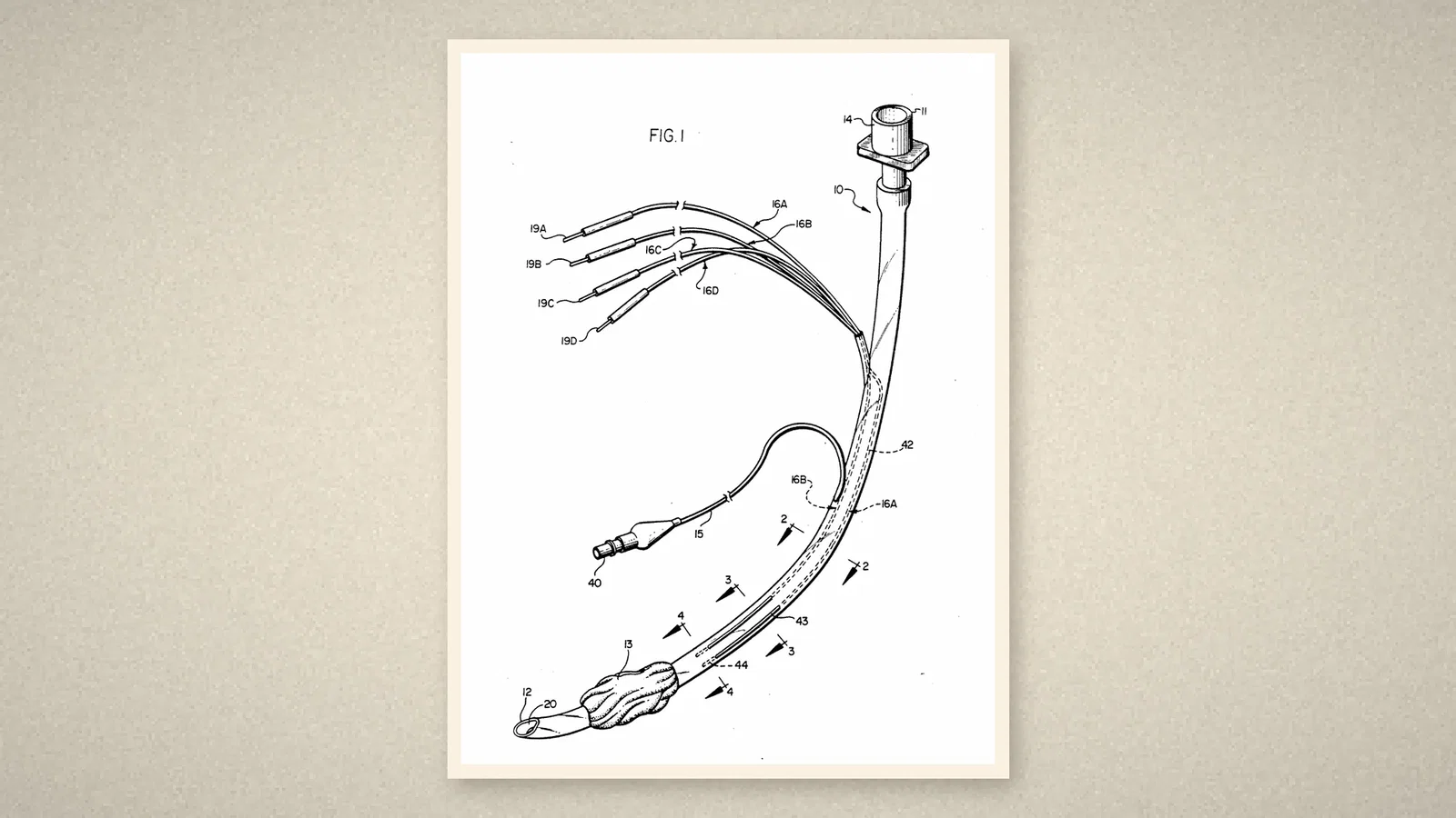
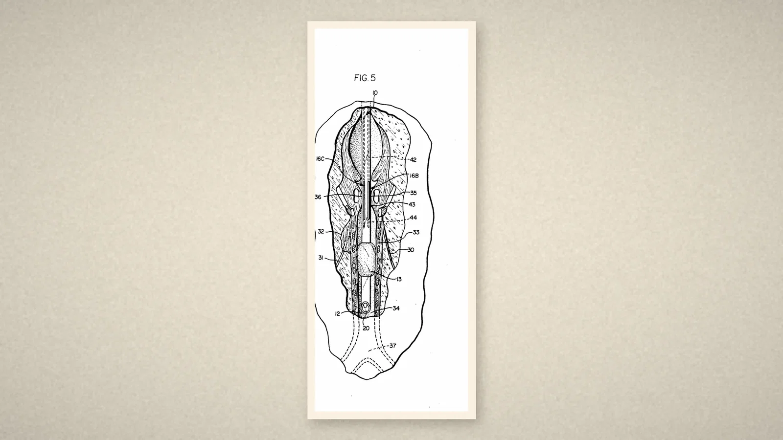
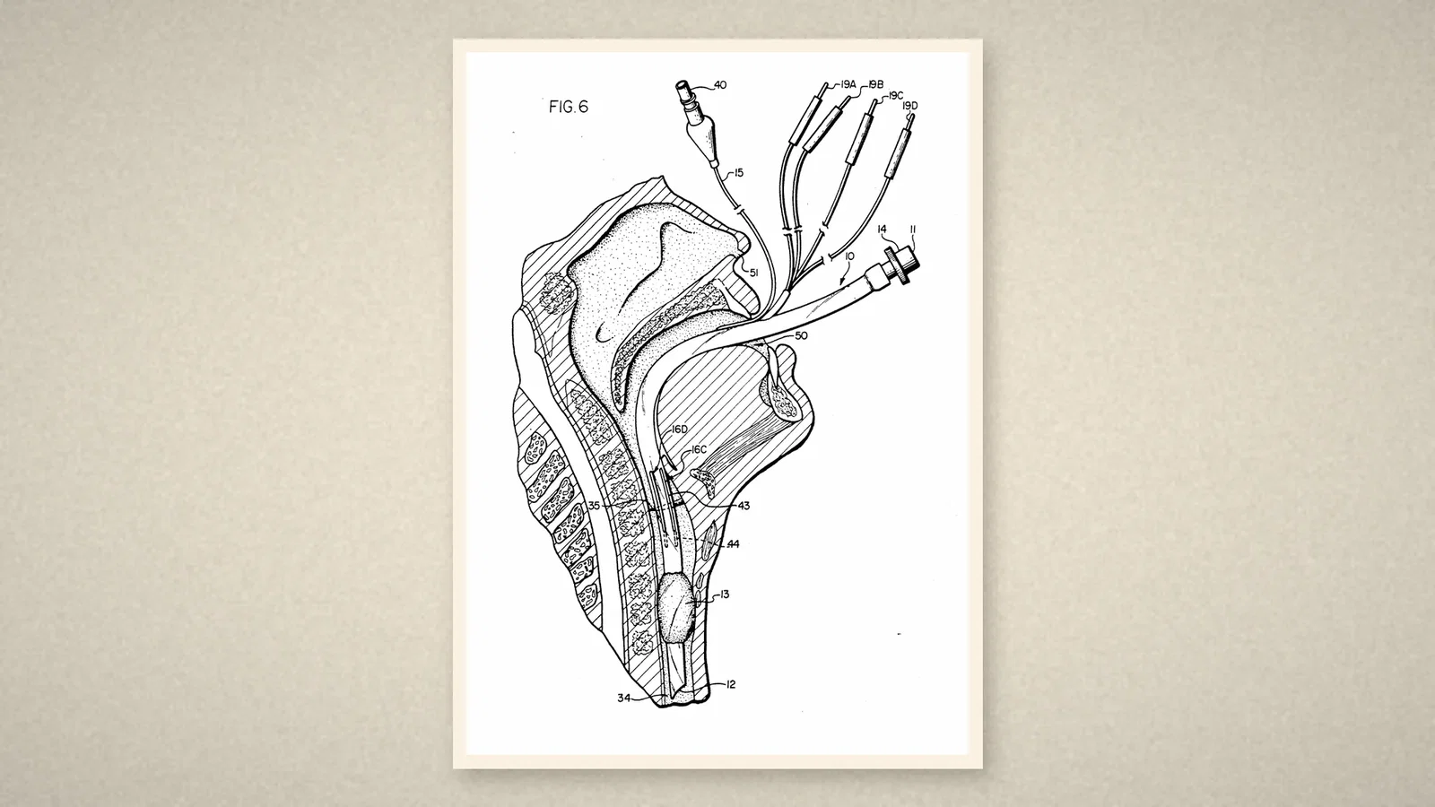
The answer was "an endotracheal tube which performs the functions of ventilating the lungs during surgery and monitoring the EMG signals of the laryngeal muscles". Electrode wires run parallel to the tube's axis, insulated inside its wall and bare only along a short stretch near the tip. With the tube correctly placed, tip above the carina and cuff below the laryngeal muscles, the bare stretches lie against the muscles on each side, "particularly a vocal cord". [1, col. 3]
Two kinds of signal could be recorded: the response when the surgeon stimulated the nerve with a probe, and the discharge that occurs when a nerve is touched or pulled, so that the tube could "alert a surgeon when potentially damaging contact with a laryngeal nerve is imminent". Under light anaesthesia, breathing-related muscle activity also showed that the tube was in the right place. And it was easy to place, because "many doctors, particularly anesthesiologists, are already skilled in the insertion of those tubes". [1, cols. 3-4]
The timeline's account
The Australian Society of Anaesthetists (ASA) timeline lists "The EMG electrode endotracheal tube invented" under 1990, credited to Drs Andrew C Goldstone and Raymond L Schettino of Baltimore, and notes that it is now made by Medtronic as the "Nerve Integrity Monitoring Electromyographic Endotracheal Tube", used to monitor the recurrent laryngeal and vagus nerves during neck surgery. [3, p. 82]
What the record shows
The moment the anaesthetist's tube became the surgeon's monitor. Nothing about the airway changed; what changed was that the one object guaranteed to sit between the vocal cords was made to report on them. It is a small idea with a large consequence: the person who manages the airway now also watches the nerve the surgeon is trying to save.
Sources and reading limits
Read: US Patent 5,024,228, front page, background, summary and list of drawings; US Patent 5,125,406, front page and related-application data; the ASA timeline 2024, p. 82.
Not covered: the inventors' paper of 25 August 1989 cited in the patents; clinical studies of the tube; the manufacturer's literature; the earlier papers on cuff, needle and oesophageal methods cited in the patents.
Limits: the two patents give the parent application's filing date as 29 November and 22 November 1989; this article uses the date printed on the parent patent. The ASA dates the invention to 1990. The patents' front pages carry the inventors' home addresses and are not reproduced. The advantages stated are the patent's own. Dimensions are not repeated. Quotations are from the English originals.
Three illustrations are crops of the drawing sheets of US Patent 5,024,228; one is drawn from the patents.
- Goldstone AC, Schettino RL. Electrode endotracheal tube. US Patent 5,024,228. 18 Jun 1991 (filed 29 Nov 1989). Google Patents
- Goldstone AC, Schettino RL. Electrode endotracheal tube. US Patent 5,125,406. 30 Jun 1992. Google Patents
- Australian Society of Anaesthetists. Anaesthesia history timeline. 2024:82. ASA
Continue exploring
Related reading connects shared questions and contexts; it does not by itself establish historical influence.
Anesthesia Epochs · The Source Room · About & Editorial Method
Published November 3, 2026. Sources and reading limits are stated above. Illustrations are labelled as original source images or AI-made. Historical accounts are educational; they are not current treatment instructions.