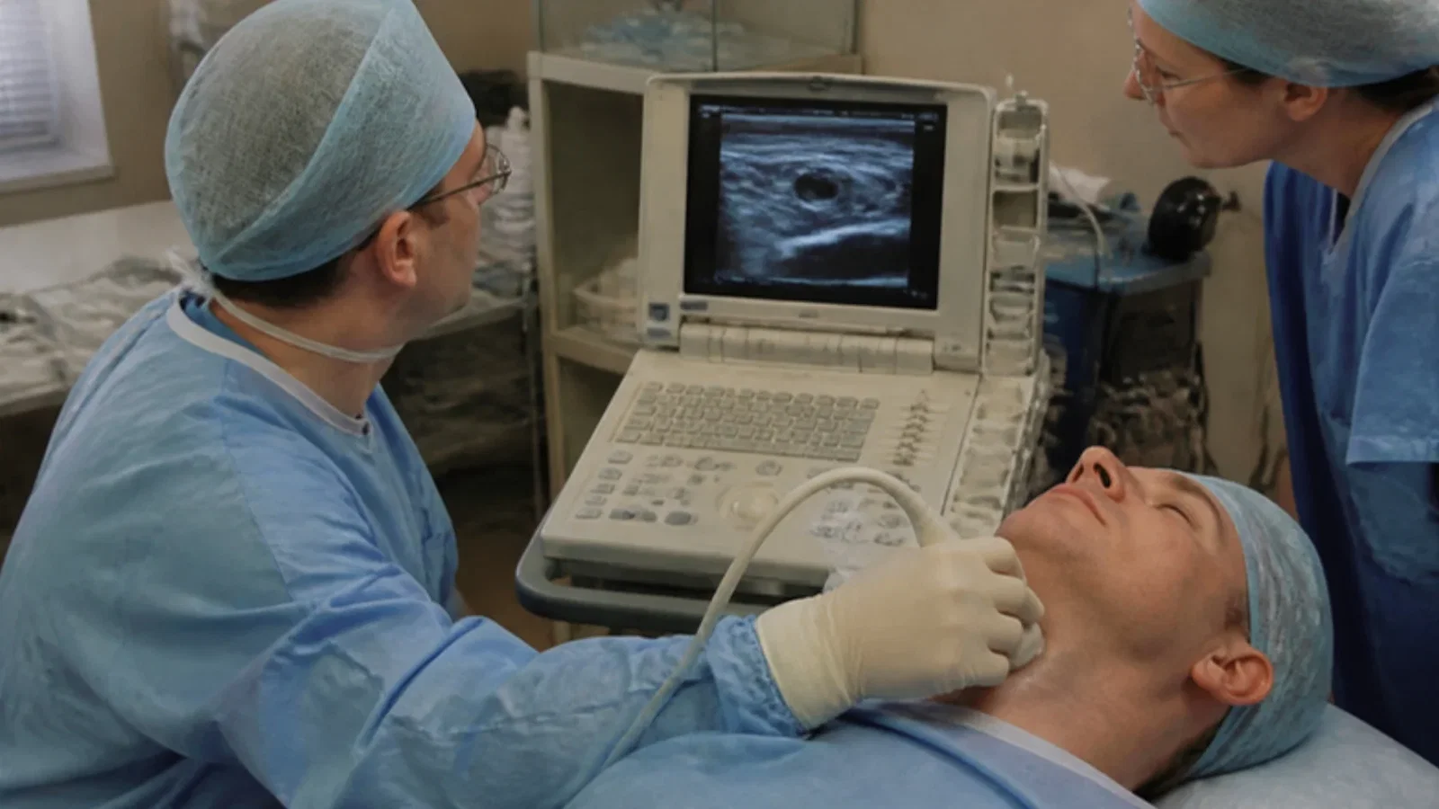
A. Hatfield and A. Bodenham of Leeds General Infirmary, in 'Ultrasound: an emerging role in anaesthesia and intensive care', observed that diagnostic and interventional ultrasound was established in medicine but rarely performed by anaesthetists or intensivists; illustrating every image with a portable machine bought with charitable funds, they discussed vascular access, pleural drainage, the airway and nerve block, hoped that seeing nerves directly would make needle placement more accurate, acknowledged that others questioned the evidence, and called for training and standards; the History of Anaesthesia Society timeline marks 1999 as the year reviews recommending routine ultrasound began to appear.
Reading Library · Anesthesia Epochs · 中文版
Watch the education video (for clinicians)
1999: Hatfield and Bodenham — Ultrasound in the Anaesthetist's Hands · Anesthesia Epochs Education Series
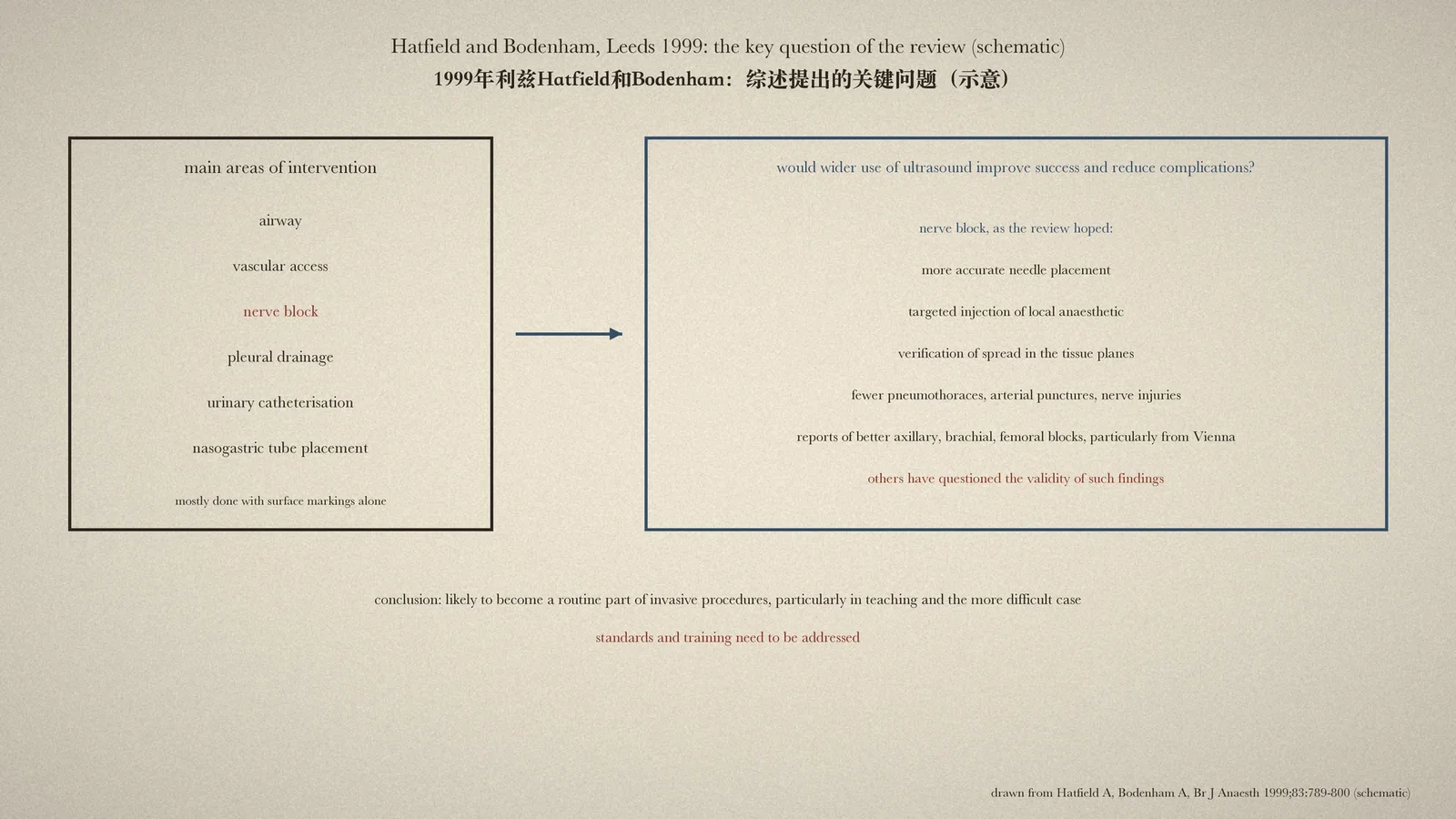
Most needles in anaesthesia and intensive care, the review discussed here observes, were placed with surface markings alone. The History of Anaesthesia Society (HAS) timeline records a turning point under 1999: "First reviews are published recommending the routine use of ultrasound for regional nerve blocks and placement of central venous lines." The entry names no review. This article reads one of that year: A. Hatfield and A. Bodenham, "Ultrasound: an emerging role in anaesthesia and intensive care", a review article in the British Journal of Anaesthesia from Leeds General Infirmary. [1, p. 789][2]
Ultrasound in the hands of the anaesthetist
The review opens with an observation: diagnostic and interventional ultrasound had "an established role in many areas of medicine but are not often performed by anaesthetists or intensive care physicians". The authors set out to discuss uses "which we envisage to be performed, in person, by such clinicians", and in particular B mode ultrasound "to improve the efficacy of interventional techniques". Their own experience had centred on vascular access, pleural drainage and the airway. They apologised in advance for being "oversimplistic", preferring that to putting readers off with physics. [1, p. 789]
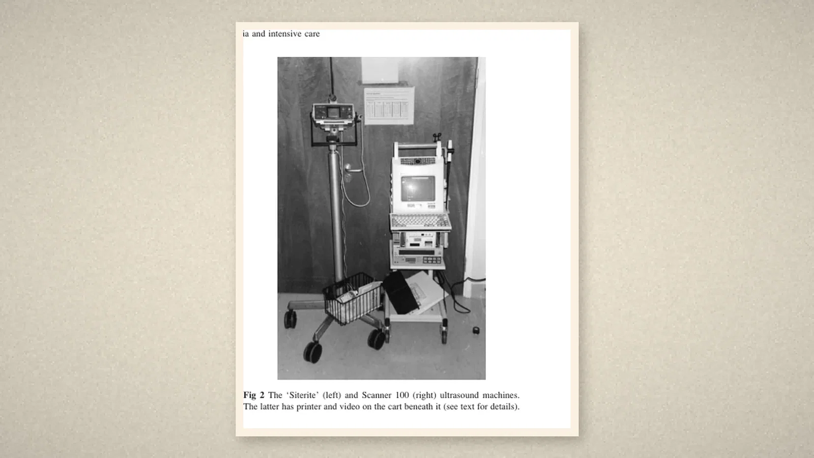
The equipment was modest. The department had bought "with charitable funds" a small machine designed for vascular access and a general purpose portable machine, the Scanner 100, and "all images in this review were generated with this device, rather than larger more sophisticated machines". The authors were also frank about their own background: "We have received no specific training in the use of ultrasound devices", only advice from radiologists and radiographers, and they knew of no training literature specific to their specialties. Radiologists had raised valid concerns that a lack of training might lead to missed or wrong diagnoses; yet clinicians from outside radiology, the review argued, could rapidly become proficient in limited areas. [1, pp. 790-1]
The key question
The main interventions of anaesthesia and intensive care, the review listed, were "airway, vascular access, nerve block, pleural drainage, urinary catheterization and nasogastric tube placement", and most were done with surface markings alone, although obscured or altered anatomy could make them difficult. "The key question is whether the wider use of ultrasound to view anatomy and guide interventions would improve the overall success and reduce complication rates of such procedures?" For vascular access the evidence was growing: the review cited a meta-analysis of randomised studies which concluded that ultrasound improved success, reduced the number of passes and decreased complications of internal jugular and subclavian catheterisation. Even so, many colleagues were unwilling to try it, seeing it as difficult to learn, time consuming, of little help to the experienced, and "an admission of defeat or inadequacy". [1, pp. 792-3]
Nerve block
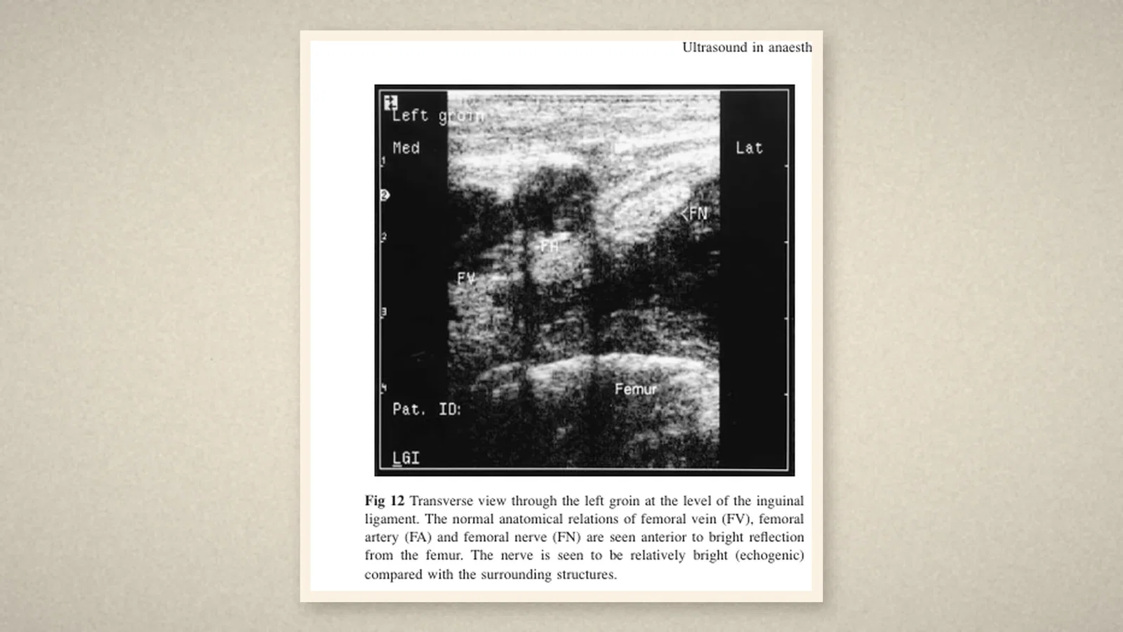
Nerves, the authors explained, reflect ultrasound much like muscle and other soft tissues, but larger nerves can be seen, especially when a neighbouring vessel shows where to look; the probe should be perpendicular to the nerve. Fig. 12 shows the femoral vein, artery and nerve in the groin. "In the context of anaesthesia, it is hoped that direct imaging of nerves allows more accurate needle placement, targeted injection of local anaesthetic agent and verification of spread in the appropriate tissue planes." There were reports that ultrasound improved the success of axillary, brachial and femoral nerve blocks, "particularly from Vienna", but "others have questioned the validity of such findings". Accurate placement should reduce pneumothorax, arterial puncture and direct nerve damage, and blocks that were hard to locate, such as the sciatic, might gain interest if the nerve could be seen. Around the spine, ultrasound might show the bony landmarks and the depth of structures; the review cited Currie's measurement of the depth to the lumbar epidural space. [1, p. 797]
The conclusion
The review ends with a forecast, not a mandate. Small, competitively priced, high resolution devices were "likely to lead to increased use", and "it is likely that, in the future, such techniques will become a routine part of invasive procedures, particularly in teaching and in the more difficult case. Issues of standards and training for ultrasound use in these areas need to be addressed." [1, p. 798]
What the record shows
The review shows a technique at the moment it crossed from one specialty to another: two clinicians with charitable money, two portable machines, no specific training, and a clear question about success and complications. It is candid about what was not yet known. For nerve block it offered hope and reported dissent side by side. The HAS reads it as a recommendation for routine use; the text itself speaks more cautiously of what was likely to come.
Sources and reading limits
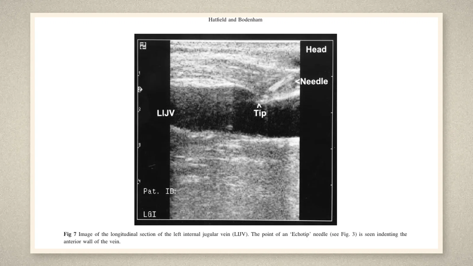
Read: Hatfield A, Bodenham A, Br J Anaesth 1999;83:789-800, in full, from the PDF (text layer), with page images of pp. 789, 791 (Fig. 2), 794 (Figs. 7-9) and 797 (Fig. 12); the HAS timeline entry for 1999.
Not covered: the other reviews that the HAS entry may mean (it speaks of "reviews" in the plural and names none); the Vienna studies of nerve block that the review cites, and the report that questioned them; the editorial and the vascular access meta-analysis that it cites. The HAS's "First reviews" is quoted as the society's; this article makes no "first" claim. The review's text dates Currie's measurement to 1989, while its reference list gives Br J Anaesth 1984; Currie's paper was not read, so the discrepancy is noted, not resolved. Machine prices, probe frequencies and needle sizes are not repeated. Quotations are from the English original.
Three illustrations are crops of pp. 791, 794 and 797, including Figs. 2, 7 and 12; one is drawn from the text.
- Hatfield A, Bodenham A. Ultrasound: an emerging role in anaesthesia and intensive care. Br J Anaesth. 1999;83(5):789-800. BJA
- History of Anaesthesia Society. Timeline. HAS
Continue exploring
Related reading connects shared questions and contexts; it does not by itself establish historical influence.
- 1978: A Doppler Probe Guides the Supraclavicular Brachial Plexus Block
- 1994: A Brachial Plexus Study Watches the Needle and Local Anesthetic
Anesthesia Epochs · The Source Room · About & Editorial Method
Published November 2, 2026. Sources and reading limits are stated above. Illustrations are labelled as original source images or AI-made. Historical accounts are educational; they are not current treatment instructions.