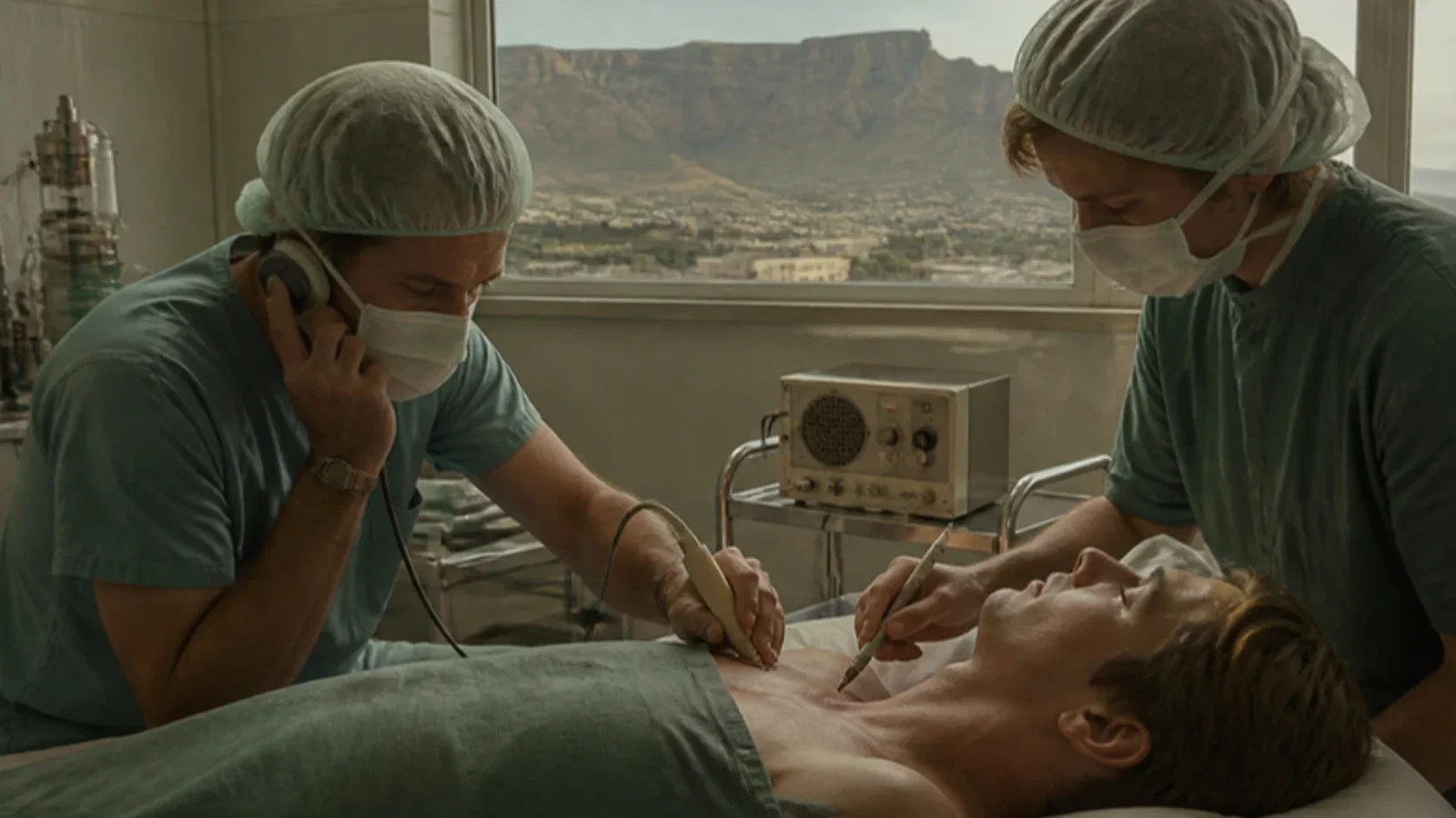
La Grange, Foster and Pretorius of Tygerberg Hospital, South Africa, reported in the British Journal of Anaesthesia that, because the plexus trunks lie close to the great vessels, a standard 9.5 MHz Doppler bloodflow detector could locate the third part of the subclavian artery, marked on the skin with water-soluble dye, before supraclavicular block; in 61 adults for orthopaedic surgery success was 98 per cent with no pneumothorax or neurological sequelae, even in obese patients or distorted anatomy.
Reading Library · Anesthesia Epochs · 中文版
Watch the education video (for clinicians)
1978: Doppler-Guided Supraclavicular Block — Tygerberg Hospital · Anesthesia Epochs Education Series
Ultrasound guidance in regional anaesthesia has a history. The Australian Society of Anaesthetists (ASA) timeline places in 1978 what it calls the "first use of Doppler ultrasound to locate nerves", by P. du P. la Grange and colleagues, for supraclavicular brachial plexus block, and links it to an echogenic needle patented in 1981 and an ultrasound-guided axillary block in 1992. This article reads the 1978 paper: "Application of the Doppler ultrasound bloodflow detector in supraclavicular brachial plexus block", by P. du P. la Grange and P. A. Foster of the department of anaesthesia and L. K. Pretorius of orthopaedics at Tygerberg Hospital, South Africa, in the British Journal of Anaesthesia. [1][2]
The problem with the supraclavicular route
The authors noted that the supraclavicular approach "produces a more extensive area of blockade than the axillary approach for the same dose of local anaesthetic", but that the axillary approach carried less risk of pneumothorax and, to a lesser extent, of arterial puncture and haematoma. "Any method which may decrease the problems of the supraclavicular approach warrants attention." [1]
Finding the artery
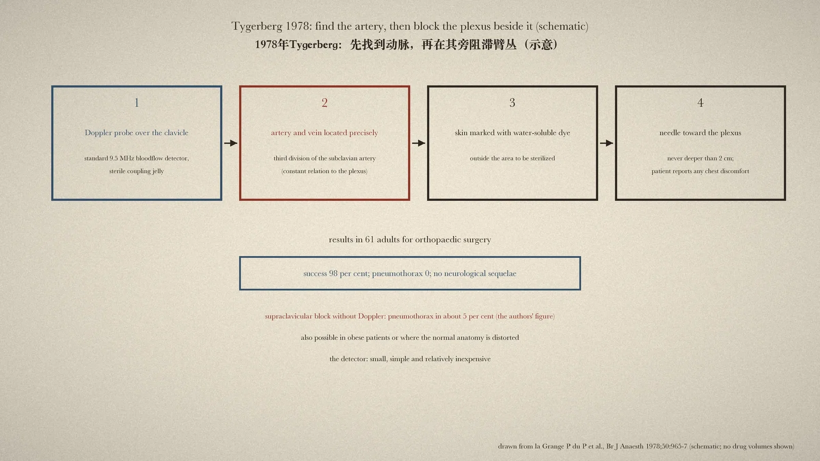
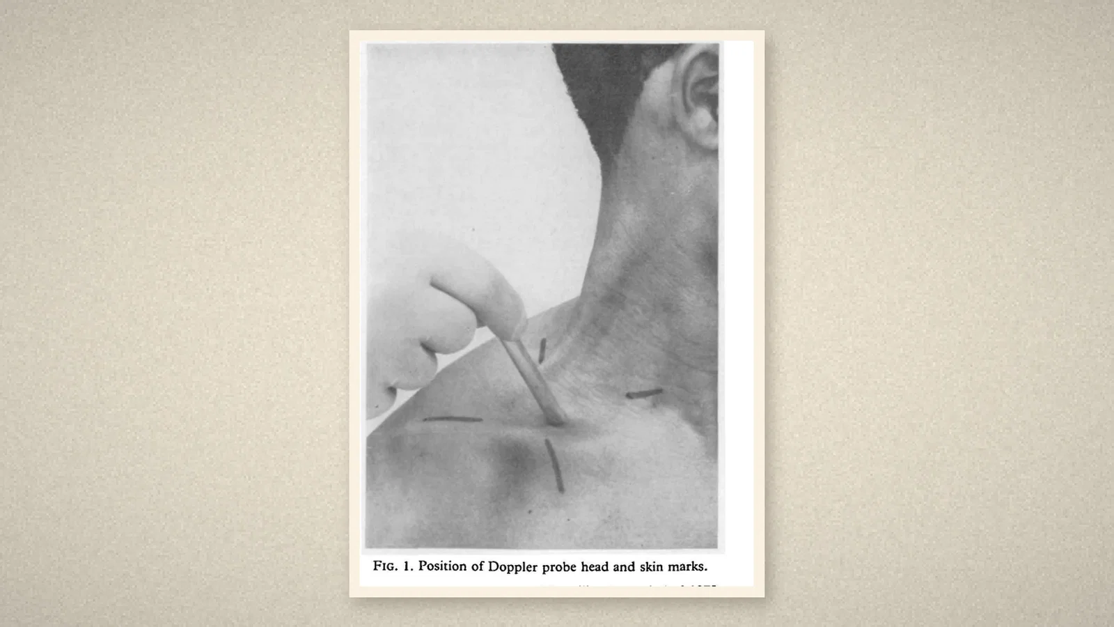
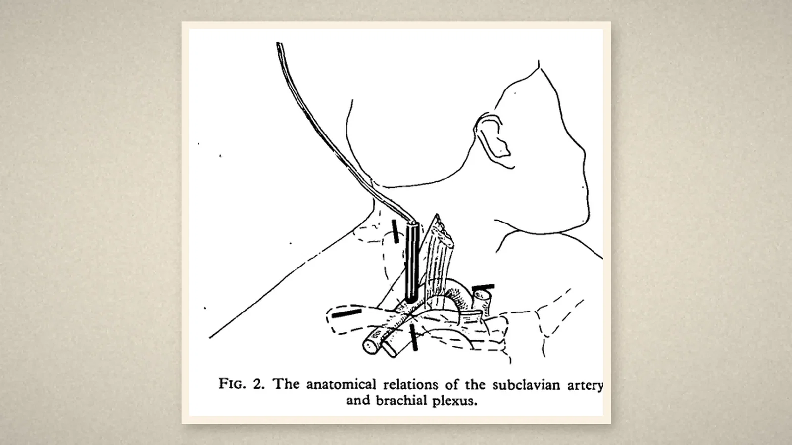
The method rested on anatomy: "The major nerve trunks lie close to the major vessels." On the basis of "a constant relationship between the third division of this artery and the six divisions of the three trunks of the brachial plexus", locating the subclavian artery should allow accurate placement of the local anaesthetic. The authors used "a standard 9.5-mHz ultrasonic Doppler bloodflow detector". With the patient supine, the arm at the side, the chin turned away and the shoulder depressed, the artery was palpated above the midpoint of the clavicle; "if there was any doubt about its location", the probe head, containing transmitting and receiving elements, was applied with sterile coupling jelly. "The artery and its accompanying vein were then located precisely and marks made with a water-soluble dye on the skin outside the area to be sterilized." [1]
The needle was then directed towards the plexus. Where paraesthesiae could not be found at once, further probing was careful: "Care was taken never to penetrate more than 2 cm and the patient was instructed to report immediately any discomfort in the chest." [1]
Results
Sixty-one adults having orthopaedic operations, such as carpal tunnel and tendon surgery, were treated. Table I, which set the series beside earlier published ones, gives the present study 98 per cent success with no pneumothorax and no other complication. "No neurological sequelae were found. In addition there were no symptoms or clinical evidence of pneumothorax." By contrast, the authors wrote, supraclavicular blocks "performed without Doppler control have been associated with pneumothorax in approximately 5% of the patients". The method also made it possible to block "the obese patient or where the normal anatomy is distorted". [1]
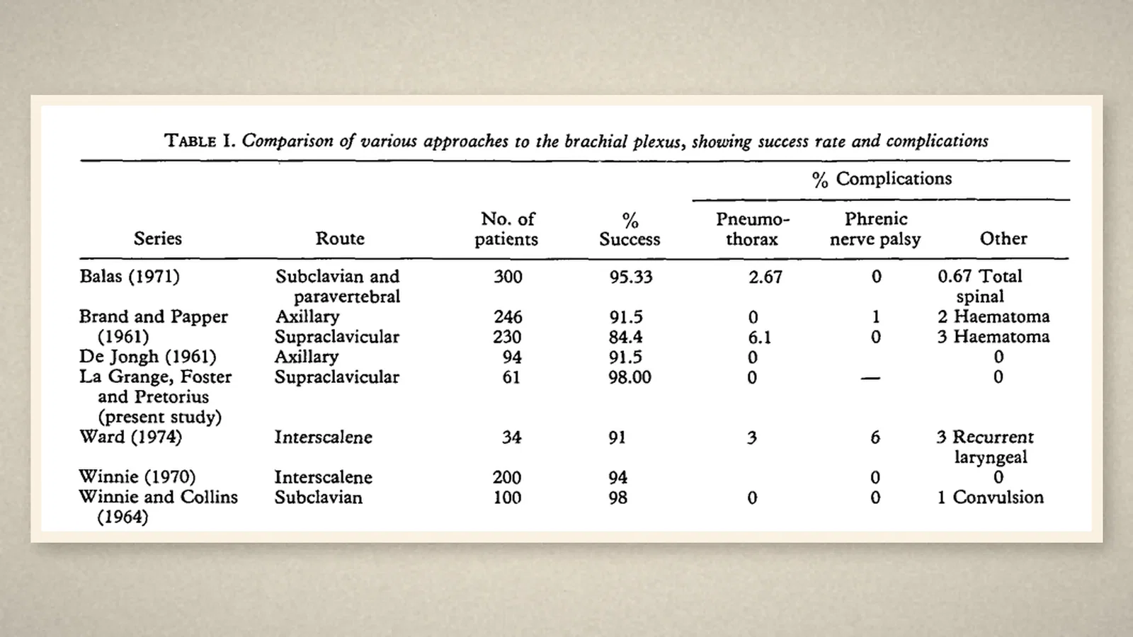
The conclusion was confident: with the Doppler detector "it is possible to block the brachial plexus as successfully by the supraclavicular approach as by any other technique, and virtually without complications". The detector was "a small, simple and relatively inexpensive apparatus". [1]
What the record shows
The Tygerberg paper did not image nerves. It used sound to find the artery and relied on the fixed anatomical relationship between artery and plexus. The idea that a portable ultrasound device could replace guesswork at the bedside, and make a feared approach safer, is already present. The ASA's later entries show the same line continuing into needles designed to reflect ultrasound and into ultrasound-guided axillary block.
Sources and reading limits
Read: la Grange P du P, Foster PA, Pretorius LK, Br J Anaesth 1978;50:965-7, in full from the PDF (text layer), with page images of pp. 965-7, including Figs. 1-2 and Table I; the ASA timeline entries for 1978, 1981 and 1992.
Not covered: the comparison series in Table I, beyond the figures printed there; the 1981 echogenic needle patent; the 1992 axillary block report; an ASA timeline item for 2006, which was not located; later ultrasound-guided block literature. The ASA's "first use of Doppler ultrasound to locate nerves" is quoted as its own; the paper located the artery, not the nerves. Table I combines other authors' series and is not a controlled comparison. Local anaesthetic volumes and concentrations are not repeated. Patients are not identified. Quotations are from the English original.
Three illustrations are crops of pp. 965-6, including Figs. 1-2 and Table I; one is drawn from the text.
- la Grange P du P, Foster PA, Pretorius LK. Application of the Doppler ultrasound bloodflow detector in supraclavicular brachial plexus block. Br J Anaesth. 1978;50(9):965-7. BJA
- Australian Society of Anaesthetists. Anaesthesia history timeline. 2024. ASA
Continue exploring
Related reading connects shared questions and contexts; it does not by itself establish historical influence.
- 1885: Halsted and the Brachial Plexus Block, an Attribution Without a Paper
- 1911: Two Needles for the Brachial Plexus
Anesthesia Epochs · The Source Room · About & Editorial Method
Published November 2, 2026. Sources and reading limits are stated above. Illustrations are labelled as original source images or AI-made. Historical accounts are educational; they are not current treatment instructions.