The open connection described in 1937 and the later work of revising and analyzing it.
Reading Library · Anesthesia Epochs · 中文版
Watch the companion film
1937: Ayre’s T-Piece—A Simpler Breathing Circuit for Babies · Watch on YouTube
Historical anchor: 1937. Date details and source qualifications are explained below.
A small strip of gauze appears in Philip Ayre’s 1937 account of anesthesia for infant cleft surgery. Attached near the open end of a short tube, it moved with the patient’s breathing. A modest detail, it points to the practical question behind the apparatus: after securing the airway, how could the anesthetist let a baby breathe through the equipment and see that breathing was continuing? [1, p. 132]
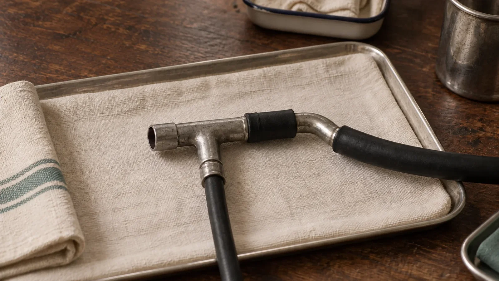
Historical concept illustration, not a photograph of an original museum object or an assembly drawing for clinical use.
The connection beyond the tracheal tube
Ayre’s report came from the Babies’ Hospital in Newcastle upon Tyne. His patients already had oral rubber tracheal catheters placed using Magill’s technique. The difficulty he described lay partly beyond that secured route: the earlier nitrous oxide and oxygen arrangement had proved unsatisfactory, even with a smaller rebreathing bag. He recorded disturbed breathing, poor color and troublesome operating conditions. [1, p. 131]
The replacement connected the catheter through a short tube and angle piece to a T junction. Oxygen and ether vapor entered one branch; another remained open to the surrounding air, with an optional short extension. A rebreathing bag and expiratory valve were absent from this arrangement. Reducing the apparatus burden was central to Ayre’s explanation. [1, pp. 131–132]
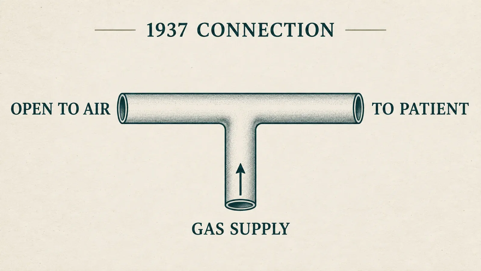
An editorial explanation of the supply branch, patient connection and opening to air, checked against figure 101 of the original paper. It omits construction details and operating settings.
It is tempting to make such a description carry a general verdict on other equipment. The paper cannot support that. Ayre was describing difficulties with a particular historical arrangement, not establishing that every breathing bag was unsuitable for children. His discussion of inadequate oxygen supply should also remain separate from much later questions about anesthetic drugs and neurodevelopment. Those are different claims requiring different evidence. [1, p. 131]
Magill’s earlier instrument design offers a useful comparison. In a 1920 note, Magill described forceps designed to grip and guide a catheter while preserving the operator’s view. Ayre’s later paper explicitly used Magill’s name for the intubation technique, then addressed the gas connection beyond the tube. The comparison is between access to the airway and the burden of the apparatus attached to it. Magill’s 1920 account concerned a nasal route; Ayre’s 1937 patients were intubated orally. The two procedures should remain distinct. [1, p. 131; 5]
These accounts show complementary kinds of airway work. They do not establish that the 1920 forceps publication itself caused the invention of the T-piece.
What the observer could see
The moving gauze gave an observer a visible indication of breathing activity. It did not measure oxygenation, carbon dioxide concentration or tidal volume. That distinction matters when reading accounts written around what a clinician could see and hear. A sign of movement is evidence of movement; adequate gas exchange is a further question.
Ayre described improved color, quieter breathing and smoother recovery with the new arrangement. His two-page report does not provide a patient denominator, a comparison group or a mortality estimate. It records a design and its author’s favorable experience. Claims of a demonstrated general reduction in pediatric surgical mortality would require evidence that this report does not supply. [1, p. 132]

Concept illustration. No invented patient series, quantitative result or 1937 experiment is depicted.
A publication year, followed by revisions
The date 1937 anchors the publication examined here. Ayre said the method had already been used locally for about 18 months. Without an established manuscript date, that interval cannot safely be converted into an exact invention date. The Royal College of Anaesthetists’ later biography also associates his T-piece with the 1937 infant cleft-surgery report, but is retrospective context rather than a contemporary record of the first procedure. [1, p. 131; 4]
His own later writing shows that the technique continued to change. In a 1952 letter to the British Medical Journal, Ayre explained revisions to the gas mixture and flow, and the importance he then gave to an extension at the open end. That extension provided space for gas to collect; its length affected the admission of room air and dilution of the anesthetic mixture. These were adjustments in the inventor’s practice, not proof that one unchanged setup had served every patient since 1937. [2, pp. 327–328]
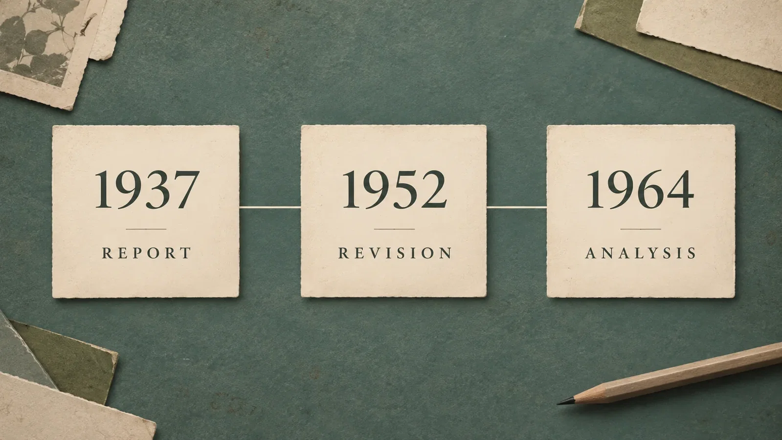
Three documents at three dates: the 1937 report, Ayre’s 1952 clarification and Harrison’s 1964 review. Their contents are not interchangeable.
By 1964, G. A. Harrison could review a family of modifications. He grouped them by the absence of an expiratory limb, or by its capacity relative to tidal volume. The review discussed arrangements with bags and ventilators as well as simpler connections. It distinguished rebreathing of expired gas from dilution by room air, and examined resistance in relation to geometry and gas flow. [3, pp. 115–119]
These distinctions limit any blanket description of a “T-piece.” Low resistance is not zero resistance. An open system does not have the same behavior under every combination of tubing, flow and breathing pattern. Harrison compared model assumptions and differing findings; his historical numerical recommendations are not used here as present-day operating instructions. [3, pp. 116–119]
Read together, the papers preserve a more useful story than an unchanging device with a single birthday. The 1937 connection simplified one infant-anesthesia problem. The 1952 letter recorded further adjustments. The 1964 review asked how the different arrangements behaved. The historical interest lies in that continuing effort to understand what a patient must breathe through. [1–3]
Sources and reading limits
- Ayre P. Anaesthesia for hare-lip and cleft palate operations on babies. Br J Surg. 1937;25(97):131–132. DOI:10.1002/bjs.1800259716. Complete original article read in the WLM reprint collection · Archived copy. The other articles in the 80-page collection were not read in full.
- Ayre P. T-piece Anaesthetic Technique. Br Med J. 1952;1(4753):327–328. Original scanned pages at PMC. Entire letter read.
- Harrison GA. Ayre’s T-piece: a review of its modifications. Br J Anaesth. 1964;36(2):115–120. DOI:10.1093/bja/36.2.115. All six pages, figures, tables and references read. Earlier papers cited within it, including Rees 1950, were not individually obtained and read for this article.
- Warde D. Dr Thomas Philip Ayre. Royal College of Anaesthetists. Biography and source note read. Its 131–3 page range differs from the actual two-page article, which controls the citation here.
- Magill IW. Forceps for Intratracheal Anaesthesia. Br Med J. 1920;2:670. The original page and complete short item were read in an archived copy. Its nasal technique is not presented as Ayre’s later oral T-piece arrangement.
Continue exploring
Related reading connects shared questions and contexts; it does not by itself establish historical influence.
The Source Room · About & Editorial Method
Sources and reading limits are stated above. Illustrations are labelled as original source images or AI-made. Historical accounts are educational; they are not current treatment instructions.