Forty-five minutes connected, twenty-six minutes of complete support.
Reading Library · Anesthesia Epochs · 中文版
Watch the companion film
1953: Gibbon — Beyond the Heart · Watch on YouTube
Historical anchor: 1953. Date details and source qualifications are explained below.
Forty-five minutes connected to the machine; twenty-six minutes entirely supported by it. Gibbon's account gives both figures for the operation of 6 May 1953. His team closed a large atrial septal defect in an eighteen-year-old patient. She recovered, and repeat catheterisation in July found the defect closed, with no remaining shunt.[1,p.176]
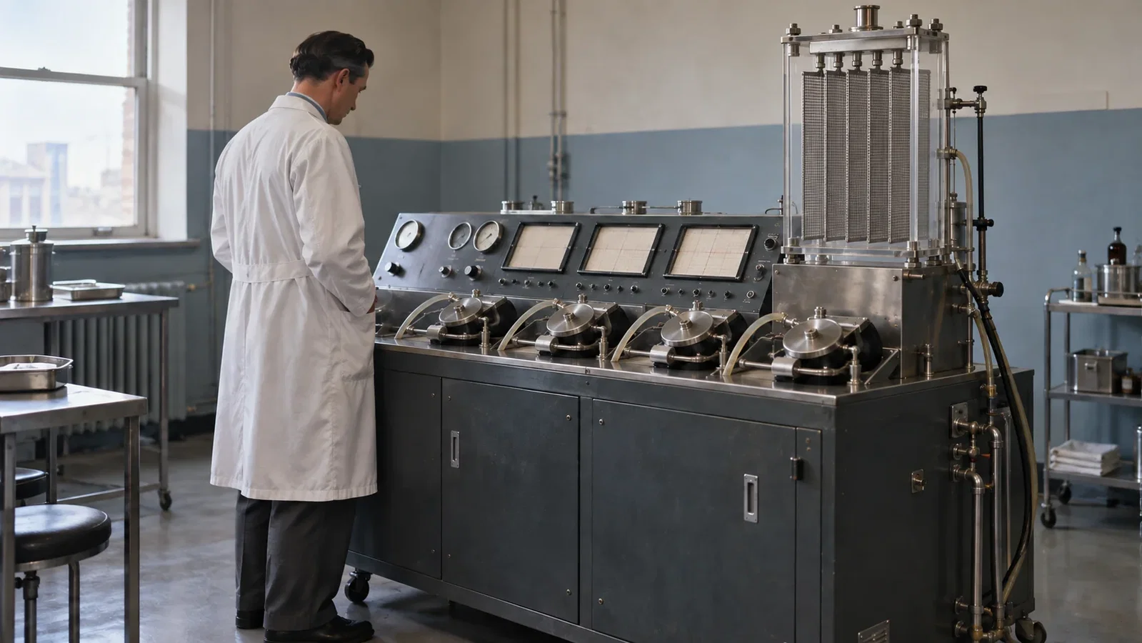
An illustration informed by contemporary cabinets, roller pumps and screen oxygenators. It does not reconstruct the May operation, an authenticated portrait or an exact machine model.
Keeping blood moving while the heart was open
An atrial defect could have an irregular margin or several openings. Miller and Gibbon argued that direct vision made complete closure easier to assess than working by touch. Their 1953 article acknowledged successful intracardiac repairs under hypothermia, which allowed a limited interval of interrupted blood flow. A pump and artificial lung offered a different condition for operating: oxygenated blood could continue to reach the brain, myocardium and other organs.[2,pp.16–17]
The machine's connection time and the period of complete support should remain distinct. The October 1953 article, signed by Bernard J. Miller and John H. Gibbon Jr., reported twenty-six minutes of complete support and a patient alive and well five months later. Gibbon presented the detailed account on 16 September 1953; it appeared in print in March 1954. The operation, presentation and publications belong to different dates.[1,pp.171,176;2,p.17]
Complete support here refers to temporary support of circulation and gas exchange. It did not require the myocardium to stop beating. In the film of another operation described by Gibbon, coronary blood flow sustained a beating heart. That film did not record the successful May case.[1,pp.176–177]

The durations come from the report. Their positions in the illustration do not specify documented moments of transition.
What the apparatus had to control
Roller pumps moved the blood. In the artificial lung, blood spread across parallel stainless-steel screens inside a transparent chamber. The thin film took up oxygen and released carbon dioxide. In this screen oxygenator, gas exchange occurred at the surface where the thin blood film met the gas.[1,pp.171,174–175;2,pp.18–19]
Blood volume also required control. Accumulation in the apparatus could deplete the patient's circulation. Gibbon credited Miller with an electronic system that sensed blood level in collecting chambers and adjusted pump speed. Further components collected blood from the operative field and dealt with entrained air. The system had to preserve circulation while allowing the surgeon to see inside the heart.[1,pp.172–175]
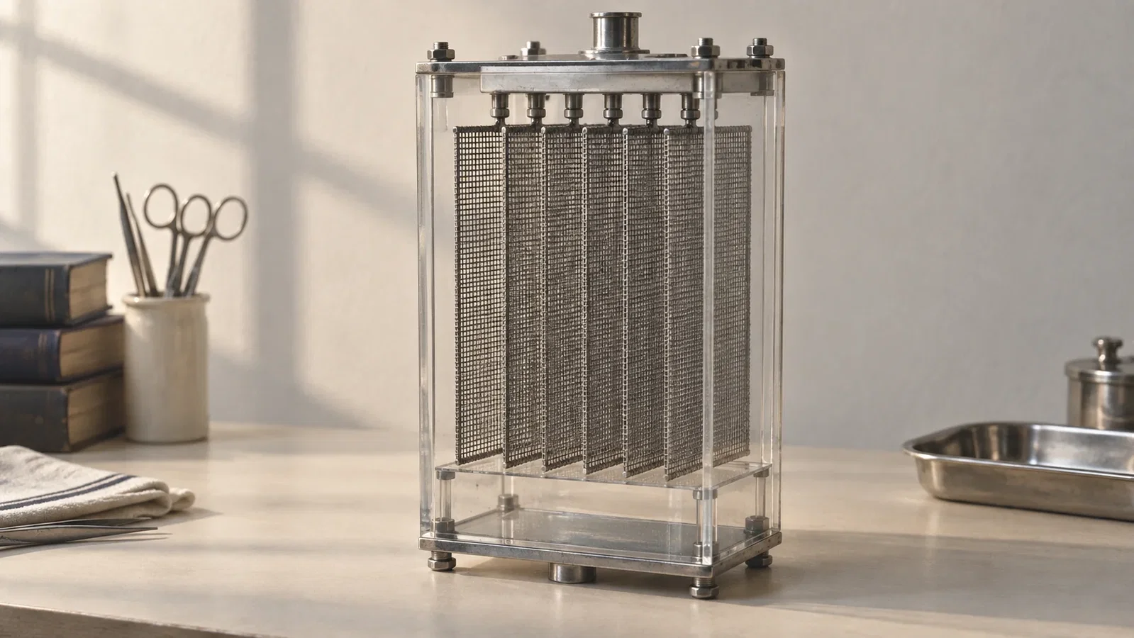
An editorial study of a screen oxygenator's structural idea, disconnected from a patient. It is neither an original object nor a complete working circuit.
Gibbon was explicit about a limit of visual observation: the colour of blood could not establish its carbon-dioxide tension. His apparatus used continuous blood-pH observation to help control CO2, under the stated condition that fixed acids did not rise substantially in those experiments. That historical approach should not be read as making pH a sufficient measure of CO2 in every setting.[1,pp.171–172]
Ibsen's Copenhagen report offers a useful comparison. Oxygen could improve blood oxygenation while CO2 continued to accumulate; respiratory-gas measurement and an oximeter distinguished the two. Ibsen assisted ventilation through the patient's airway and lungs, whereas Gibbon's screen lung exchanged gases in an extracorporeal blood circuit. The shared issue is that oxygenation does not establish adequate CO2 removal. This is an editorial comparison of mechanisms and measurements, not evidence that one team taught the other.[4,p.72;1,pp.171–172]
Reading the survivor alongside the other cases
Gibbon reported four human applications, with one surviving patient at the time of his account. In the first infant, a suspected atrial defect was not found; a large, unrecognised patent ductus arteriosus was identified after death. Two girls operated on in July 1953 also died. Gibbon attributed the three deaths to human error. His explanation should remain attributed, rather than becoming independent proof that the apparatus was safe.[1,p.176]
Jefferson's archive identifies the surviving patient as Cecelia Bavolek, the hospital as Jefferson and the apparatus as Model II. Its priority claim concerns a successful intracardiac operation of this kind. Here, that means repair of an intracardiac defect under complete mechanical heart-lung support, followed by survival. It does not mean the first heart-lung device or the first use of any mechanical bypass. In the accompanying discussion, Dodrill described bypass procedures retaining the patient's own lungs. Dennis recalled artificial heart-lung attempts in 1951 in which the patients did not survive.[3;1,pp.177–180]
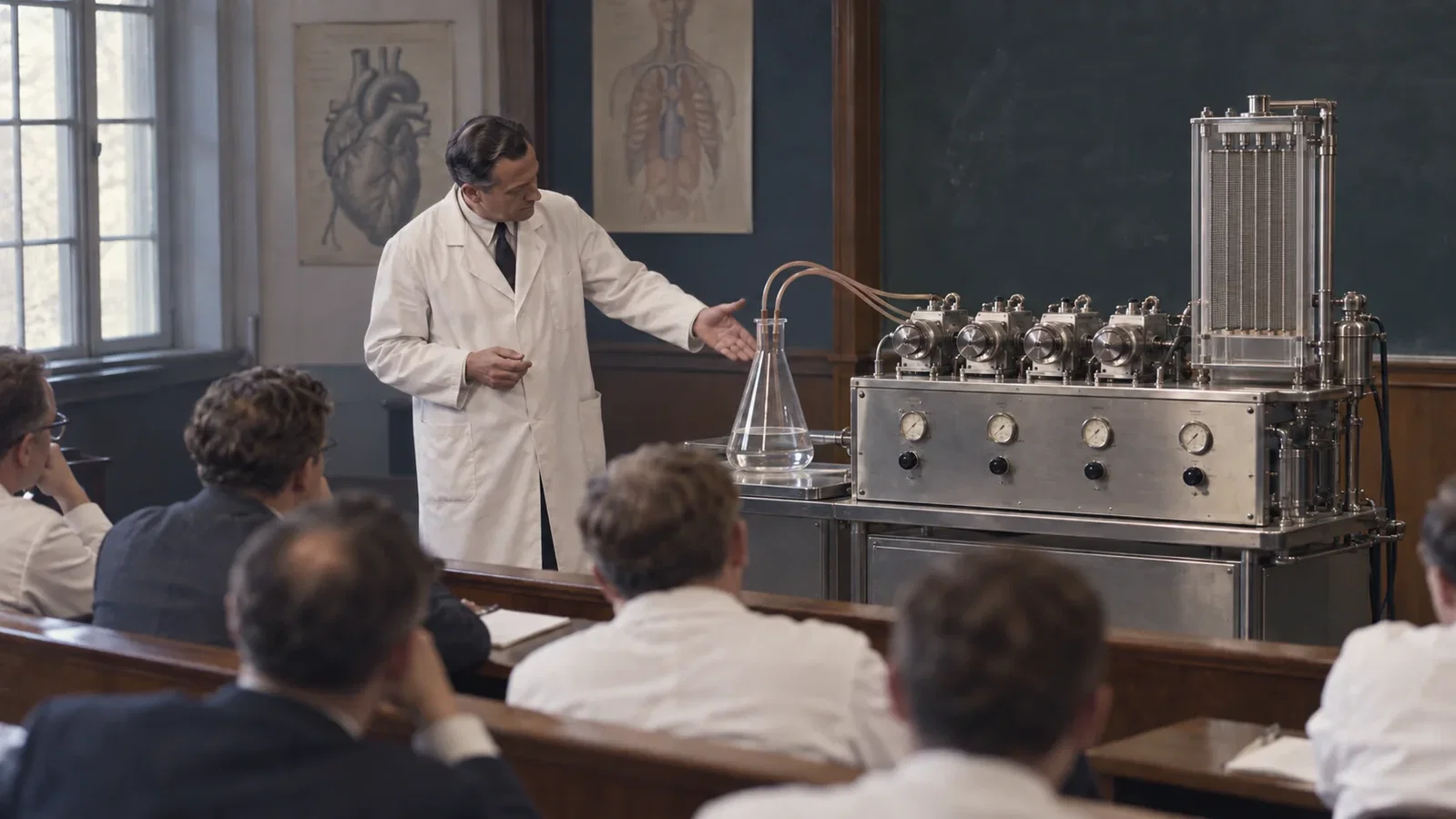
Contemporary photographs show a large flask used as a stand-in for the subject. This illustration adopts that teaching theme with imagined people and surroundings; it is not an original television frame.
The October article's photographs also show Gibbon explaining flow to and from the machine using that flask. The photographs illustrated the direction of flow; the clinical report preserved both the successful repair and the deaths of the other patients. The surviving patient showed what complete mechanical heart-lung support could make possible. The accounts also show how much remained unresolved before it could become routine.[2,p.19;1,pp.176–180]
Sources and reading limits
The first two sources share authorship and a research team. They should not be counted as two independent clinical validations.
- Gibbon JH Jr. Application of a mechanical heart and lung apparatus to cardiac surgery. Minnesota Medicine. 1954;37(3):171–180,185, as cited by the reprint. WLM authorized reprint collection. PubMed 13154149. Main text and accompanying Dodrill/Dennis discussion on pp.171–180 read in full, with page images (collection PDF pp.104–113). The supplied p.185 belongs to a different article; the advertised reference continuation was not available. The rest of the collection was not read in full.
- Miller BJ, Gibbon JH Jr. The present status of the mechanical heart and lung apparatus. Jefferson Medical College Alumni Bulletin. 1953;8(4):16–19. Original issue, October 1953. All four article pages, including diagrams and captions, read in the original scan (PDF pp.19–22). This is a contemporary institutional publication, not an independent clinical study.
- Thomas Jefferson University Archives & Special Collections. Dr. John H. Gibbon, Jr. and Jefferson’s Heart-Lung Machine. Institutional history. Entire accessible page text archived and read on 13 September 2026. A retrospective source for the hospital, patient's name and Model II; its account of the first infant is less specific and differs in the timing of death from [1].
- Ibsen B. The Anaesthetist's Viewpoint on the Treatment of Respiratory Complications in Poliomyelitis During the Epidemic in Copenhagen, 1952. Proc R Soc Med. 1954;47(1):72–74. Original article and scans. DOI:10.1177/003591575404700120. The original p.72 scan was read for this comparison. Pages 73–74 were read for the earlier Ibsen article, but were not reread for this article.
Continue exploring
Related reading connects shared questions and contexts; it does not by itself establish historical influence.
The Source Room · About & Editorial Method
Sources and reading limits are stated above. Illustrations are labelled as original source images or AI-made. Historical accounts are educational; they are not current treatment instructions.