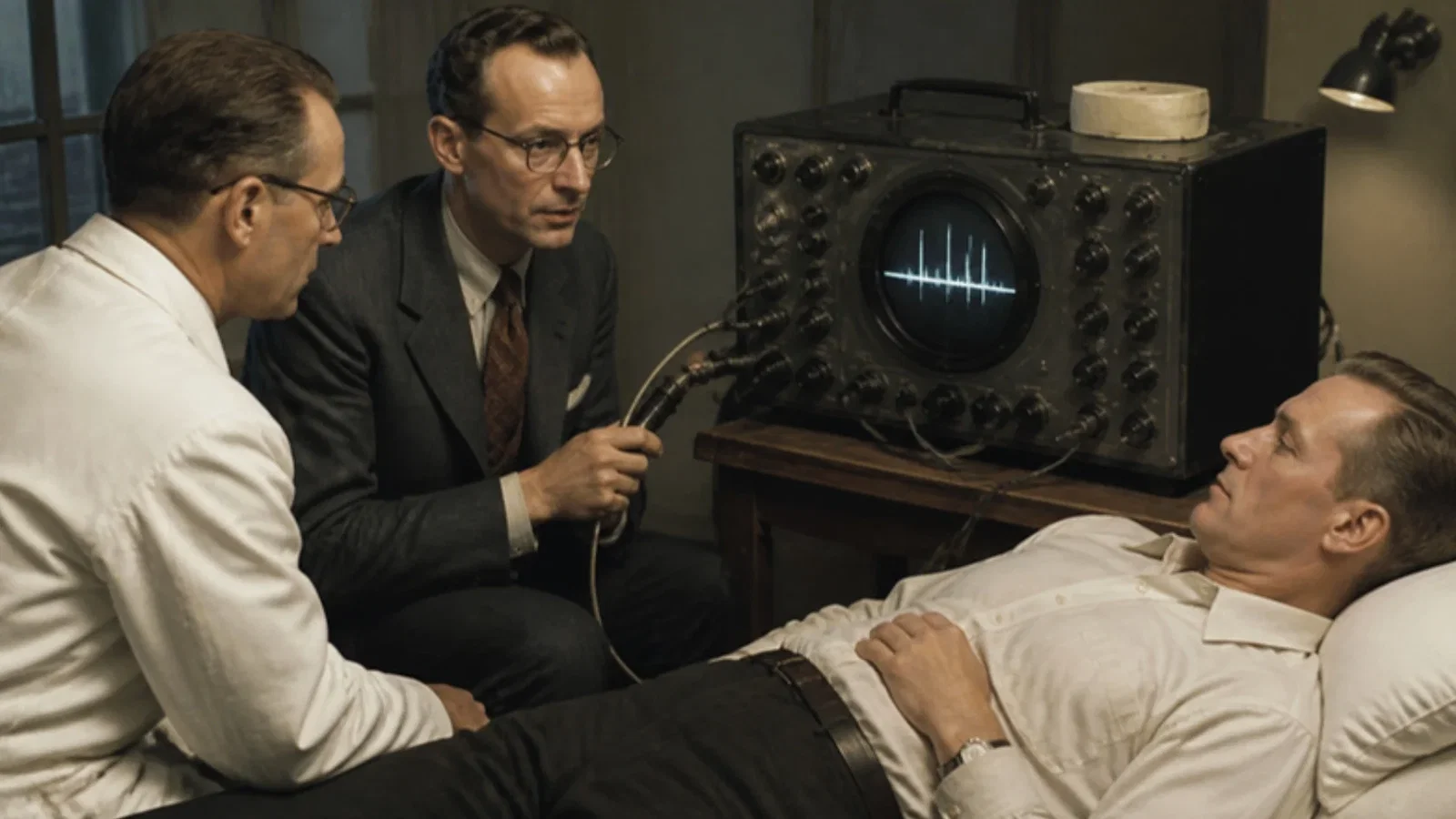
The authors' 1956 paper in Acustica and a 2007 history: a shipyard's materials-testing instrument, a 2.5 MHz quartz crystal, film drawn past a slit, and an oesophageal probe they gave up.
Reading Library · Anesthesia Epochs · 中文版
Watch the education video (for clinicians)
1953: Edler and Hertz — Recording the Moving Heart · Anesthesia Epochs Education Series
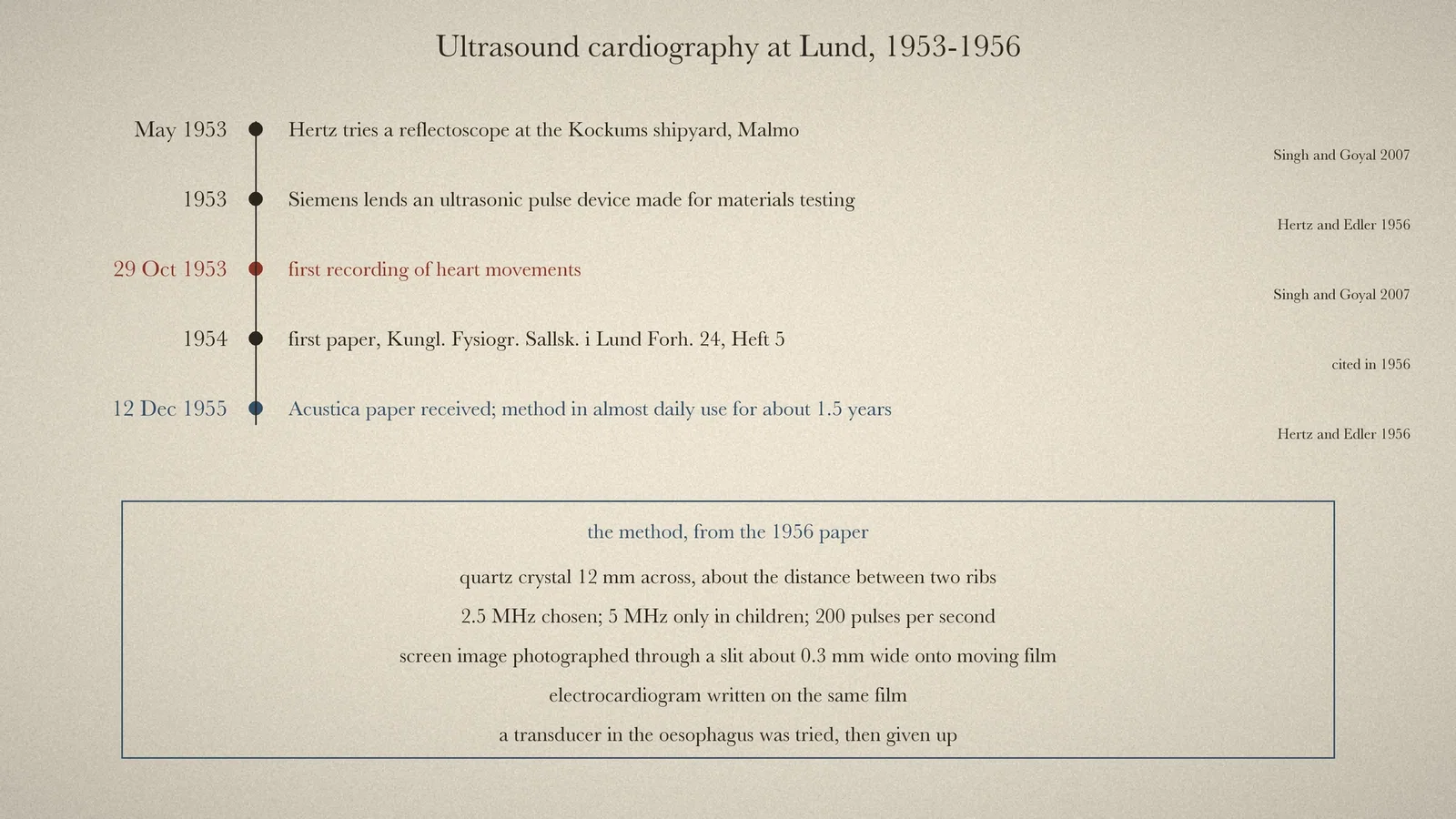
An existing article in this series follows the 1996 practice guideline for perioperative transoesophageal echocardiography. The method's ancestry runs back to Lund in 1953, where a cardiologist and a physicist turned an instrument built for testing materials on the human chest. This article tells that beginning from the two researchers' own account of 1956 and from a 2007 history. It also records one detail from the 1956 paper that anaesthetists may find striking: they tried putting the transducer in the oesophagus.
How it started
The story of 1953 comes from a 2007 tribute to Inge Edler (1911-2001) by Singh and Goyal in the Texas Heart Institute Journal. The first ultrasonic reflectoscope in the region had gone to a non-destructive testing company at the Kockums shipyard in Malmö. In May 1953 the physicist Hellmuth Hertz visited the company and "applied the transducer" to the chest, seeing echo signals that pulsed. [2] Through Hertz's father, Gustav Hertz, who had directed a Siemens laboratory before the war, they contacted Wolfgang Gellinek of the Siemens medical branch in Erlangen. [2] Then, "on 29 October 1953", with the Siemens ultrasonic pulse device, Edler and Hertz recorded the first moving pictures of the heart, beginning what was called "ultrasound cardiography". [2] Their first paper appeared in Lund in 1954. [2] [4]
The method, in the authors' words
The fullest account by the two authors available here is a German paper in Acustica, received on 12 December 1955 and published in 1956. Its English summary states the idea: with the ultrasonic reflectoscope, "the position of the heart walls in the human body can be located" by placing the quartz crystal on the chest "in such a way that the sound passes between the ribs and falls upon the heart region", and with a special camera the movements of the heart walls could be recorded continuously. The curves "permit an exact diagnosis of certain cardiac diseases which hitherto were hard to distinguish". [1, p. 361] Hertz signed from the Physics Institute and Edler from the Medical Clinic of the University of Lund. [1, p. 361]
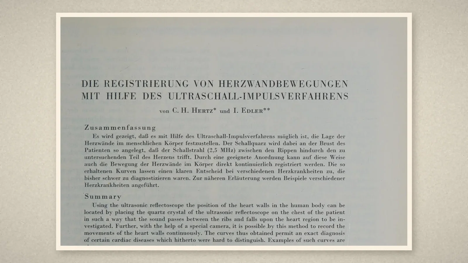
The problem they set out to solve was surgical. Mitral stenosis could now be operated on, but it often came with mitral regurgitation, and if regurgitation was strong no operation should be done. Telling the two apart was difficult, and in some cases impossible, with existing methods. [1, p. 361] The instrument was an ultrasonic pulse device that Siemens-Reiniger-Werke made for materials testing. A round quartz crystal 12 mm across, about the width of the gap between two ribs, sent the beam. Frequencies of 0.5, 1, 2.5 and 5 MHz were tried; 2.5 MHz suited best, and 5 MHz could be used only in children, whose hearts lie nearer the chest wall. The pulse rate was 200 per second. [1, p. 362] Earlier experiments on dead hearts had shown a strong echo at the boundary between heart muscle and blood. [1, p. 362]
To record movement, the screen image was photographed through a slit about 0.3 mm wide onto film drawn past at constant speed. A still echo drew a straight line and a moving one drew a curve, and an electrocardiogram was written on the same film. [1, p. 363] The patients noticed no pain or discomfort during or after the examination. [1, p. 364]
What the curves showed
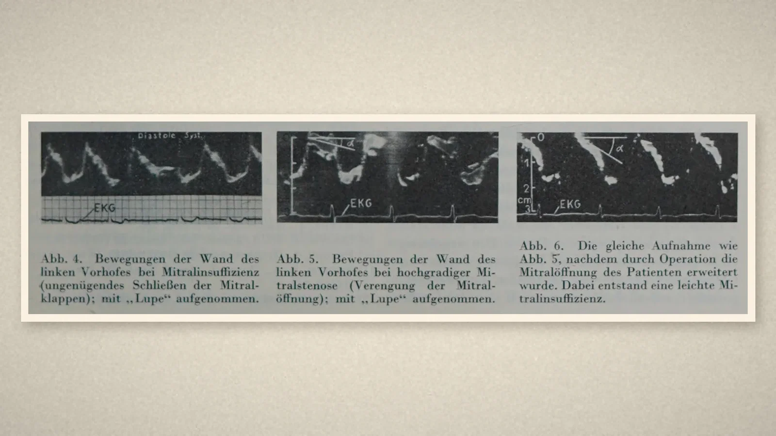
The paper's figures show a heart swinging inside a fluid-filled pericardium and lying back against the chest wall once the fluid had been drawn off; the left atrial wall in mitral regurgitation and in mitral stenosis, whose curves differed in a typical way; and the same stenotic patient after the surgeon had widened the mitral opening. [1, p. 364] Clots in the left atrium could also be detected. [1, p. 364] The method, they wrote, had been in almost daily use at the university clinic in Lund for about a year and a half. [1, p. 364] They thanked Siemens-Reiniger-Werke for lending the instrument. [1, p. 364]
The probe in the oesophagus
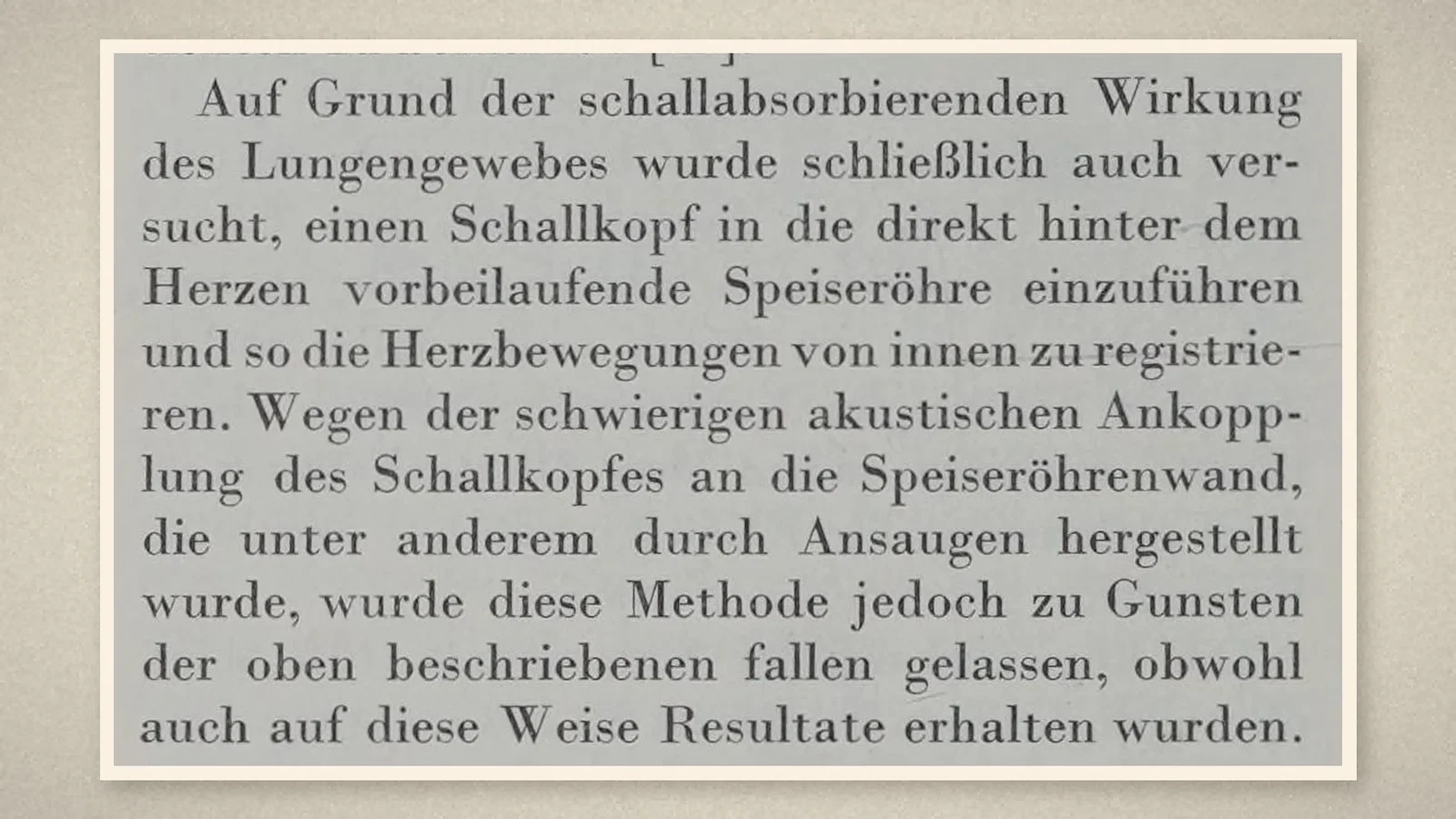
Because lung absorbs sound so strongly, the authors also tried to insert a transducer into the oesophagus, which runs directly behind the heart, and record the heart's movements from inside. Acoustic coupling to the oesophageal wall was difficult, achieved among other ways by suction, and the method was dropped in favour of the chest approach, "although results were obtained in this way too". [1, p. 364]
Later accounts
Singh and Goyal write that Edler used the technique mainly "for the preoperative study of mitral stenosis and diagnosis of mitral regurgitation", and that others later developed Doppler, two-dimensional, contrast and transoesophageal echocardiography. [2] The Australian Society of Anaesthetists' timeline lists "Echocardiography developed" under 1953 and notes that Edler first used M-mode to assess mitral valve disease before operation. [3, PDF p. 61]
What the record shows
Echocardiography began as a preoperative question: whether a mitral valve should be operated on. A borrowed industrial instrument and a strip of moving film answered it. In their 1956 paper the inventors described, as a side attempt, looking at the heart from the oesophagus. They gave it up as impractical with the equipment they had.
Sources and reading limits
Read: Hertz and Edler, Acustica 1956;6:361-364, in full; Singh and Goyal 2007, full text on PubMed Central; the ASA timeline entry for 1953; the PubMed record of the 2004 reprint of the 1954 paper.
Not covered: the original 1954 paper from Lund is not covered, and its 2004 reprint on Wiley could not be accessed. Edler's 1954 supplement in Acta Medica Scandinavica was not read. The dates and details of 1953 (May at Kockums, the Siemens contact, 29 October) come only from Singh and Goyal, a later secondary account; the 1956 paper confirms the Siemens loan and the period of daily use but gives no dates for 1953. Singh and Goyal give the 1954 title as "heart valves", while PubMed and the 1956 paper speak of heart walls. The paper does not name the patients or give their number.
One illustration is a timeline drawn from the sources; three are crops of the 1956 paper.
- Hertz CH, Edler I. Die Registrierung von Herzwandbewegungen mit Hilfe des Ultraschall-Impulsverfahrens. Acustica. 1956;6(4):361-4. Internet Archive
- Singh S, Goyal A. The origin of echocardiography: a tribute to Inge Edler. Tex Heart Inst J. 2007;34(4):431-8. PMC
- Australian Society of Anaesthetists. Anaesthesia History Timeline. 2024. PDF
- Edler I, Hertz CH. The use of ultrasonic reflectoscope for the continuous recording of the movements of heart walls. 1954 [reprint]. Clin Physiol Funct Imaging. 2004;24(3):118-36. PubMed
Continue exploring
Related reading connects shared questions and contexts; it does not by itself establish historical influence.
Anesthesia Epochs · The Source Room · About & Editorial Method
Published November 2, 2026. Sources and reading limits are stated above. Illustrations are labelled as original source images or AI-made. Historical accounts are educational; they are not current treatment instructions.