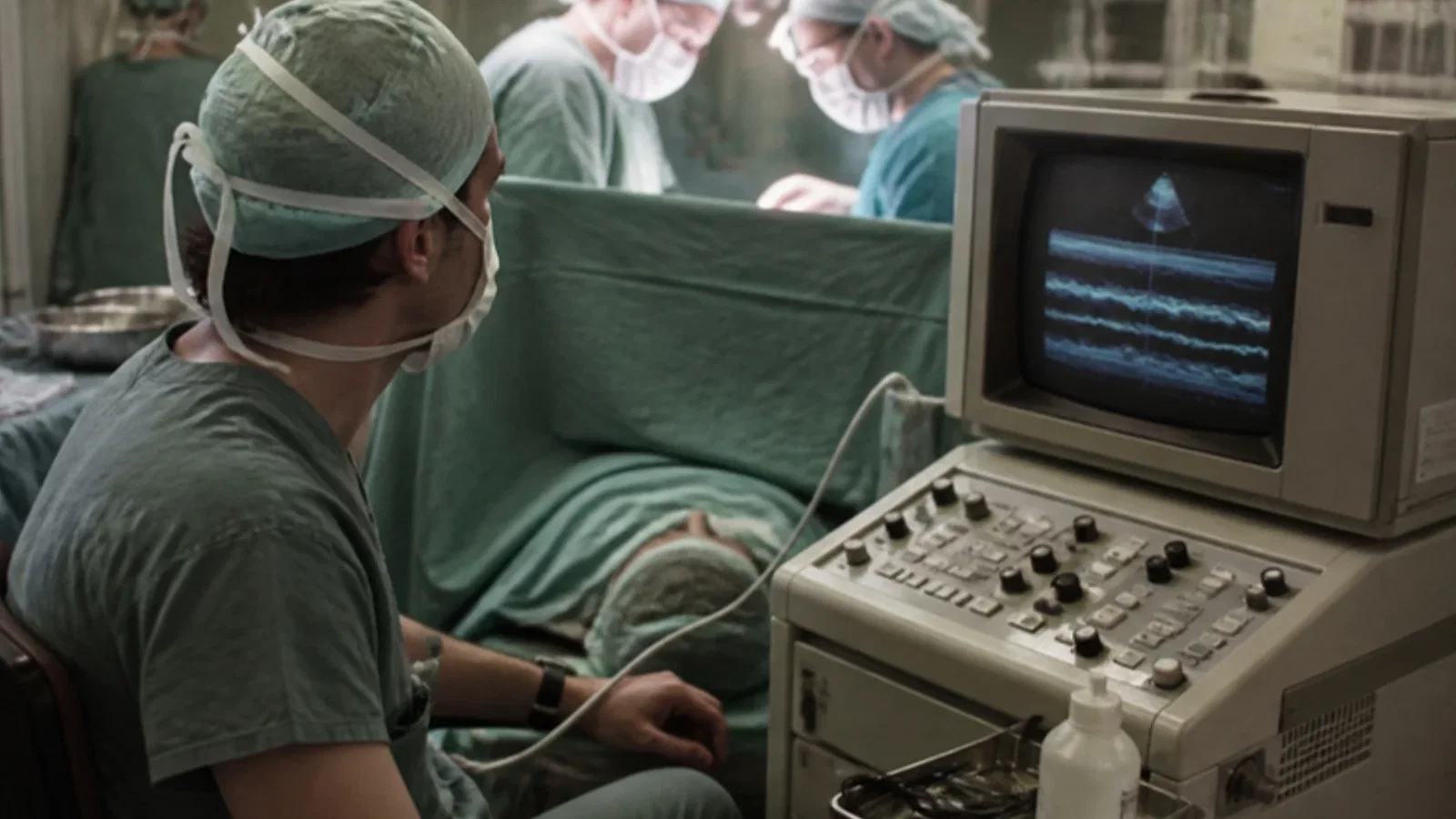
From Frazin's diagnostic oesophageal echocardiography in 1976 to Matsumoto and Oka's use of transoesophageal M-mode echo to watch the left ventricle continuously during open-heart surgery, 1979-80.
Reading Library · Anesthesia Epochs · 中文版
Watch the education video (for clinicians)
1976–1980: The Oesophageal Echo Probe Enters the Operating Room · Anesthesia Epochs Education Series
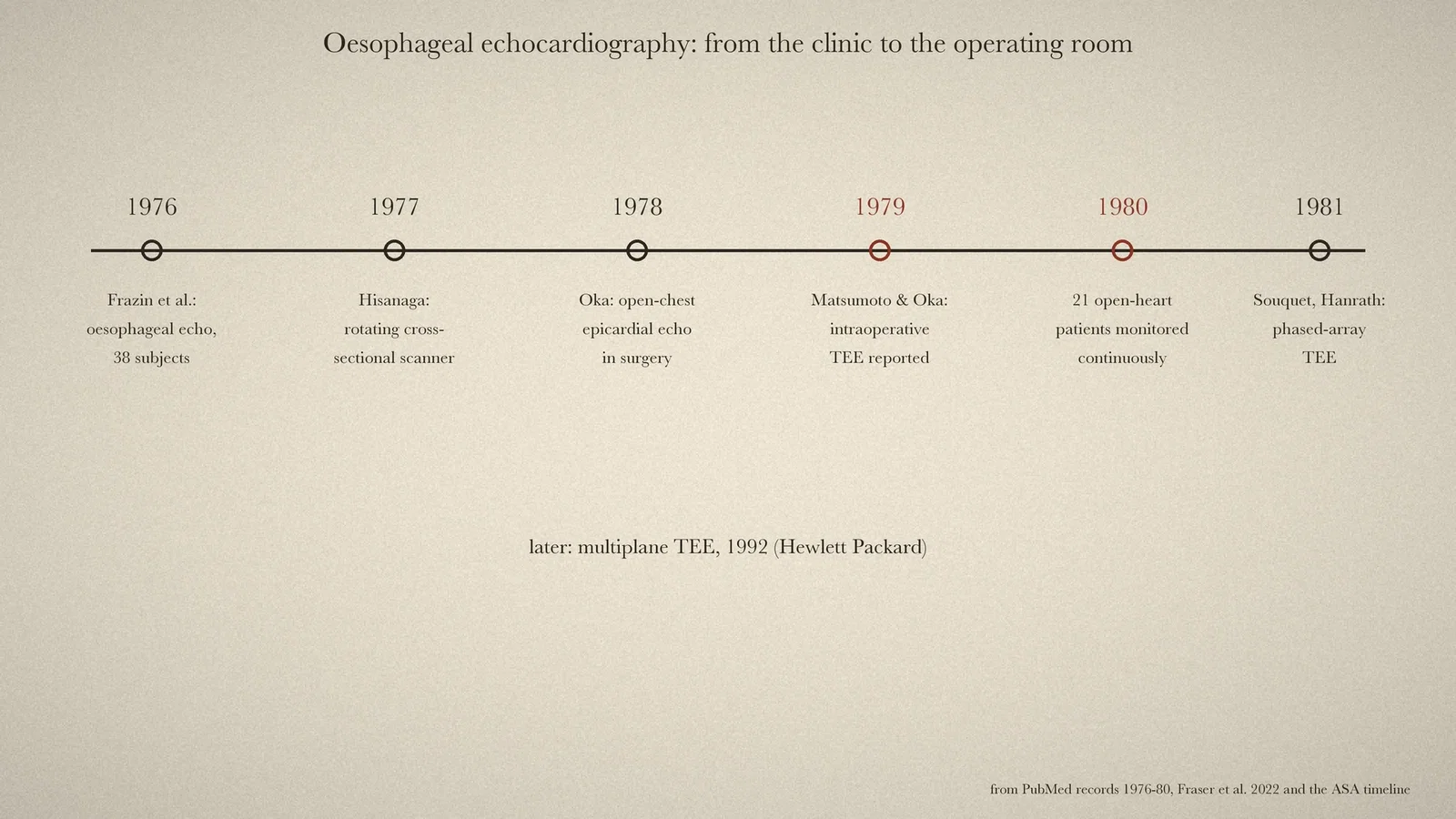
Transoesophageal echocardiography (TEE) is now one of the cardiac anaesthetist's main tools. An earlier article in this series followed the first heart ultrasound, by Edler and Hertz, including their abandoned attempt with an oesophageal probe. This article follows the step that made the probe an operating-room monitor: a diagnostic oesophageal echo described in 1976, then its use by Matsumoto, Oka and colleagues in New York to watch the left ventricle during open-heart surgery in 1979-80. None of the original papers could be read in full. The account rests on their PubMed records, an open-access history of echocardiography and the Australian Society of Anaesthetists' timeline.
A diagnostic probe, 1976
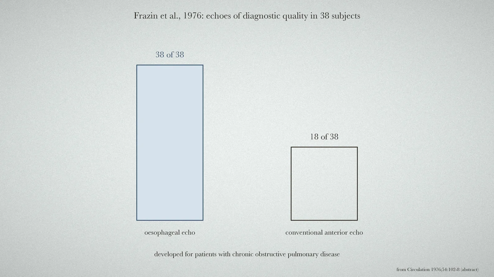
In July 1976 Frazin and colleagues reported in Circulation that "Esophageal echocardiography has been developed for use in patients" "with chronic obstructive pulmonary disease". It was safe and gave "high resolution mirror image echoes" of many cardiac structures. Conventional anterior and "esophageal echocardiograms were performed in 38 subjects". The oesophageal echoes were of diagnostic quality in all 38, the "anterior echoes were of diagnostic quality in only 18". [1] The 2022 history by Fraser and colleagues lists it under 1976 as "single-element transoesophageal echocardiography". [4] The ASA timeline describes it as a non-operative report: "Trans-Oesophageal Echocardiography reported (non-operative)". [5, p. 75]
Into the operating room
The ASA timeline names the next steps in New York. In 1978 came "Open intra-operative epicardial echocardiography used", by "Dr Yasu Oka in New York", with the probe on the surface of the heart while the chest was open. In 1979: "Intra-operative Trans-Oesophageal Echocardiography study reported", by Oka and Matsumoto. [5, p. 77] Their first paper, in the New York State Journal of Medicine in January 1979, carried the title "Transesophageal echocardiography; for assessing ventricular performance". PubMed holds no abstract for it. [2]
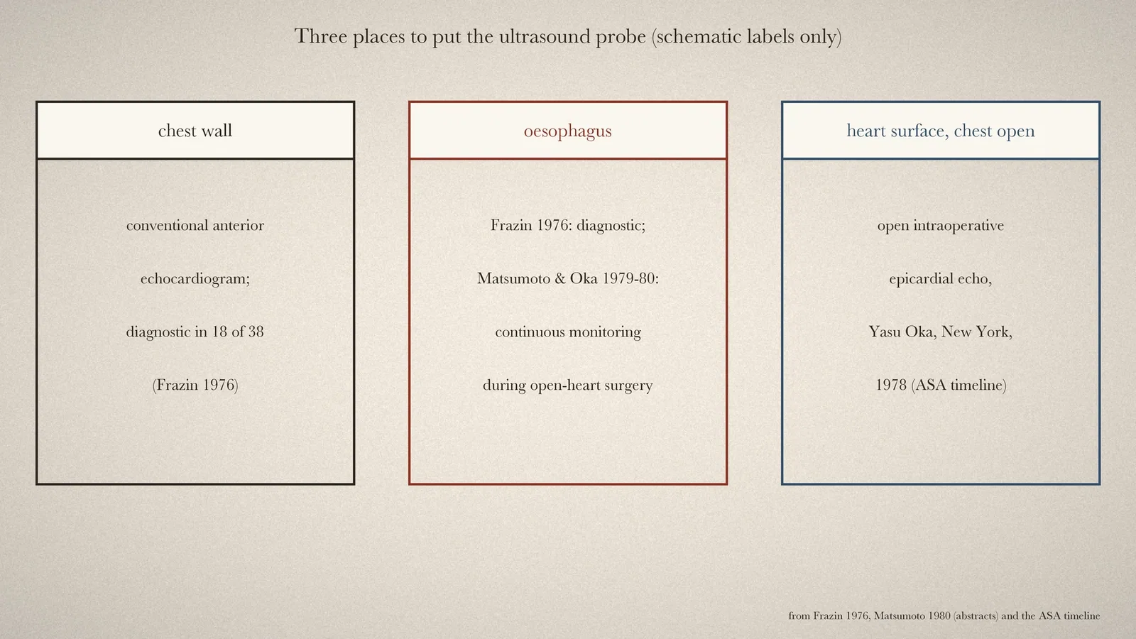
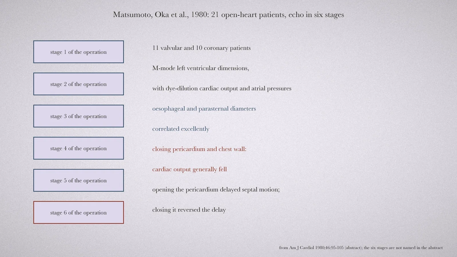
The fuller report appeared in the American Journal of Cardiology in July 1980, titled "Application of transesophageal echocardiography to continuous intraoperative monitoring of left ventricular performance". [3] Transoesophageal M-mode echo was used to follow "left ventricular dimensions in 21 patients", "11 with valvular and 10 with" coronary heart disease, undergoing open-heart surgery. Echocardiograms were "recorded in six stages of the procedure", with cardiac output (by dye dilution) and atrial pressures measured at the same time. The oesophageal measurements of left ventricular diameter "correlated excellently" with standard parasternal ones. [3] The study also showed what the operation itself did to the heart. "Pericardial and chest wall closures generally caused a significant decrease" in cardiac output, which the authors linked to mechanical restriction of the ventricle. Opening the pericardium delayed septal motion, and closing it reversed the delay. They concluded that the study confirmed the validity of the technique and "its usefulness in monitoring changes in ventricular function during cardiac surgery". [3]
The probe improves
The 2022 history traces the probe's development in the same years. In 1977 Kohzoh Hisanaga built a "high-speed rotating cross-sectional transoesophageal scanner". In 1981 Souquet and Hanrath introduced "transoesophageal phased-array echocardiography". In 1992 came "Multiplane transoesophageal echocardiography" from Hewlett Packard. [4] The M-mode recordings of 1979-80 followed a single line through the ventricle; the cross-sectional, phased-array and multiplane probes that followed gave the images used in operating rooms later.
What the record shows
TEE came into the operating room as a way to see what the surgeon and anaesthetist were doing to the left ventricle, stage by stage, in a patient whose chest was open and whose heart was being handled. A probe first developed for patients with chronic lung disease turned out to keep a view of the heart throughout an open-chest operation.
Sources and reading limits
Read: the PubMed records of Frazin et al. 1976 (abstract), Matsumoto, Oka et al. 1979 (title only) and Matsumoto, Oka et al. 1980 (abstract); the timeline in Fraser et al.'s 2022 history of echocardiography; the ASA history timeline (2024).
Not covered: none of the three primary papers was read in full. Oka's 1978 open-chest epicardial echocardiography is known only from the ASA timeline, and no paper for it was identified. The authors' institutions are not given in the records read, beyond the timeline's "New York". The six stages of the 1980 study are not named in the abstract and are not named here. The earlier history of oesophageal ultrasound is left to the article on Edler and Hertz. All four illustrations are drawn from the sources.
- Frazin L, Talano JV, Stephanides L, Loeb HS, Kopel L, Gunnar RM. Esophageal echocardiography. Circulation. 1976;54(1):102-8. PubMed
- Matsumoto M, Oka Y, Lin YT, Strom J, Sonnenblick EH, Frater RW. Transesophageal echocardiography; for assessing ventricular performance. N Y State J Med. 1979;79(1):19-21. PubMed
- Matsumoto M, Oka Y, Strom J, Frishman W, Kadish A, Becker RM, Frater RW, Sonnenblick EH. Application of transesophageal echocardiography to continuous intraoperative monitoring of left ventricular performance. Am J Cardiol. 1980;46(1):95-105. PubMed
- Fraser AG, Monaghan MJ, van der Steen AFW, Sutherland GR. A concise history of echocardiography: timeline, pioneers, and landmark publications. Eur Heart J Cardiovasc Imaging. 2022;23(9):1130-43. DOI
- Australian Society of Anaesthetists. Anaesthesia History Timeline 2024. PDF
Continue exploring
Related reading connects shared questions and contexts; it does not by itself establish historical influence.
Anesthesia Epochs · The Source Room · About & Editorial Method
Published November 2, 2026. Sources and reading limits are stated above. Illustrations are labelled as original source images or AI-made. Historical accounts are educational; they are not current treatment instructions.