ITL-7920f967ea18
Juan Li, MD · 更新于2026年11月18日
核心要点
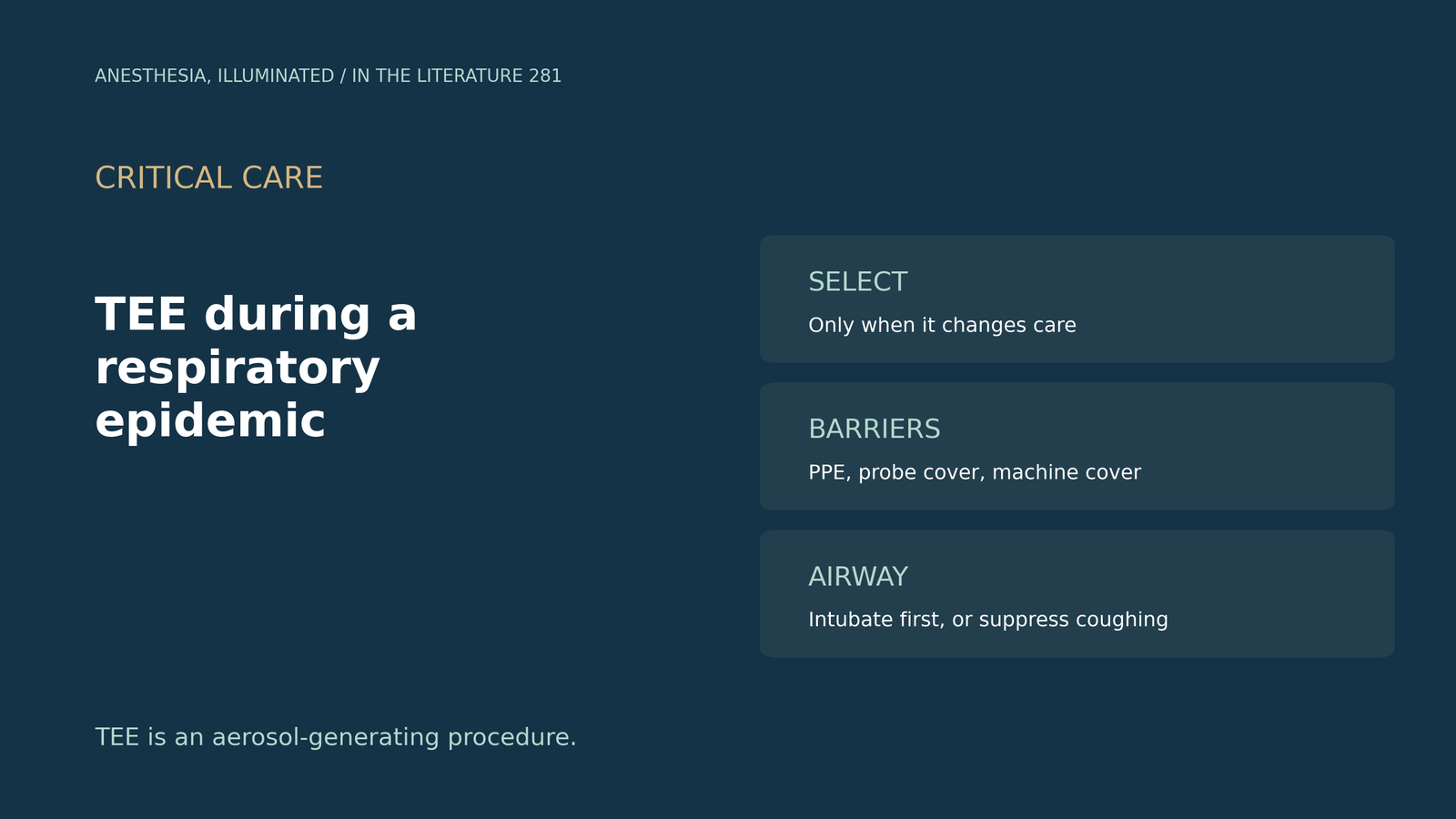
- 先选择,再聚焦。TEE 应根据风险获益只用于选定的患者,并采用聚焦、分步的检查。[1]
- 先插管,再置入探头。带套的探头只在气管插管后置入,检查后对探头和机器进行消毒。[1]
- 简单的探头套就有用。在人体模型试验中,普通超声探头套使模拟分泌物没有沾到操作者手上。[2]
- 清醒 TEE 需要控制咳嗽。抑制咽反射,预期拔出探头时会咳嗽,可考虑使用内镜给氧面罩。[3], [4]
- 保护医护人员和其他患者。缩短在病房内的时间,按气道操作级别防护,并筛查急诊心脏病患者。[5], [6], [7]
一位疑似呼吸道感染的患者需要急诊心脏手术,团队希望术中做 TEE。探头、医护人员和下一位患者应该怎样防护?
谁做 TEE,怎么做
EACTA/SCA 共识(Guarracino 等,J Cardiothorac Vasc Anesth 2021)指出,TEE 应根据风险获益只用于选定的患者,并应考虑聚焦、分步的 TEE 检查。带套的 TEE 探头只在气管插管后置入,并必须按厂家要求对探头和机器进行适当消毒。对于支气管镜和 TEE 等产生气溶胶的操作,共识列出了具体的防护装备,包括贴合良好的过滤口罩或 PAPR。TEE 机和麻醉机可以用一次性透明塑料膜覆盖,检测应尽量靠近手术时间,最好在 48 小时内。共识中未注明出处的条目属于专家意见。[1]
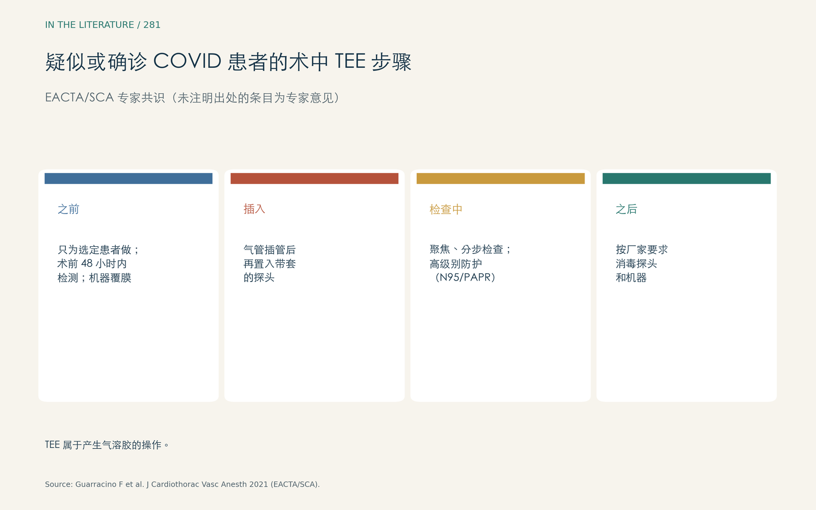
图 1. 选择患者、插管后置入探头、聚焦检查、事后消毒。数据来自 EACTA/SCA 共识。[1] 查看大图。

探头套
一个团队在急诊手术中把普通超声探头套用胶带固定在 TEE 手柄根部(Moulthrop 等,J Cardiothorac Vasc Anesth 2021)。在人体模型试验中,使用探头套时操作者手上没有检出荧光粉,而不用探头套时,操作者手部及周围大片区域都可见污染。人体模型无法模拟人体的湿度和软组织;定制的 TEE 探头套价格可达普通超声探头套的两倍。[2]
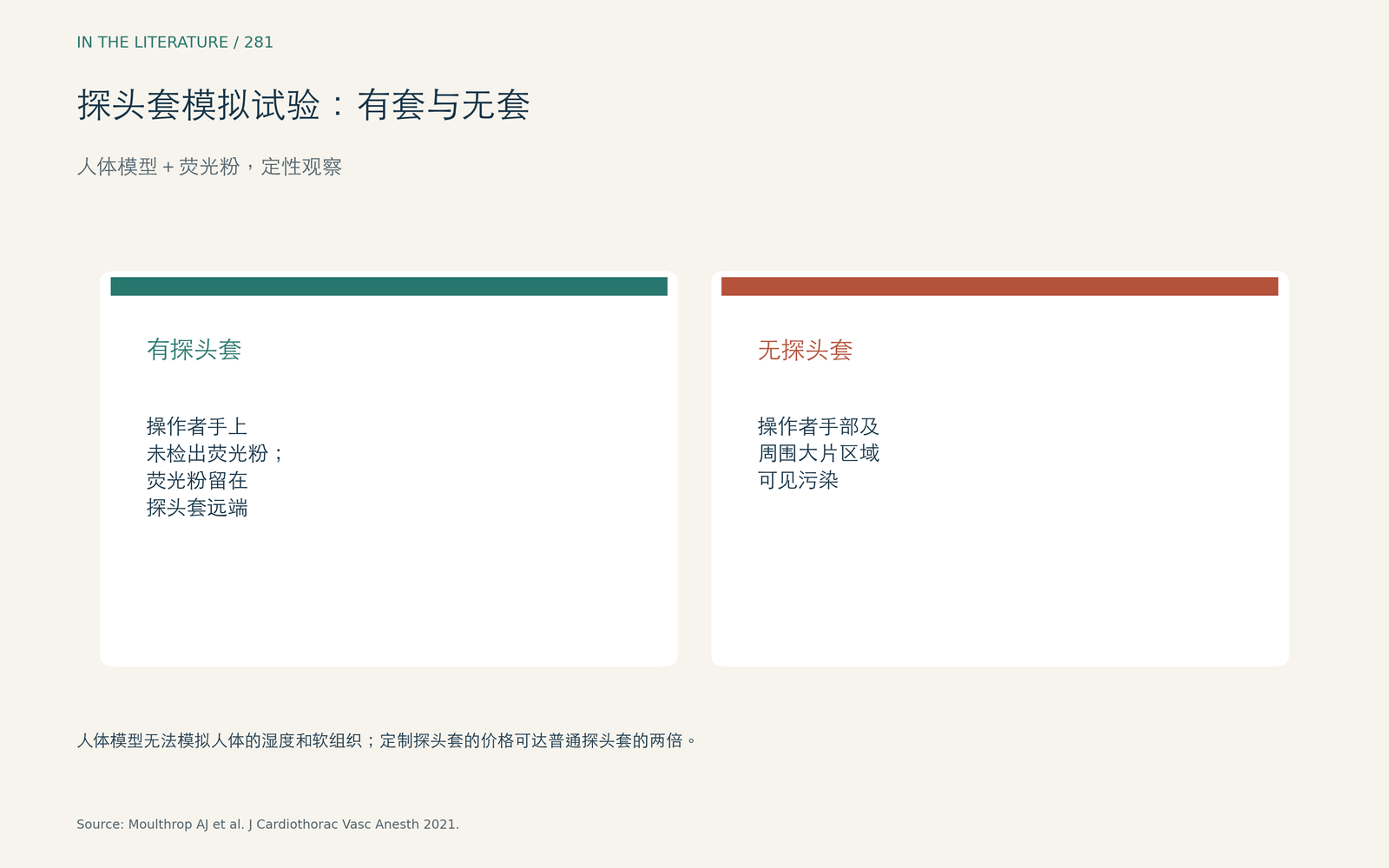
图 2. 在定性的人体模型试验中,普通探头套把荧光标记物留在了套内。数据来自 Moulthrop 等。[2] 查看大图。

清醒或镇静患者的 TEE
置入 TEE 探头会刺激咽反射,可能引发咳嗽和喷嚏;建议用局麻药喷雾和静脉镇静抑制咽反射。一封超声室来信的作者观察到,拔出探头时咳嗽比置入时更多(Behjati 和 Tohidi,Caspian J Intern Med 2024)。[3] 带自封式插入口的内镜给氧面罩配合储气袋可提供 0.88–0.95 的 FiO2,面罩还能作为咳嗽的物理屏障(Gonzalez,J Am Soc Echocardiogr 2021);作者是该面罩厂家的教育顾问。[4]
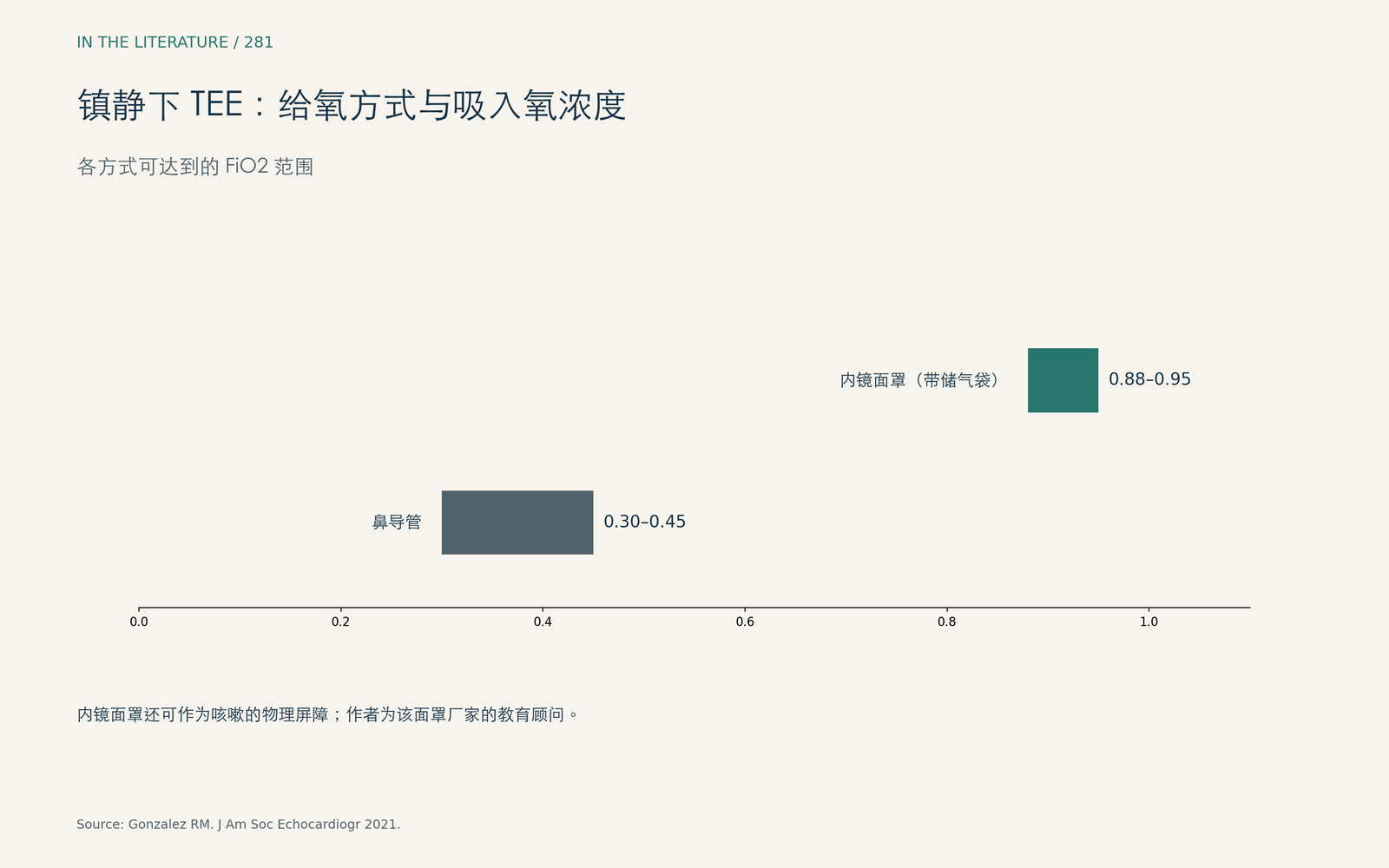
图 3. 内镜面罩的 FiO2 远高于鼻导管,并能阻挡咳嗽。数据来自 Gonzalez。[4] 查看大图。

保护医护人员和患者
一项在 38 例 COVID ICU 患者中使用的聚焦检查方案,把应变分析移到病房外完成,以减少超声医师的暴露(Kaminski 等,J Am Soc Echocardiogr 2020)。[5] 对疑似或确诊感染者,最低防护是隔离衣、手套、N95 口罩和护目镜,而涉及鼻、口或气道的操作需要更高级别的防护(Maracaja 等,J Cardiothorac Vasc Anesth 2020)。[6] 在患者方面,每一位急诊入院的心脏病患者可能都应先隔离并筛查,在病毒学结果出来之前应保持独立通道(Pisano 等,J Cardiothorac Vasc Anesth 2020)。[7]
临床带走的三点
- 把 TEE 当作产生气溶胶的操作:只在会改变处理时才做,并保持聚焦。[1]
- 麻醉患者先插管,再置入带套探头;覆盖机器,事后消毒。[1], [2]
- 清醒患者要抑制咽反射,准备好应对拔出探头时的咳嗽,并考虑使用有屏障作用的给氧面罩。[3], [4]
参考文献
- Guarracino F, Shernan SK, El Tahan M, et al. EACTA/SCA recommendations for the cardiac anesthesia management of patients with suspected or confirmed COVID-19 infection: an expert consensus from the European Association of Cardiothoracic Anesthesiology and Society of Cardiovascular Anesthesiologists with endorsement from the Chinese Society of Cardiothoracic and Vascular Anesthesiology. J Cardiothorac Vasc Anesth. 2021;35(7):1953–1963. doi:10.1053/j.jvca.2021.02.039
- Moulthrop AJ, Kolarczyk LM, Teeter EG. Preventing contamination during transesophageal echocardiography in the face of the coronavirus disease 2019 (COVID-19) pandemic: a Glo Germ experience. J Cardiothorac Vasc Anesth. 2021;35(9):2824–2826. doi:10.1053/j.jvca.2021.02.051
- Behjati M, Tohidi F. Transesophageal echocardiography probe intubation or extubation: which side is generating more aerosols associated with spreading COVID-19 contamination? Caspian J Intern Med. 2024;15(4):748–749. doi:10.22088/cjim.15.4.748
- Gonzalez RM. Maximizing patient and staff safety during transesophageal echocardiography in the COVID-19 era by use of a new oxygen delivery system: endoscopy oxygen masks with self-sealing endoscope insertion ports. J Am Soc Echocardiogr. 2021;34(4):453–454. doi:10.1016/j.echo.2020.12.022
- Kaminski A, Payne A, Roemer S, Ignatowski D, Khandheria BK. Answering to the call of critically ill patients: limiting sonographer exposure to COVID-19 with focused protocols. J Am Soc Echocardiogr. 2020;33(7):902–903. doi:10.1016/j.echo.2020.05.006
- Maracaja L, Blitz D, Maracaja DLV, Walker CA. How 3D printing can prevent spread of COVID-19 among healthcare professionals during times of critical shortage of protective personal equipment. J Cardiothorac Vasc Anesth. 2020;34:2847–2849. doi:10.1053/j.jvca.2020.04.004
- Pisano A, Landoni G, Zangrillo A. Protecting high-risk cardiac patients during the COVID-19 outbreak. J Cardiothorac Vasc Anesth. 2020;34:1698. doi:10.1053/j.jvca.2020.03.043