Within three months of Koller's report on cocaine drops, Knapp injected six minims close behind the globe to remove an eye, and warned at once about the orbit's blood supply.
Reading Library · Anesthesia Epochs · 中文版
Watch the education video (for clinicians)
1884: Knapp's Retrobulbar Cocaine — Anaesthesia for Eye Removal · Anesthesia Epochs Education Series
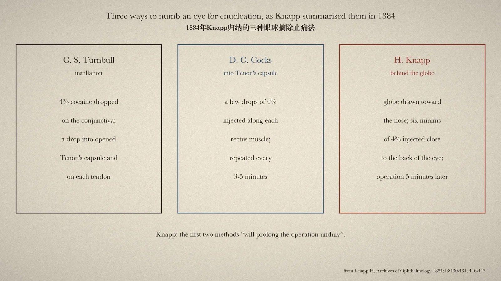
In the autumn of 1884 cocaine drops changed eye surgery almost overnight. H. Knapp opened a long review in Archives of Ophthalmology by saying that no modern remedy had been received "with such general enthusiasm" as cocaine, "the local anaesthetic recently introduced by Dr. C. Koller, of Vienna". [1] Drops on the surface worked well for operations on the cornea and conjunctiva. Removing a whole eye was harder: the nerves that matter run behind it. By early December three surgeons had tried three different answers, and Knapp's article sets them side by side. [1]
Drops, then injections
C. S. Turnbull, oculist to the German Hospital of Philadelphia, relied on instillation. Two drops of four per cent cocaine were put in every three minutes until eight had been used; when Tenon's capsule was opened a drop was allowed into the cut; each tendon received a drop before it was divided; and two or three large drops were run down the blades of the scissors on either side of the optic nerve before it was cut. The patient "remained entirely quiet". [1]
D. C. Cocks of New York injected instead. After anaesthetising the cornea and conjunctiva and opening the conjunctiva, Cocks used an Anel's syringe to put "a few drops of a 4% solution over the tendon of the external rectus", then pushed the syringe as far back as possible near the globe and injected again, repeating for each rectus. The operation took about half an hour, with drops or injections every three to five minutes; the patient "occasionally felt some pain, but not enough to interfere with the operation". [1]
Behind the globe
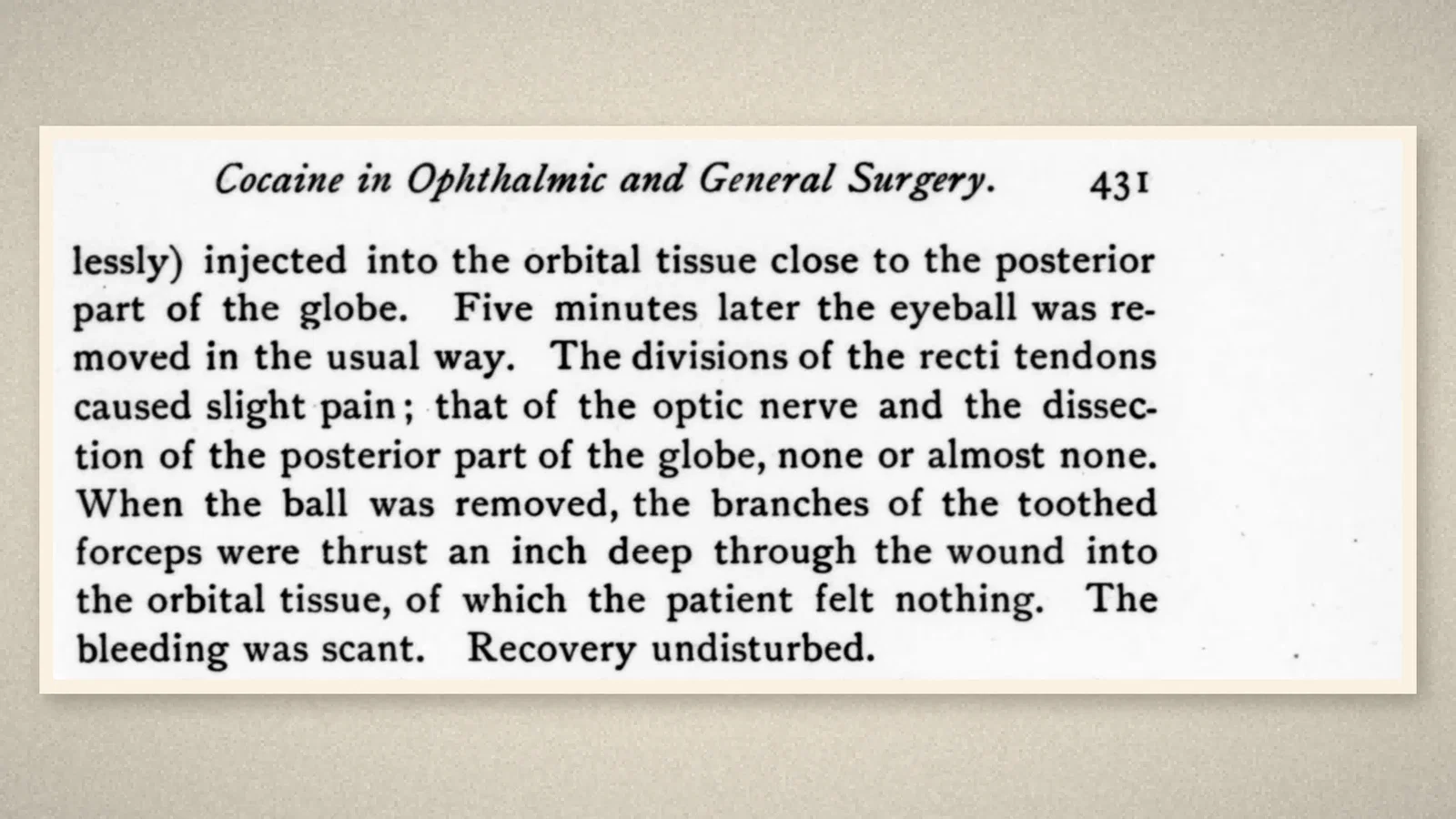
Knapp's own case went further back. After the conjunctiva was anaesthetised, the globe was "strongly drawn toward the nose" with forceps and six minims of a 4 per cent solution were injected, painlessly, "into the orbital tissue close to the posterior part of the globe". Five minutes later the eye was removed in the usual way. Cutting the muscle tendons caused slight pain; cutting the optic nerve and freeing the back of the globe, "none or almost none". When the toothed forceps were pushed an inch into the orbit afterwards, "the patient felt nothing". Bleeding was scant and recovery undisturbed. [1]
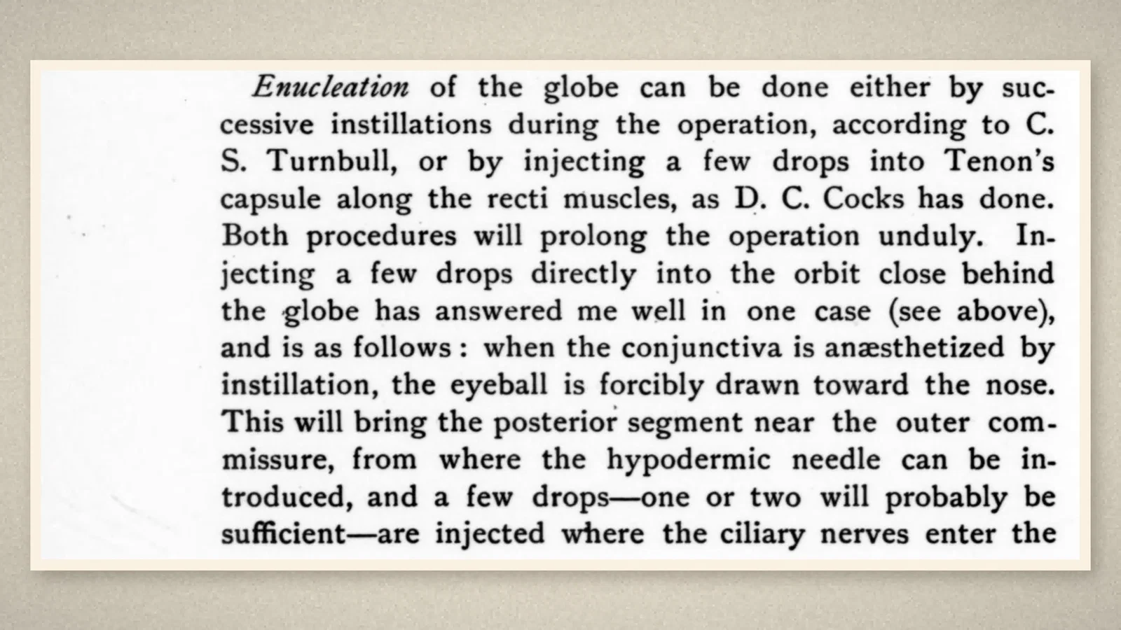
In the summary at the end of the article Knapp compared the methods. Enucleation could be done "by successive instillations ... according to C. S. Turnbull, or by injecting a few drops into Tenon's capsule along the recti muscles, as D. C. Cocks has done. Both procedures will prolong the operation unduly." Injecting "a few drops directly into the orbit close behind the globe" had answered well "in one case", and Knapp described the route: with the eye pulled toward the nose, the back of the globe comes near the outer corner of the lids, where the needle enters, and one or two drops "are injected where the ciliary nerves enter the globe". [1]
A caution in the same breath
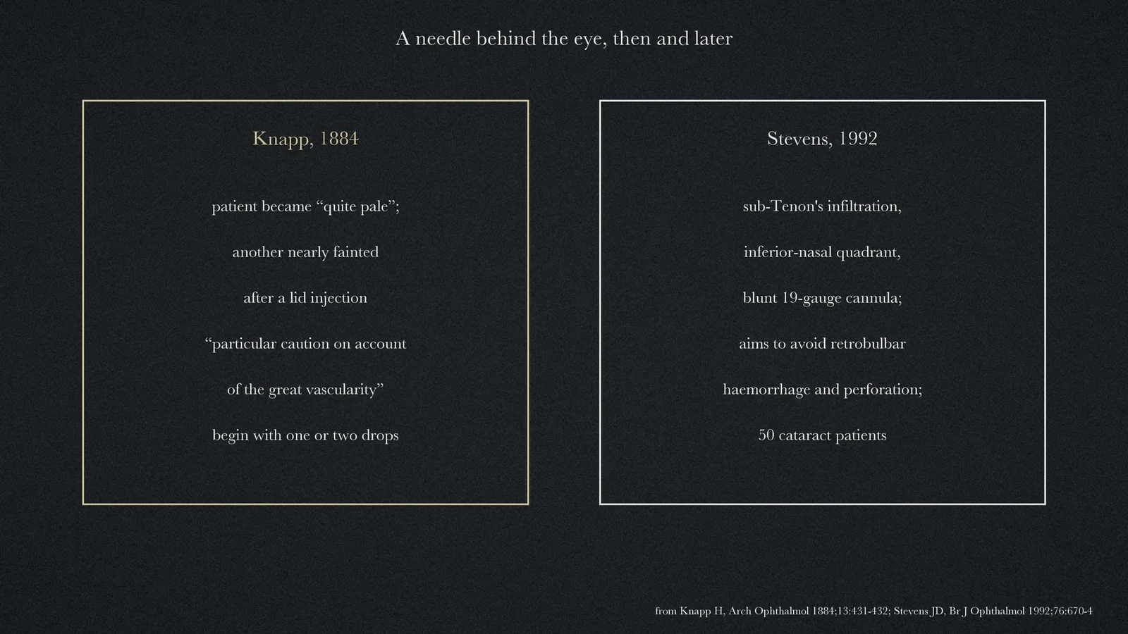
Knapp did not present the injection as harmless. The enucleation patient "had become quite pale". A week later, after five minims were injected under a lid tumour, a strong man who had never fainted "became as pale as a corpse", felt faint and broke into a cold sweat for about fifteen minutes. Knapp sent the case to the Medical Record "as a word of caution", wrote that injections into the orbit "should be made with particular caution on account of the great vascularity of the tissue", and proposed starting with no more than one or two drops. [1]
A century later
The concern did not go away. In 1992 J. D. Stevens of Moorfields Eye Hospital described a technique of injecting local anaesthetic through the conjunctiva into the sub-Tenon's space with a blunt cannula, used in 50 patients for cataract surgery, designed "to avoid the risks of retrobulbar haemorrhage, perforation of the globe, damage to the optic nerve, and injection into the subarachnoid space" and so that no "sharp instrument" is "passed into the orbit". [2] The space Cocks had injected in 1884, and the needle Knapp had pushed behind the eye, mark the two directions that ophthalmic regional anaesthesia would keep exploring.
What the record shows
The 1884 reports record, within three months of topical cocaine, an injection placed deliberately behind the eye to block the nerves entering it, tested in a single enucleation and published together with a warning about the orbit's blood supply. They also show three methods developing in parallel, which makes single "firsts" hard to assign.
Sources and reading limits
Read: Knapp's article in Archives of Ophthalmology, vol. 13 (1884): the opening, the sections on enucleation and orbital injection, and the closing summary (pp. 402, 427–432, 446–447); the abstract of Stevens 1992.
Not covered: the original notes by Knapp, Turnbull and Cocks in the Medical Record, Medical and Surgical Reporter and Medical News, which are known here through Knapp's summaries; Koller's paper; later retrobulbar and peribulbar techniques; the full text of Stevens 1992. Some timelines credit Turnbull with the first sub-Tenon's route; in Knapp's account Turnbull dropped cocaine into the opened capsule, while Cocks injected into it. This article assigns no "first". Names are given as printed.
Two illustrations are crops of Knapp's article; two are drawn from the sources.
- Knapp H. On cocaine and its use in ophthalmic and general surgery. Arch Ophthalmol. 1884;13:402–48. Available from: archive.org
- Stevens JD. A new local anesthesia technique for cataract extraction by one quadrant sub-Tenon's infiltration. Br J Ophthalmol. 1992;76(11):670–4. doi:10.1136/bjo.76.11.670
Continue exploring
Related reading connects shared questions and contexts; it does not by itself establish historical influence.
- 1884: Koller Brings Cocaine to the Surface of the Eye
- 1884: Halsted and Hall Report a Jaw Nerve Block in a Postscript
Anesthesia Epochs · The Source Room · About & Editorial Method
Published November 2, 2026. Sources and reading limits are stated above. Illustrations are labelled as original source images or AI-made. Historical accounts are educational; they are not current treatment instructions.