
An idea of parting the dural fibres instead of cutting them, carried from office lumbar puncture to spinal anaesthesia.
Reading Library · Anesthesia Epochs · 中文版
Watch the education video (for clinicians)
1926: Greene's Round-Pointed Spinal Needle — Point or Size? · Anesthesia Epochs Education Series
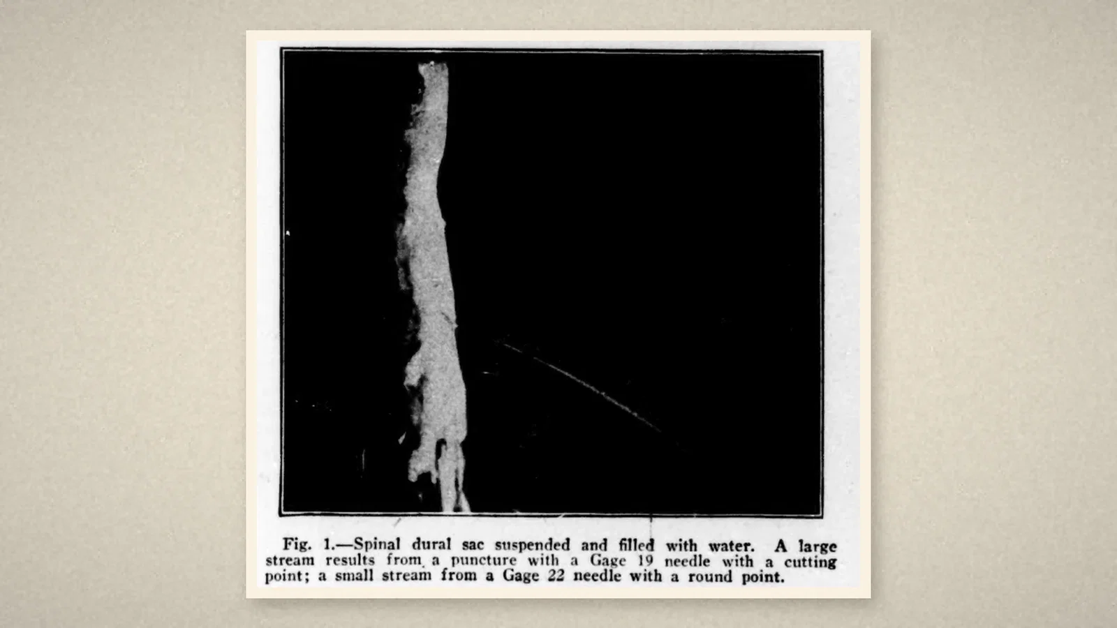
Any anaesthetist who gives spinal anaesthetics today chooses between a cutting needle and a pencil-point needle, and knows the second is chosen to spare the patient a headache. One root of that choice is a two-page paper in the Journal of the American Medical Association of 6 February 1926, "Lumbar Puncture and the Prevention of Postpuncture Headache", by H. M. Greene, a member of the teaching staff of the University of Oregon Medical School in Portland. [1, p. 391]
A dural sac filled with water
Greene began with the conclusion. As a result of experiments made during 1923 and experience since then with a small needle "having a round, sharp, tapering point", Greene was convinced that "postpuncture headache is caused by trauma to the spinal dura" sufficient to let cerebrospinal fluid leak until the brain was left without its water cushion. [1, p. 391] The experiments showed that "a greater trauma was produced by the use of a needle with a blunt cutting point" than by one of the same calibre with a rounded, tapering, sharp point, that leakage followed every puncture of a dural sac suspended and filled with water, and that "the amount of leakage was in direct relation to the size of the needle used". [1, p. 391] Greene's Fig. 1 shows the result: a large stream from a 19-gauge cutting needle, a small one from a 22-gauge round point.
The method was practical. The patient should lie still; local anaesthetic should be used; the needle should be sharpened "so as to resemble an ordinary cambric needle" and polished before each use; "Gage 22 is the most suitable". [1, p. 391] Then the figure: "Two hundred and fifteen consecutive punctures have been done, with a headache incidence of two in the series." [1, p. 391] These were diagnostic lumbar punctures, "nearly all done in the office"; patients went home to lie down, and some "resumed his work for the remainder of the day". [1, p. 391]
Fibres that run lengthwise
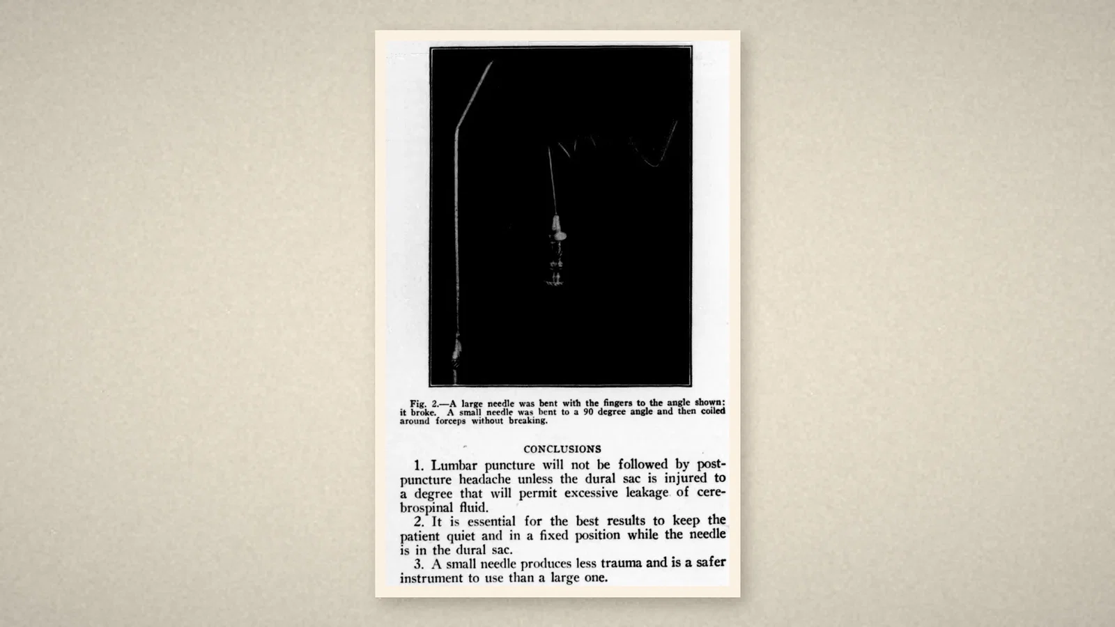
Why should the point matter? The spinal dura, Greene wrote, consists of fibrous and elastic tissue in bands that are mostly parallel and "have a longitudinal arrangement". With no circular fibres, "it is possible to pass a small sharp, round, tapering pointed instrument between the fibers" without cutting any, leaving "a longitudinal slit". [1, p. 391] On dissection Greene saw that the sac moves in the canal as the head bends, so a large rigid needle would see-saw in the dura and enlarge the hole. A small needle was also safer "because it will bend before it will break". [1, p. 392] A footnote gives credit to Becton, Dickinson & Co. for experimental work on a needle of small calibre. [1, p. 391]
Twenty-five years later: the pencil point
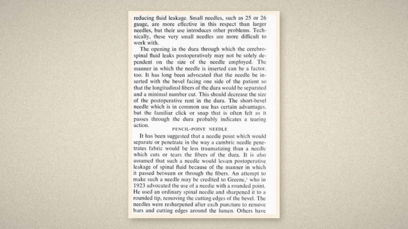
In 1951 James R. Hart and R. J. Whitacre of the Department of Anesthesiology, Huron Road Hospital, Cleveland, published "Pencil-Point Needle in Prevention of Postspinal Headache" in the same journal. [2, p. 657] The majority opinion, they wrote, was that headache follows leakage of fluid through the dural hole, whose longitudinal fibres let the aperture gape. Turning the bevel sideways was an old advice; the familiar click of the short-bevel needle through the dura "probably indicates a tearing action". [2, p. 657]
"An attempt to make such a needle may be credited to Greene", they wrote, who in 1923 advocated a rounded point. Following that reasoning, "we asked the Becton, Dickinson Company to make a 20 gauge" needle with a solid end drawn to a point like a finely sharpened pencil — "hence we commonly refer to it as a pencil-point needle" — with the opening on the side, just proximal to the solid tip. [2, p. 657] They were candid: "Such deductive reasoning is not supported by any direct evidence." [2, p. 657]
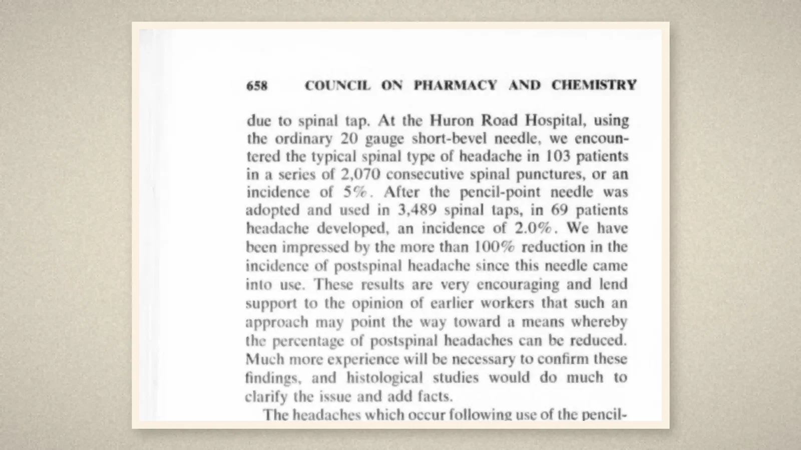
Their numbers came from their own hospital. With the ordinary 20-gauge short-bevel needle, 103 of 2,070 consecutive spinal punctures were followed by the typical headache, 5%; with the pencil-point needle, 69 of 3,489, 2.0%. [2, p. 658] They added, "much more experience will be necessary to confirm these findings". Their rules were simple: the hole as small as possible, the patient still, and "there should be only one hole in the dura". For treatment, "time still is the surest cure". [2, p. 658]
What the record shows
The idea of parting the dural fibres instead of cutting them began with a physician doing lumbar punctures in an office, testing needles on a water-filled dural sac. It reached spinal anaesthesia when two Cleveland anaesthetists, crediting Greene, had a manufacturer build a solid-tipped needle with a side hole, and counted their headaches before and after.
Sources and reading limits
Read: Greene, JAMA 6 February 1926, pp. 391–392, with Figs. 1–2; Hart and Whitacre, JAMA 13 October 1951, pp. 657–658.
Not covered: Greene's 1923 paper in Northwest Medicine is not covered. Greene's figure is 2 headaches in 215 diagnostic punctures, and Greene's are not spinal anaesthetics; Greene does not say the experiments used cadavers. Hart and Whitacre's "more than 100% reduction" is their wording for a fall from 5% to 2.0%.
All four illustrations are crops of the 1926 and 1951 journal pages.
- Greene HM. Lumbar puncture and the prevention of postpuncture headache. JAMA. 1926;86(6):391-2. Internet Archive
- Hart JR, Whitacre RJ. Pencil-point needle in prevention of postspinal headache. JAMA. 1951;147(7):657-8. Internet Archive
Continue exploring
Related reading connects shared questions and contexts; it does not by itself establish historical influence.
Anesthesia Epochs · The Source Room · About & Editorial Method
Published November 2, 2026. Sources and reading limits are stated above. Illustrations are labelled as original source images or AI-made. Historical accounts are educational; they are not current treatment instructions.