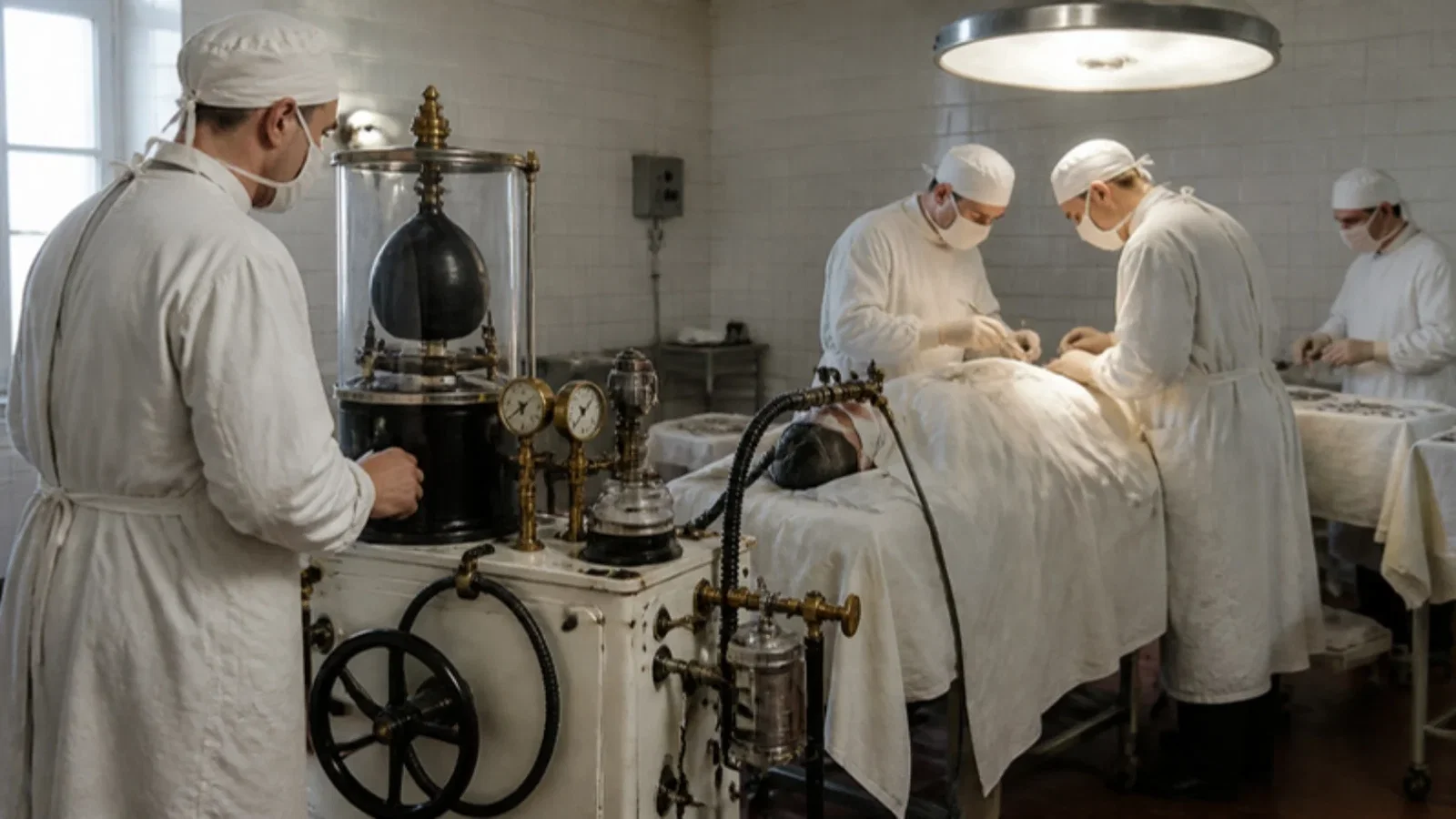
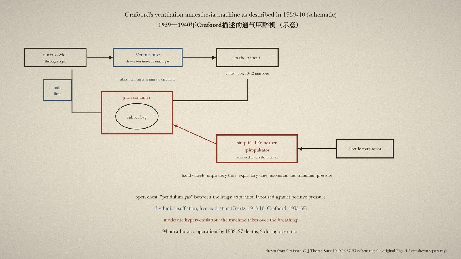
At Sabbatsberg Hospital, Stockholm, Clarence Crafoord judged that constant-flow positive pressure caused 'pendulum gas' between the lungs and laboured expiration, and with the engineer Anderson coupled a modified Frenckner spiropulsator to a nitrous oxide circuit: the breathing bag sat in a glass container whose pressure the compressor-driven spiropulsator raised and lowered, with hand wheels for the inspiratory and expiratory times and the maximum and minimum pressures; after moderate hyperventilation the machine took over, in 94 intrathoracic operations by 1939.
Reading Library · Anesthesia Epochs · 中文版
Watch the education video (for clinicians)
1938: Crafoord's Spiropulsator — Stockholm · Anesthesia Epochs Education Series
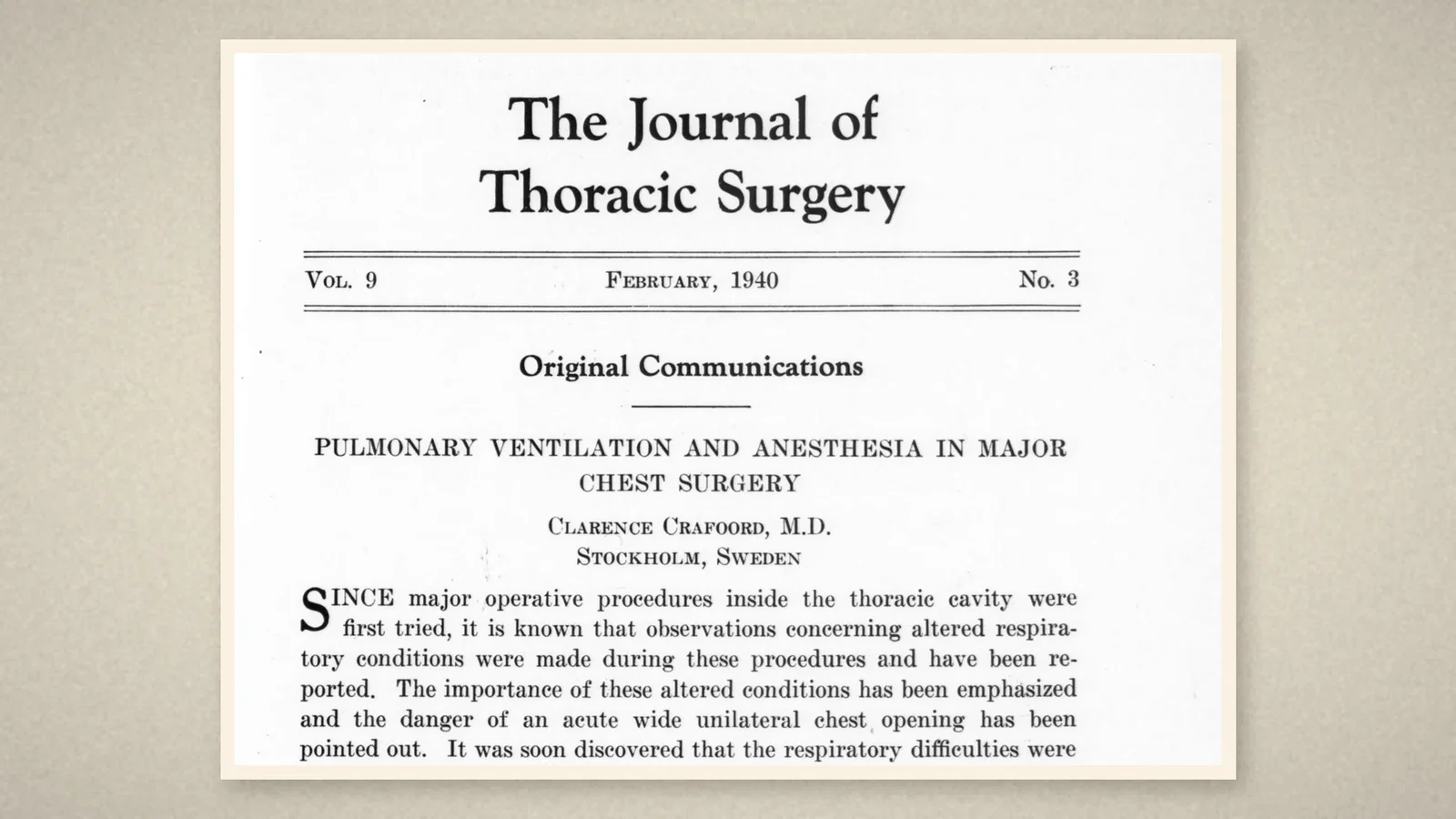
The anaesthesia ventilator is now so ordinary that it is hard to imagine an operating theatre without one. The Australian Society of Anaesthetists (ASA) timeline places in 1938 the use during surgery of a mechanical ventilator, the "Spiropulsator", by Clarence Crafoord in Sweden. This article reads Crafoord's own account, "Pulmonary ventilation and anesthesia in major chest surgery", read before the American Association for Thoracic Surgery at Los Angeles in July 1939 and published in The Journal of Thoracic Surgery in February 1940; one of its anaesthesia charts carries the case number 815/1938. [1][2]
What goes wrong in an open chest
Crafoord, of Sabbatsberg Hospital in Stockholm, explained the physiology. With one side of the chest wide open, inspiration drew gas not only from the trachea into the intact lung but also from the collapsed lung into it; on expiration part of the gas flowed back. This "so-called pendulum gas" diluted each breath with stale gas. In one lobectomy for a lung abscess Crafoord watched the cavity, of at least 75 c.c., empty with each inspiration and refill with each expiration, while the tidal volume was only 150 to 200 c.c. [1]
The usual remedy, a constant-flow positive pressure machine, steadied the mediastinum but did not stop the pendulum gas, and it made expiration hard work. In dogs with the chest opened and a positive pressure of 4 to 8 cm of water, arterial carbon dioxide rose over about three hours from a normal of about 40 to about 82 and 95 volumes per cent, despite pure oxygen. Crafoord also noted how hard patients' respiratory muscles worked during open-chest operations elsewhere, a strain that itself consumed oxygen and produced carbon dioxide. [1]
Rhythmic insufflation
Crafoord's chief, K. H. Giertz, had met the same problem while working with Sauerbruch in Munich in 1912 and in experiments from 1913 to 1916 had concluded that "rhythmic insufflation of gas was the safest method", through an airtight tracheal tube, with physiological breaths at a normal rate, warm and moist gas, and pressures up to 15 cm of water. Crafoord took up the work from 1933. With the engineer Anderson, Crafoord built a machine that gives "physiologic quantities of gas during the inspiratory phase" and "allows the lungs to empty themselves without any resistance during the expiratory phase", with the rate and the maximum and minimum pressures adjustable. A moderate hyperventilation lowered the irritability of the respiratory centre, and then the patient's own respiratory movements subsided. [1]
The machine
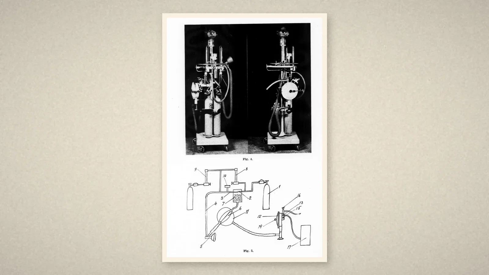
The apparatus combined "a modified Frenckner spiropulsator and a continuously flowing gas narcosis apparatus". Nitrous oxide passed through a jet into a Venturi tube, which drew about ten times as much gas round through the soda lime container, so that some ten litres a minute circulated for one litre of fresh gas, reducing dead space practically to zero; oxygen, cyclopropane or ether could be added. "When operating with a wide-open chest, the rubber bag is enclosed in a glass container." The spiropulsator, supplied with compressed air from an electric compressor, "rhythmically increases and decreases pressures inside the glass container", compressing and releasing the bag, and four hand wheels set "the length of the inspiratory phase, the length of the expiratory phase, the maximum pressure, and the minimum pressures". [1]
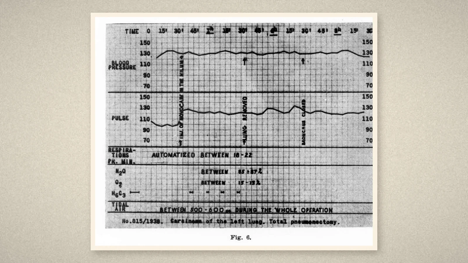
In a typical pneumonectomy the diseased bronchus was blocked with a gauze tampon through a bronchoscope, a cuffed tube of at least 10 to 12 mm bore placed in the trachea, and nitrous oxide-oxygen given; after about ten minutes rhythmic ventilation began, at first following the patient's own breaths by hand, until "the machine takes over the breathing of the patient". The chart in Fig. 6, numbered 815/1938, records respirations "automatized between 18-22" and a tidal air of 500 to 600 c.c. throughout the operation. [1]
Results and cautions
Crafoord reported 94 intrathoracic operations with this rhythmic ventilation, including pneumonectomies, lobectomies, oesophagectomies and pericardial and heart operations, with 27 deaths, two during operation; one was from air embolism through a ruptured pulmonary vein. Crafoord also described running nitrous oxide with oxygen as low as 13 to 25 per cent, occasionally about 10 per cent, under hyperventilation, supplemented by intercostal and vagal local anaesthesia and small amounts of cyclopropane. These are historical practices and not guidance. [1]
What the record shows
Crafoord's paper sets out the logic of intraoperative mechanical ventilation: the open chest destroys spontaneous breathing; positive inspiration with free expiration restores tidal exchange; mild hyperventilation lets a machine take over; and the anaesthetist sets inspiratory and expiratory times and pressure limits. The bag-in-a-bottle design, in which a driving gas squeezes a bag of anaesthetic mixture, remained the basis of many later anaesthesia ventilators.
Sources and reading limits
Read: Crafoord C, J Thorac Surg 1940;9:237-53, in full; the ASA timeline entry for 1938.
Not covered: Crafoord's 1938 thesis on the technique of pneumonectomy; Frenckner's 1934 description of the spiropulsator; Giertz's Swedish papers; later ventilators. The date 1938 is the ASA's and is consistent with the case number on Fig. 6; the full description dates from 1939-40. The ASA spells the inventor "Frecken"; the paper prints Frenckner. The ASA's "first use" is quoted as its own; Crafoord credits Giertz's earlier experiments. Results and practices are Crafoord's. Premedication doses are not repeated.
Three illustrations are crops of the February 1940 issue, including Figs. 4-6; one is drawn from the text.
- Crafoord C. Pulmonary ventilation and anesthesia in major chest surgery. J Thorac Surg. 1940;9:237-53. Internet Archive
- Australian Society of Anaesthetists. Anaesthesia history timeline. 2024. ASA
Continue exploring
Related reading connects shared questions and contexts; it does not by itself establish historical influence.
- 1936: Rovenstine's Anaesthesia for Chest Surgery
- 1908: The Draeger Pulmotor Blows Air In and Sucks It Out
Anesthesia Epochs · The Source Room · About & Editorial Method
Published November 2, 2026. Sources and reading limits are stated above. Illustrations are labelled as original source images or AI-made. Historical accounts are educational; they are not current treatment instructions.