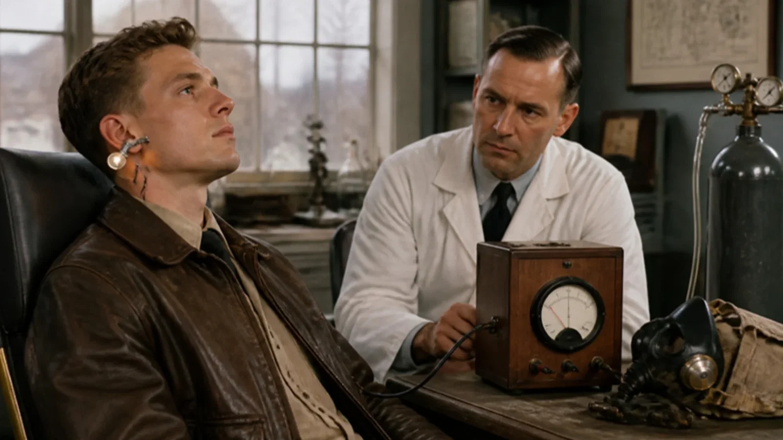
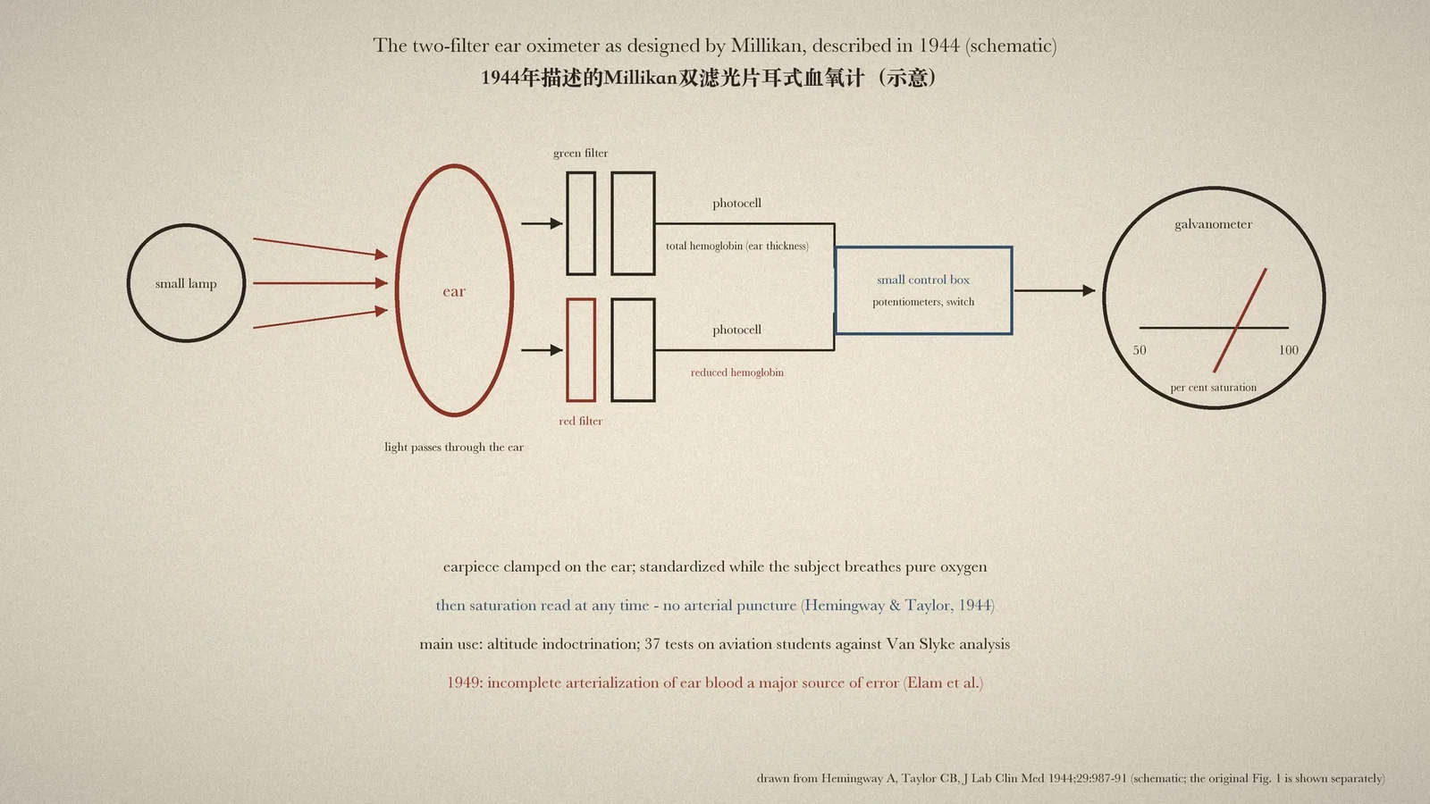
Millikan's two-filter ear oximeter put a small lamp on one side of the ear and two photocells with filters on the other; standardised while the subject breathed pure oxygen, it then gave saturation directly on a galvanometer; mainly used in altitude training, it was checked against arterial blood on aviation students by the Army Air Forces in 1944, analysed for sources of error by Elam and colleagues in 1949, and used in an improved form in some 20 intrathoracic operations, Gibbon reported.
Reading Library · Anesthesia Epochs · 中文版
Watch the education video (for clinicians)
1942: Millikan's Ear Oximeter — From Altitude to the OR · Anesthesia Epochs Education Series
A number on a monitor now tells every anaesthetist how saturated the patient's arterial blood is. Before that number existed, colour was the guide, and colour misleads. The New Zealand Society of Anaesthetists (NZSA) places in 1939-42 the development of "an ear oxygen meter" and the coining of the term oximeter; the Australian Society of Anaesthetists (ASA) gives 1942 for the "original oximeter machine". The 1942 paper itself, by G. A. Millikan, is not covered here. What follows reads two papers that describe and test the instrument: a 1944 calibration by two officers of the United States Army, and a 1949 analysis of its errors read before the American Surgical Association. [1][2][3][4]
Light through the ear
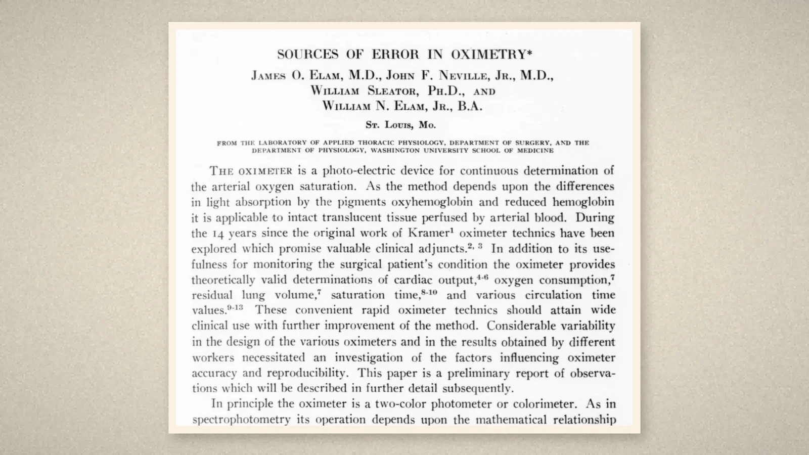
The principle is set out at the opening of the 1949 paper by James O. Elam, John F. Neville Jr, William Sleator and William N. Elam Jr of Washington University, St Louis: "The oximeter is a photo-electric device for continuous determination of the arterial oxygen saturation." Because oxyhemoglobin and reduced hemoglobin absorb light differently, the method "is applicable to intact translucent tissue perfused by arterial blood", and the ear is such a tissue. "In principle the oximeter is a two-color photometer or colorimeter." Besides measuring saturation, the authors wrote, it was useful "for monitoring the surgical patient's condition". [2]
The 1949 authors also gave the instrument a history. "Original contributions to oximetry should be credited to Kramer and to Matthes." Kramer's one-colour unit fitted around an intact artery of the dog and agreed "within 1 per cent" with chemical analysis; Matthes adapted the method to the transilluminated human ear and added a second, near infra-red measurement as an index of the total hemoglobin in the light path. Goldie in 1942 introduced a correction for the light absorbed by the bloodless ear tissue. Millikan's oximeter, also of 1942, gave the saturation as a simple difference between the red and infra-red readings; that simplification was chosen "to obtain a simple qualitative instrument for use in altitude physiology". [2]
The instrument in wartime use
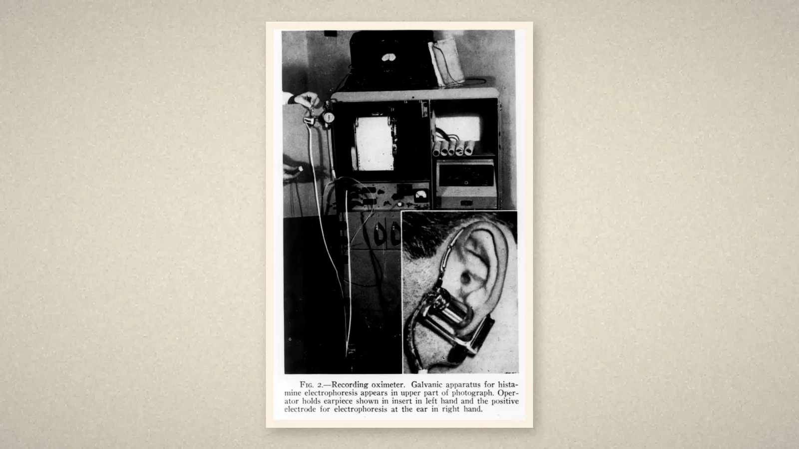
In September 1944 Major Allan Hemingway of the Air Corps and Captain Charles B. Taylor of the Medical Corps published laboratory tests of "the two-filter oximeter as designed by Millikan". The apparatus "consists of an earpiece, a small control box, and a galvanometer." The earpiece held "a small lamp placed on one side of the ear and two photoelectric cells on the other", each behind a Wratten filter: a "green" filter passing light absorbed by both forms of hemoglobin, an index of ear thickness, and a "red" filter passing light absorbed by reduced hemoglobin. The earpiece was clamped on the ear and the apparatus standardised while the subject breathed pure oxygen; after that the saturation could be read "at any time". It needed "no arterial punctures" and no wait of twenty or thirty minutes for a chemical result. [1]
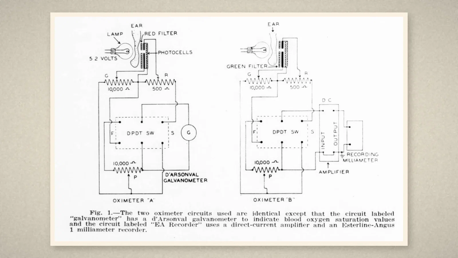
Its main use then was "the altitude indoctrination program", but the authors added that it could be extended "to operating room procedures". Older instruments had five galvanometer scales and the operator sometimes had to change scale when the ear's vessels constricted or dilated; a newer circuit used a single scale with automatic adjustment for these vasomotor changes. The scale read from 50 to 100 per cent. The apparatus needed twenty minutes to warm up, and the earpiece fifteen minutes on the ear to reach thermal equilibrium. [1]
To check the approximations in the new circuit, aviation students breathed pure oxygen and then 8 per cent oxygen in nitrogen until the reading fell to 65-70 per cent, when arterial blood was drawn and analysed by the Van Slyke method. In 37 tests, about nine readings in ten agreed with the chemical values within 5 per cent. [1]
Sources of error
The 1949 paper asked why different oximeters gave different results. The authors built an ear spectrophotometer and recorded the transmission of the bloodless and perfused ear from 4,000 to 11,000 Angstroms. Their central finding was physiological: "Failure to obtain complete arterialization of the ear blood represents a major source of error and variability." With a Millikan earpiece on the heated pinna they recorded "the prolonged saturation time of 120 seconds" on changing from air to oxygen, while a better-flushed ear lobe responded in about 20 seconds. They also judged that the need to adjust the reading to the operator's estimate limited the Millikan device clinically: without arterial calibration for each procedure, it "served merely to indicate changes in the patient's arterial oxygen saturation". Other sources of error included the placement of the two photocells and the lamp voltage. [2]
In the operating room
The discussion that followed brought the instrument to surgery. John H. Gibbon Jr of Philadelphia said that a colleague, Dr Miller, had built an oximeter that the department thought better than the Millikan, and that it had been used "in some 20 intrathoracic operations". With patients breathing ether and oxygen, the blood was "completely saturated with oxygen during the course of the operation", so the instrument was of little value except during induction, when saturation might fall, and immediately after the anaesthetic was stopped. Deflections also occurred when the patient's position on the table was changed. Gibbon wished instead for an instrument that could continuously record the carbon dioxide tension of arterial blood. Elam replied that the Millikan scale was not linear: the range from 90 to 100 per cent was about three times that from 50 to 60 per cent. In the discussion of the preceding paper, Julian Johnson noted that judging saturation from the patient's colour was inaccurate and that an oximeter on the ear, read on air and on oxygen, showed whether oxygen therapy helped. [2]
What the record shows
The ear oximeter put three lasting ideas in place: shine light through living tissue, compare two colours so that thickness cancels out, and read oxygen saturation continuously without drawing blood. The 1949 papers also show the instrument's limits as monitors: venous blood in the light path, motion, a non-linear scale, and readings that showed trends more reliably than absolute values. In the NZSA timeline the next oximetry entry is a prototype pulse oximeter used clinically in 1973.
Sources and reading limits
Read: Hemingway A and Taylor CB, J Lab Clin Med 1944;29:987-91, in full; Elam JO and colleagues, Ann Surg 1949;130:755-73, read before the American Surgical Association on 22 April 1949, in full, including the bibliography and discussion; the NZSA timeline entries for 1939-42 and 1973; the ASA timeline entry for 1942.
Not covered: Millikan's paper "The oximeter, an instrument for measuring continuously the oxygen saturation of arterial blood in man" (Rev Sci Instrum 1942;13:434); Goldie's 1942 paper; the German papers of Kramer and Matthes; Wood's papers on the modified oximeter. The instrument is therefore described from the 1944 calibration and the 1949 review. The ASA names the maker of the 1942 machine as "Robert Andrews Millikan - American physicist and Nobel Laureate"; both papers read here cite the 1942 paper as by "G. A. Millikan", the name used here. The NZSA's coining of the term is quoted as its own; the 1942 paper's title is "The oximeter". The 1949 authors credit Kramer and Matthes with the original work. Gibbon's operating-room use is reported as said in discussion, without case data. Doses used to flush the ear are not repeated. Quotations are from the English originals.
Three illustrations are crops of the 1944 and 1949 pages, including Fig. 1 of 1944 and Fig. 2 of 1949; one is drawn from the 1944 text.
- Hemingway A, Taylor CB. Laboratory tests of the oximeter with automatic compensation for vasomotor changes. J Lab Clin Med. 1944;29:987-91. Internet Archive
- Elam JO, Neville JF Jr, Sleator W, Elam WN Jr. Sources of error in oximetry. Ann Surg. 1949;130:755-73. Internet Archive
- New Zealand Society of Anaesthetists. History of anaesthesia. NZSA
- Australian Society of Anaesthetists. Anaesthesia history timeline. 2024. ASA
Continue exploring
Related reading connects shared questions and contexts; it does not by itself establish historical influence.
- 1938: Crafoord's Spiropulsator Breathes for Open-Chest Surgery
- 1924: Berger Records the Human Brain's Electrical Rhythm
Anesthesia Epochs · The Source Room · About & Editorial Method
Published November 2, 2026. Sources and reading limits are stated above. Illustrations are labelled as original source images or AI-made. Historical accounts are educational; they are not current treatment instructions.