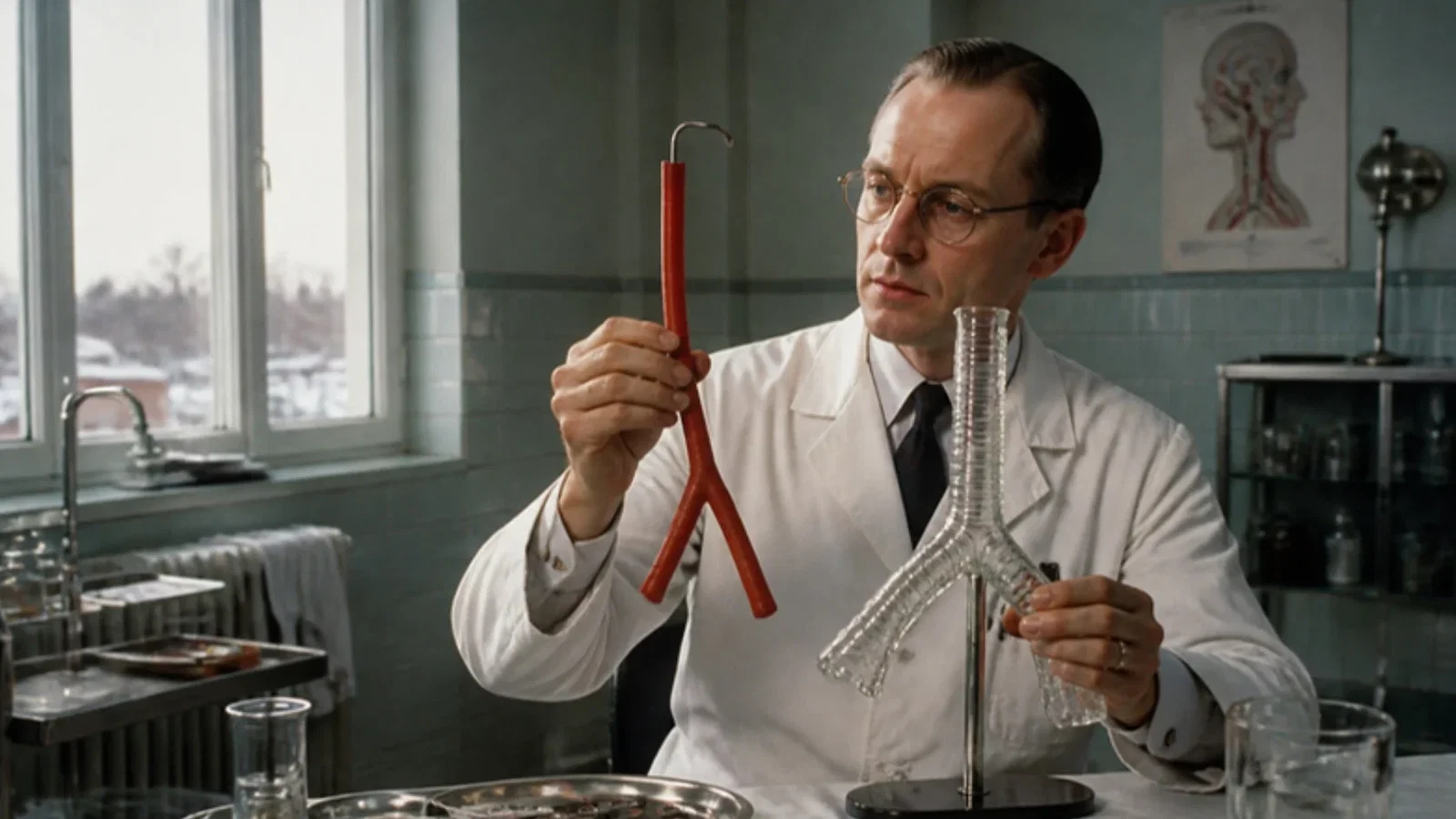
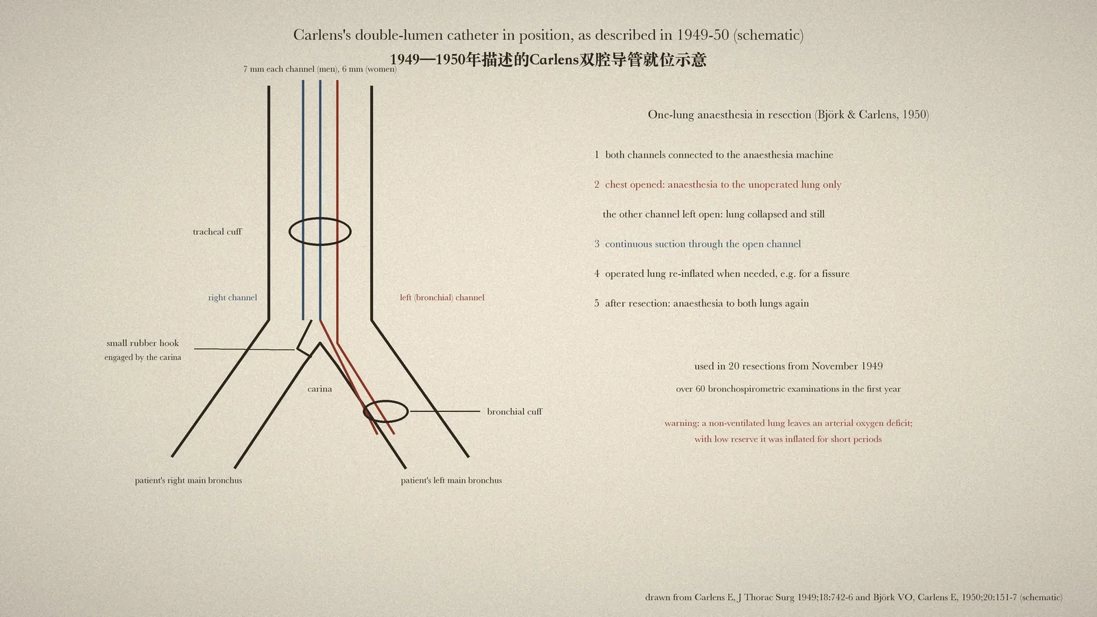
Eric Carlens of the oto-laryngologic department at Sabbatsberg Hospital, Stockholm, designed a soft rubber double-lumen catheter for measuring the function of each lung: about as stiff as a urethral catheter, its bronchial limb slid by itself into the left main bronchus and a small rubber hook caught on the carina, so no fluoroscopy was needed; control bags showed when the two cuffs were inflated; in 1950 Carlens and the thoracic surgeon Björk reported using it since November 1949 in 20 lung resections to anaesthetise and suction each lung separately, collapsing the operated lung and keeping pus out of the other.
Reading Library · Anesthesia Epochs · 中文版
Watch the education video (for clinicians)
1949: Carlens's Double-Lumen Catheter — Stockholm · Anesthesia Epochs Education Series
Thoracic anaesthetists now isolate one lung as a matter of routine: a double-lumen tube lets one lung be collapsed for the surgeon while the other keeps breathing. The Australian Society of Anaesthetists (ASA) timeline records under 1949 the Carlens double-lumen endobronchial tube, designed by Eric Carlens at the Sabbatsberg Hospital in Sweden for bronchospirometry, and under 1950 its use for one-lung anaesthesia by Carlens and the thoracic surgeon Viking Olov Björk. This article reads both papers in the Journal of Thoracic Surgery: Carlens's description of October 1949 and Björk and Carlens's report of July 1950. [1][2][3]
Measuring each lung
The catheter was made to measure, not to anaesthetise. Bronchospirometry recorded the ventilation and oxygen uptake of each lung separately. Carlens, of the oto-laryngologic department at Sabbatsberg, began with its history: the first such measurements in human subjects, by Jacobaeus, Frenckner and Björkman in 1932, used "a rigid double-lumen bronchoscope", and the rigid instruments were hard to place and "very disagreeable to the patient". In 1939 Gebauer, and independently Zavod in 1940, described soft rubber double-lumen catheters. These were easier, but they had to be placed under fluoroscopy, and their small channels caused "stenosis breathing" that could make patients short of breath and distort the readings; viscous sputum blocked them. A single-lumen catheter described by Norris and colleagues in 1948 avoided the narrowing, but it was hard to tell under the screen whether its cuff sat just between the carina and the left upper lobe bronchus. [1]
A hook for the carina
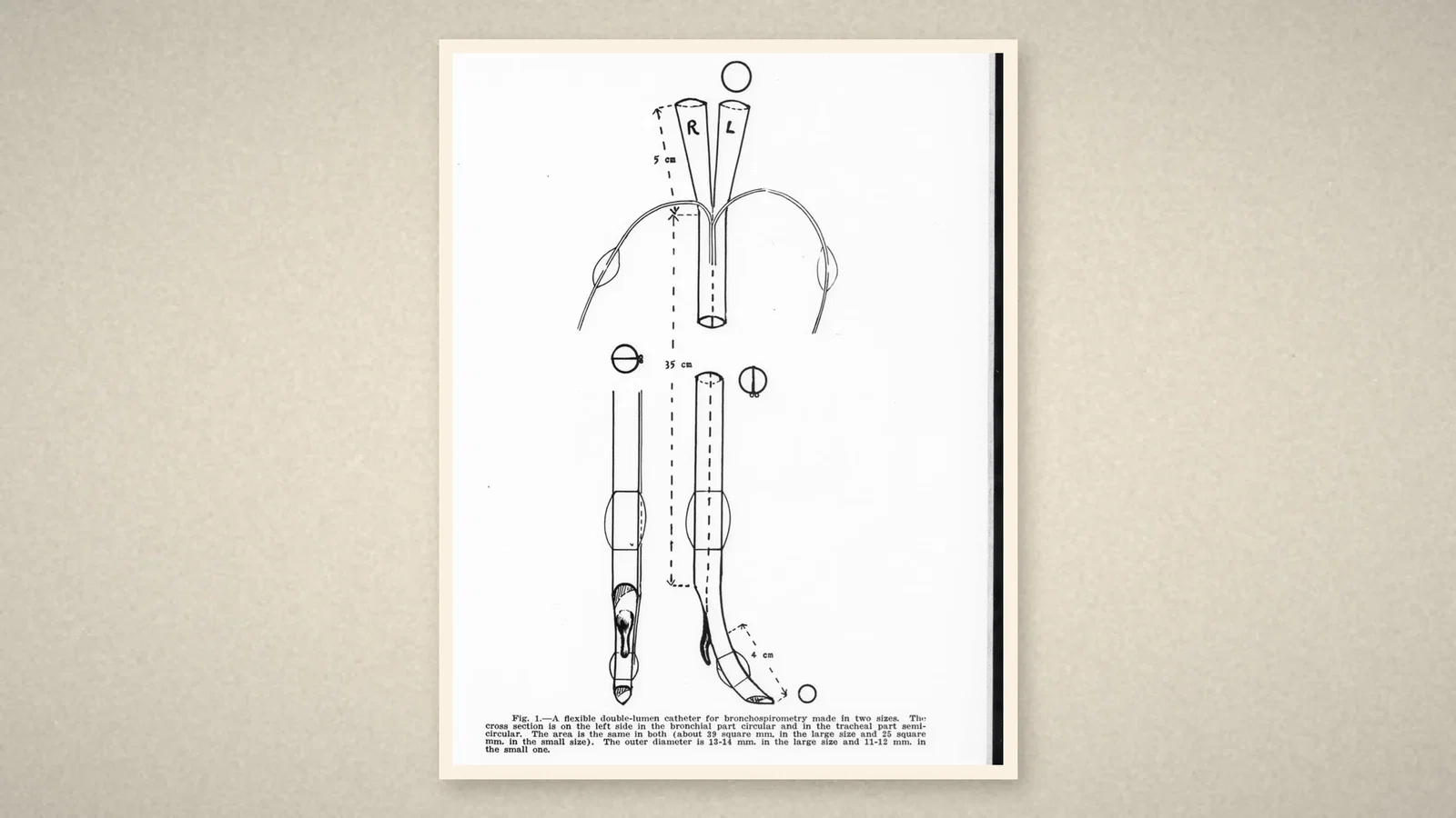
Carlens's answer was "a flexible double-lumen rubber catheter of approximately the same rigidity as an ordinary urethral catheter", made by Trelleborgs Gummifabrik, and "provided with a little hook that is automatically engaged by the carina". Each channel in the model for men was 7 mm wide, the same as the single-lumen catheter of Norris; the model for women had 6 mm channels. The outer diameter was 13-14 mm in the large size and 11-12 mm in the small. [1]
Placement needed no screen. A curved metal stylet went into the bronchial channel, and the rubber hook was tied down with a silk thread. The catheter was introduced "under mirror guidance"; once the hook had passed the cords, the stylet was removed and the slipknot pulled free. "The catheter is now turned 90 degrees to the left" and pushed gently down until the hook caught; "it is very easy to feel when the catheter is in correct position." Because the rubber pressed lightly on the left tracheal wall, the bronchial end "automatically enters the left main bronchus", even when the bronchus left the trachea at nearly a right angle. The two cuffs were inflated with a syringe until small control bags on their pilot lines filled, which "obviated the necessity for manometric control". [1]
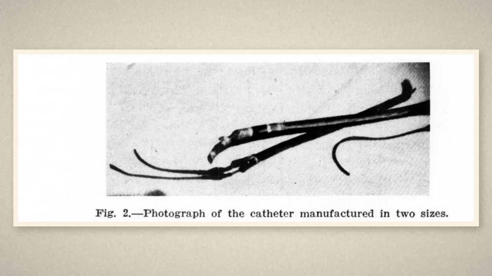
In over 60 examinations in the first year there had been no difficulty in placing it and no untoward effect, and some tests continued for an hour. In seven consecutive cases, the vital capacities of the two lungs measured through the catheter added up closely to the vital capacity measured by ordinary spirometry, which earlier double catheters had not achieved. The channels were wide enough for tests during exercise on a cycle ergometer. [1]
From the laboratory to the operating table
Björk was a thoracic surgeon in Clarence Crafoord's surgical clinic at the same hospital. In July 1950 Björk and Carlens addressed a different problem: pus or blood flooding the healthy lung during resection for suppurative disease. In one series by Moody, cited in their paper, spread had occurred in 21.7 per cent of resections. Earlier answers included endobronchial tubes with an inflatable cuff (Gale and Waters, 1931; Magill, 1936) and blockers of the diseased bronchus (Crafoord's gauze tamponade, Moody's balloon). At Sabbatsberg the tamponade had been used in about 500 cases, but it had sometimes "slipped out of place and obstructed both main bronchi", and "no instrument so far has been devised to prevent the spread of purulent secretions from the right upper lobe". [2]
"Since November, 1949, the double-lumen catheter has been used in 20 cases of pulmonary resection." In the first case, a right upper lobectomy for a lung abscess, both channels were connected to the anaesthesia apparatus after topical anaesthesia; general anaesthesia followed. Once the chest was open, "controlled respiration was maintained to the left lung only by the use of the spiropulsator". The tube to the right lung was disconnected and left open; the lung collapsed, giving more room. As the abscess was handled, "a copious amount of purulent material" was removed by continuous suction through the open right channel, and the right lung "did not move at all". For a difficult fissure the right lung was reinflated, then collapsed again for the hilum. [2]
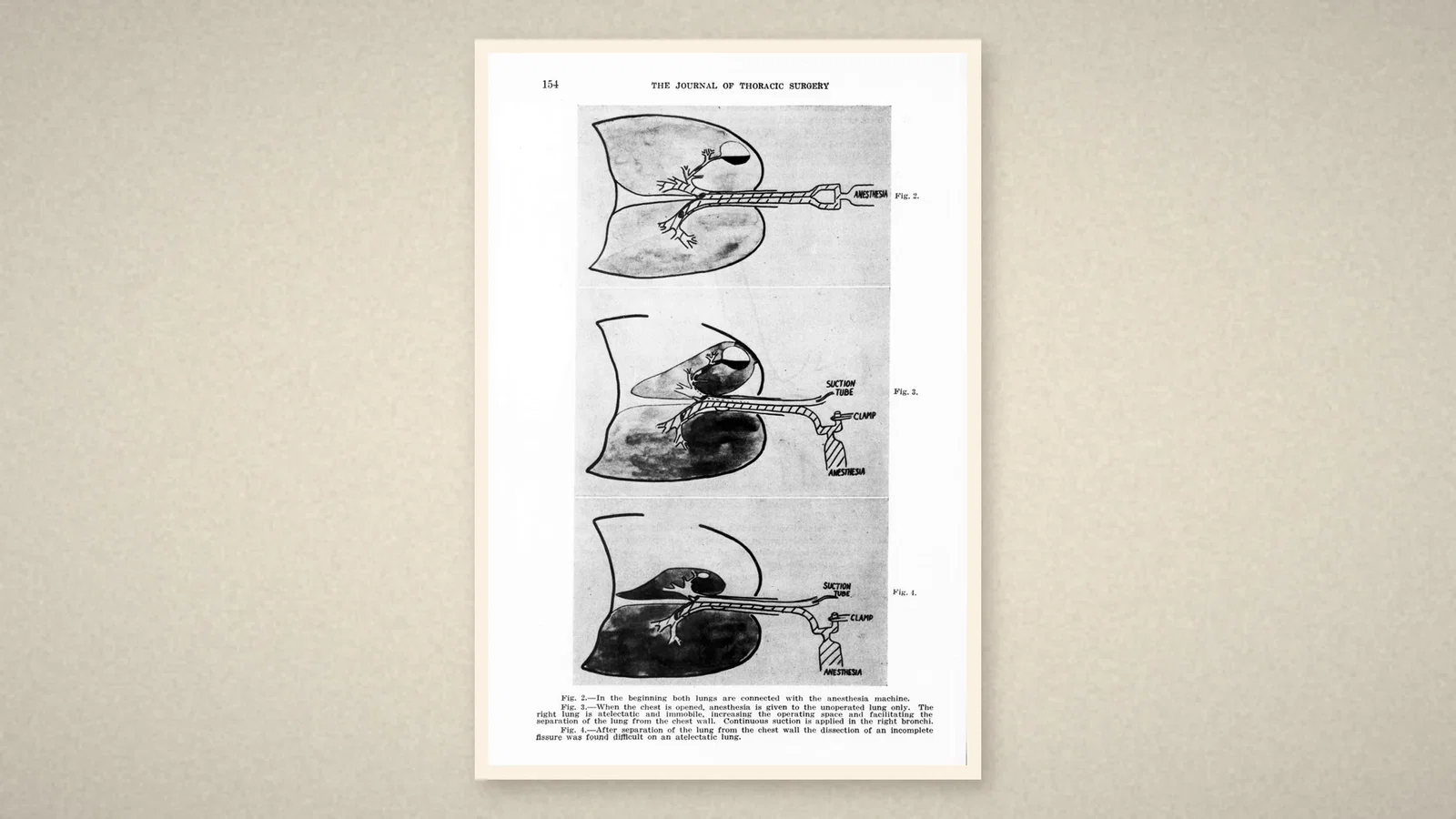
The paper also recorded the price. That patient had such a low pulmonary reserve that "prolonged one-lung anesthesia produced a pronounced cyanosis", so the right lung was inflated for short periods at intervals. The authors explained it plainly: with the lung collapsed, a considerable amount of blood passes through non-ventilated lung tissue, and "an arterial oxygen deficit will result". The catheter stayed in place for six and a half hours, and the patient recovered. In pneumonectomies the catheter was drawn back a little before the bronchus was clamped. Björk and Carlens judged the method "the best way to prevent spilling over" and the only one usable for disease of the right upper lobe. [2]
What the record shows
Two papers, one year apart, mark the passage of a physiologist's catheter into the operating theatre. The carinal hook gave a position that could be felt, the cuffs and control bags gave a seal without a manometer, and two wide channels gave each lung its own airway for ventilation and suction. The 1950 paper already states the central problem of one-lung ventilation, shunt through the collapsed lung, and the answer of intermittent reinflation.
Sources and reading limits
Read: Carlens E, J Thorac Surg 1949;18:742-6, in full; Björk VO and Carlens E, J Thorac Surg 1950;20:151-7, in full; the ASA timeline entries for 1949 and 1950.
Not covered: the papers of Gebauer, Zavod and Norris and colleagues; the endobronchial methods of Gale and Waters and of Magill; Moody's series; later double-lumen tubes. These are described only as Carlens and Björk summarised them. The ASA's "first popularized" is quoted as its own. The judgements on the method are the authors'. Premedication and topical anaesthetic doses are not repeated. Patients are not identified. Quotations are from the English originals.
Three illustrations are crops of the 1949 and 1950 pages, including Fig. 1 and Fig. 2 of 1949 and Figs. 2-4 of 1950; one is drawn from the text.
- Carlens E. A new flexible double-lumen catheter for bronchospirometry. J Thorac Surg. 1949;18:742-6. Internet Archive
- Björk VO, Carlens E. The prevention of spread during pulmonary resection by the use of a double-lumen catheter. J Thorac Surg. 1950;20:151-7. Internet Archive
- Australian Society of Anaesthetists. Anaesthesia history timeline. 2024. ASA
Continue exploring
Related reading connects shared questions and contexts; it does not by itself establish historical influence.
- 1938: Crafoord's Spiropulsator Breathes for Open-Chest Surgery
- 1936: Rovenstine's Anaesthesia for Chest Surgery
Anesthesia Epochs · The Source Room · About & Editorial Method
Published November 2, 2026. Sources and reading limits are stated above. Illustrations are labelled as original source images or AI-made. Historical accounts are educational; they are not current treatment instructions.