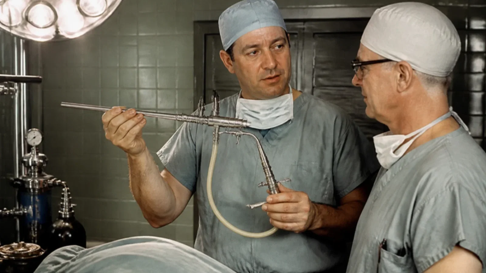
In July 1967 the Delaware Medical Journal published 'Two ventilating attachments for bronchoscopes' by R. Douglas Sanders, head of anesthesiology at the Wilmington Medical Center: under thiopental and succinylcholine for bronchoscopy, patients lived on 'diffusion respiration' and some respiratory acidosis always occurred, while earlier ventilating methods were ineffective or made the surgeon stop and close the scope with a thumb. Sanders's attachments fitted unaltered bronchoscopes and drove regulated compressed oxygen through a small injection nozzle, switched by an on-off valve; the paper reported 200 cases and a second attachment that also entrained volatile anaesthetic. The ASA timeline dates the introduction of jet ventilation to 1967.
Reading Library · Anesthesia Epochs · 中文版
Watch the education video (for clinicians)
1967: Sanders Jet Ventilation — Through an Open Bronchoscope · Anesthesia Epochs Education Series
Rigid bronchoscopy is a fight over a single tube. The surgeon needs it open to look, suck and take biopsies; the anaesthetist needs it to get gas into the lungs. In the summer of 1967 an anaesthetist in Wilmington, Delaware, described in the state medical journal a small brass fitting that let both happen at once, by blowing short bursts of oxygen down the open scope. [1]
The problem
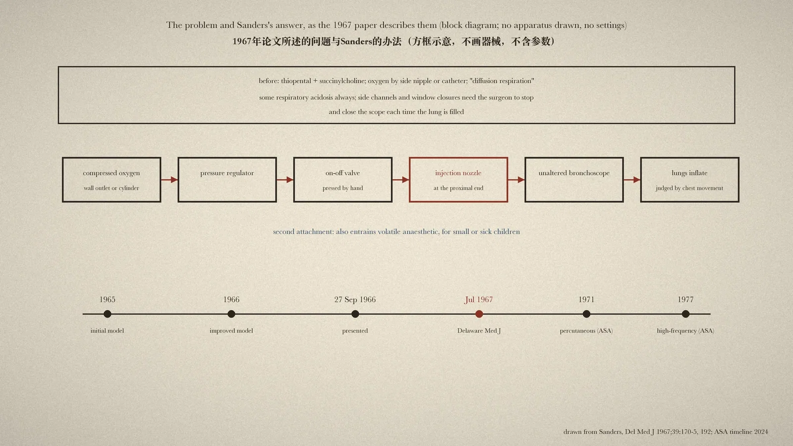
"Two Ventilating Attachments for Bronchoscopes", by R. Douglas Sanders, head of the Department of Anesthesiology at the Delaware Division of the Wilmington Medical Center, appeared in the Delaware Medical Journal in July 1967; it had been presented to the Academy of Anesthesiology on 27 September 1966. Anaesthesia for bronchoscopy with thiopental and succinylcholine, Sanders wrote, had been widely used for about fourteen years. While the surgeon worked, oxygen was given through a nipple at the side of the scope or a catheter beside it, and "life is sustained by 'diffusion respiration'". However quick or skilful the operator, "some degree of respiratory acidosis always occurs". [1, p. 170]
Sanders listed what had been tried. Compressing the lower chest was ineffective; cuirass and wrap-around respirators were cumbersome and frightening; channels in the side of the scope connected to an anaesthetic machine worked only if the surgeon closed the proximal end with a thumb; and special bronchoscopes with proximal window closures needed the surgeon to swing the window across the lumen. These last two worked, but the surgeon's attention was "divided between ventilation and operation", and the instruments had to come out each time the lung was filled. [1, p. 170]
The attachment
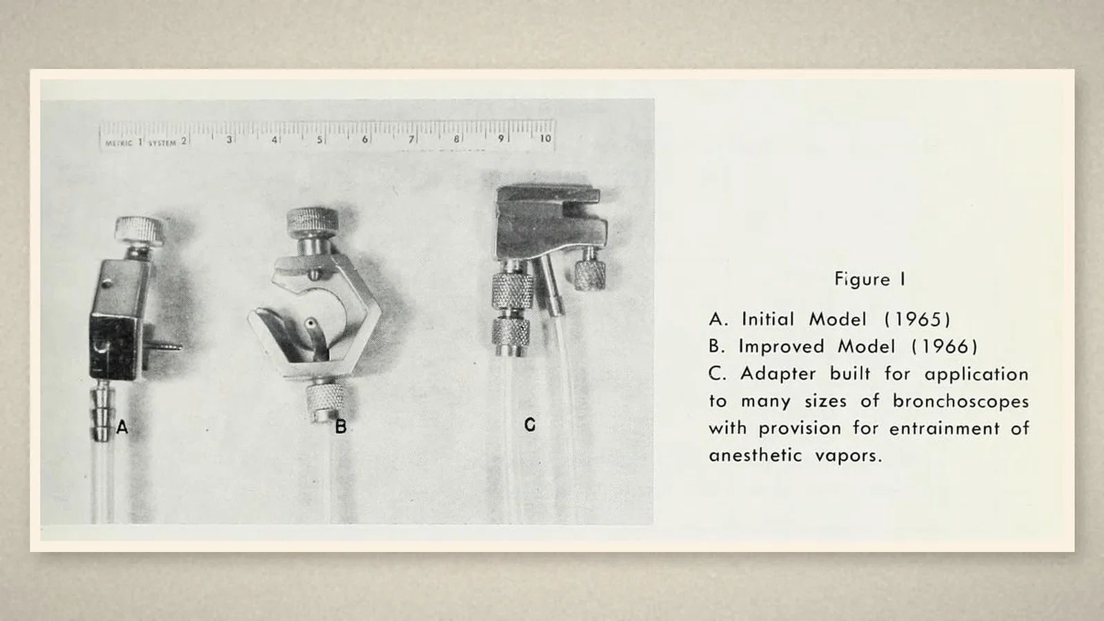
"The developments reported here are of attachments which are applied to the proximal ends of otherwise unaltered bronchoscopes, utilizing the energy of compressed oxygen exerted through an injection nozzle." Pressure was regulated and flow controlled with valves available in industry. Figure I shows the initial model of 1965, an improved model of 1966 and an adapter for many sizes of bronchoscope. The attachments were designed to work at the pressure of hospital wall oxygen outlets or less; and because different bronchoscopes needed different nozzle sizes, and smaller scopes of the same model gave much higher pressures at their tips, "any new combination" had to be studied carefully before clinical use. A hand-held on-off valve between regulator and attachment gave the intermittent flow. [1, pp. 171-3]
Two hundred cases
Sanders reported 200 cases, half of them outpatients. Ventilation, "as judged by movement of the chest and abdomen", was usually more than the patient's normal exchange, and the bronchi moved as in deep normal breathing. "The surgeons have been able to proceed with instrumentation, suction, and biopsy in a more precise and leisurely way." Arterial blood gases at the end of the procedure were much better than when patients had not been ventilated. Pulse rates were slower and irregularities few; before, "one often had to call time on the surgeon". Two patients, through too little thiopental or poor timing, were aware during the procedure, frightened though not short of breath. [1, pp. 173-4]
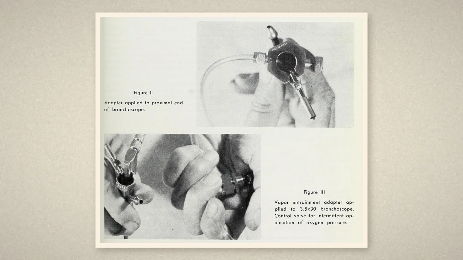
For very young or sick children, and for adults who were better given a volatile anaesthetic, Sanders built a second attachment that both ventilated and entrained anaesthetic vapour; a new adapter-bronchoscope combination designed with Frank T. O'Brien had passed its first clinical trials. [1, pp. 174-5, 192]
The timeline's account
The Australian Society of Anaesthetists (ASA) timeline lists the "Introduction of 'jet ventilation'" under 1967, credited to Richard Douglas Sanders, with technical details that match the paper. It records percutaneous intratracheal jet ventilation under 1971 and high-frequency jet ventilation under 1977. [2, pp. 70, 72, 76]
What the record shows
The moment a shared airway stopped being a turn-taking arrangement. Sanders's paper never uses the words "jet ventilation"; it speaks of an injection nozzle and the energy of compressed oxygen. But the principle is the one later named: let a narrow high-pressure stream carry gas into an open tube, so the surgeon never has to stop.
Sources and reading limits
Read: Sanders, Delaware Medical Journal 1967;39:170-5 with the continuation on p. 192, in full; the ASA timeline 2024, pp. 70, 72 and 76.
Not covered: the 1966 presentation to the Academy of Anesthesiology; any later paper by Sanders; the jet ventilation papers of 1969, 1971 and 1977, which belong to later articles.
Limits: the ASA dates jet ventilation to 1967; the paper itself dates its first adapter to 1965. The 200-case results are the author's own, and many observations went unrecorded. Driving pressures, nozzle sizes, drug doses and vapour concentrations in the paper are not repeated. Quotations are from the English original.
Three illustrations are crops of the Delaware Medical Journal (pp. 170-1); one is drawn from the paper.
- Sanders RD. Two ventilating attachments for bronchoscopes. Del Med J. 1967;39(7):170-5, 192. Internet Archive
- Australian Society of Anaesthetists. Anaesthesia history timeline. 2024:70, 72, 76. ASA
Continue exploring
Related reading connects shared questions and contexts; it does not by itself establish historical influence.
Anesthesia Epochs · The Source Room · About & Editorial Method
Published November 3, 2026. Sources and reading limits are stated above. Illustrations are labelled as original source images or AI-made. Historical accounts are educational; they are not current treatment instructions.