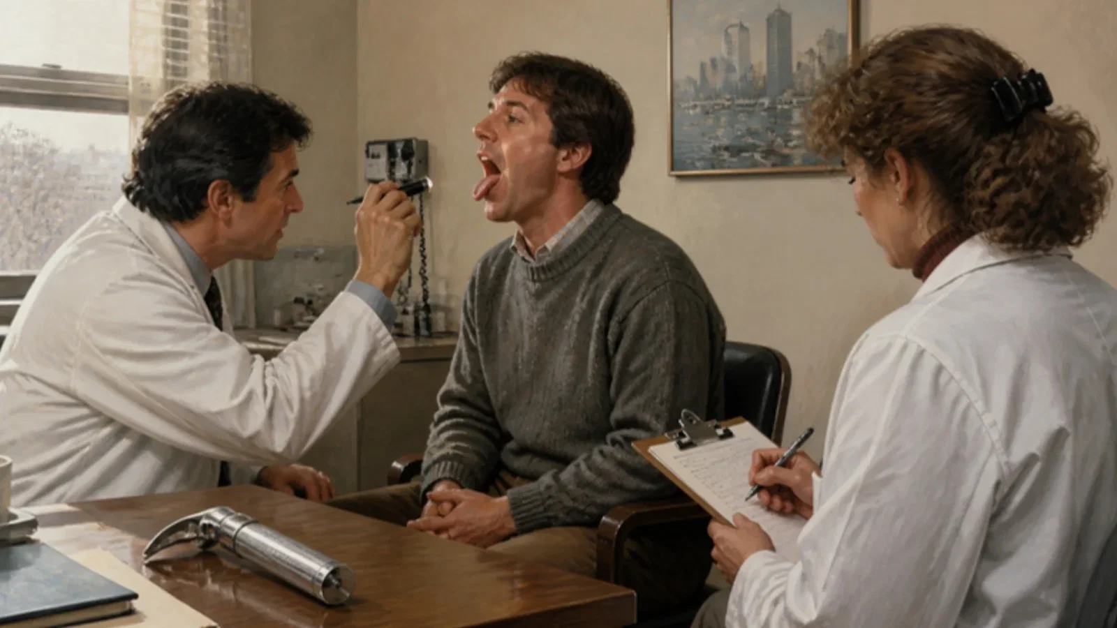
In July 1985 the Canadian Anaesthetists' Society Journal (vol. 32, no. 4, pp. 429-34) printed 'A clinical sign to predict difficult tracheal intubation: a prospective study' by S. Rao Mallampati and six colleagues of the Department of Anaesthesia, Brigham and Women's Hospital, Harvard Medical School, Boston. The principal author had earlier proposed that a base of the tongue disproportionately large for the oropharynx overshadows the larynx and makes laryngoscopy difficult; its size cannot be measured, but whether it hides the faucial pillars and uvula can be seen. In 210 adult surgical patients of ASA status 1 or 2, twenty-two anaesthetists and nurse anaesthetists asked each seated patient to open the mouth and protrude the tongue maximally, at least twice, and assigned three classes: class 1, faucial pillars, soft palate and uvula visible; class 2, uvula masked; class 3, only the soft palate. The same person later graded the laryngoscopic view. All 155 class 1 patients had adequate exposure; 14 of 40 in class 2 and nearly all 15 in class 3 had difficult views (p < 0.001). The authors noted that only the principal author knew the method beforehand and that assessment was not blinded.
Reading Library · Anesthesia Epochs · 中文版
Watch the education video (for clinicians)
1985: The Mallampati Classification — Open Wide · Anesthesia Epochs Education Series
At the preoperative visit an anaesthetist asks the patient to "open wide and stick your tongue out", glances at the back of the throat and writes down a class. Nearly every anaesthetist does it today. It began as a hypothesis about the size of the tongue, tested in a prospective study of 210 patients in Boston in 1985. [1, p. 429]
A tongue base that hides the larynx
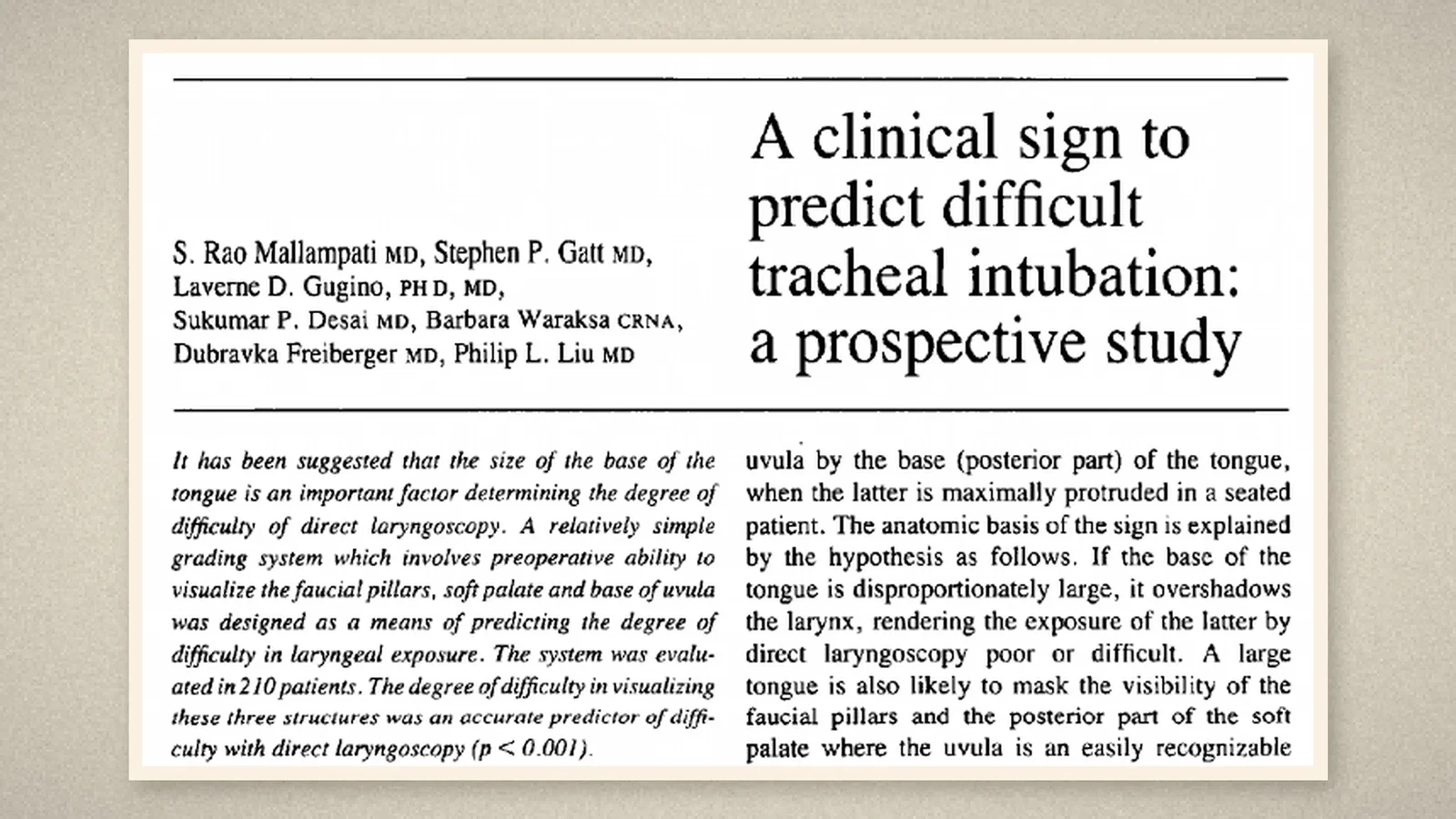
In July 1985 the Canadian Anaesthetists' Society Journal (vol. 32, no. 4) printed a paper by S. Rao Mallampati, Stephen P. Gatt, Laverne D. Gugino, Sukumar P. Desai, Barbara Waraksa, Dubravka Freiberger and Philip L. Liu of the Department of Anaesthesia, Brigham and Women's Hospital, Harvard Medical School, Boston. [1, p. 429]
Unexpected difficulty with intubation, the introduction said, can be a significant cause of morbidity and mortality, yet usual clinical observation cannot always predict which patient will be difficult, particularly when the orofacial anatomy is unremarkable. The principal author had earlier proposed a "clinical sign to predict difficult tracheal intubation (hypothesis)": concealment of the faucial pillars and uvula by the base of the tongue when it is maximally protruded in a seated patient. [1, p. 429]
The anatomical reasoning ran like this. If the base of the tongue is disproportionately large, "it overshadows the larynx", making exposure at direct laryngoscopy poor or difficult. A large tongue is also likely to hide the faucial pillars and the back of the soft palate, where the uvula is an easily recognised landmark. Since the size of the tongue base relative to the oropharynx cannot be measured, it is logical to infer that it is disproportionately large when it can mask the pillars and uvula. [1, p. 429]
Three classes
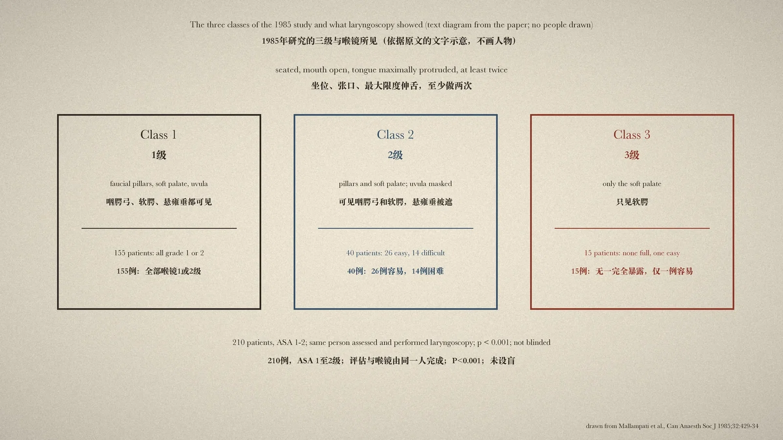
The study, approved by the institution's committee on human research, included 210 consecutive adult surgical patients of ASA status 1 or 2 who needed general endotracheal anaesthesia; patients with known cardiovascular or respiratory disease were excluded. [1, p. 429] Twenty-two people with at least two years' experience in clinical anaesthesia, six staff physicians, ten residents and six nurse anaesthetists, carried out the assessments. At the preoperative visit, the person scheduled to give the anaesthetic asked the patient to sit, open the mouth and protrude the tongue as far as possible, at least twice to reduce errors, and assigned a class: [1, p. 430]
- Class 1: faucial pillars, soft palate and uvula can be seen.
- Class 2: faucial pillars and soft palate can be seen, but the uvula is masked by the base of the tongue.
- Class 3: only the soft palate can be seen.
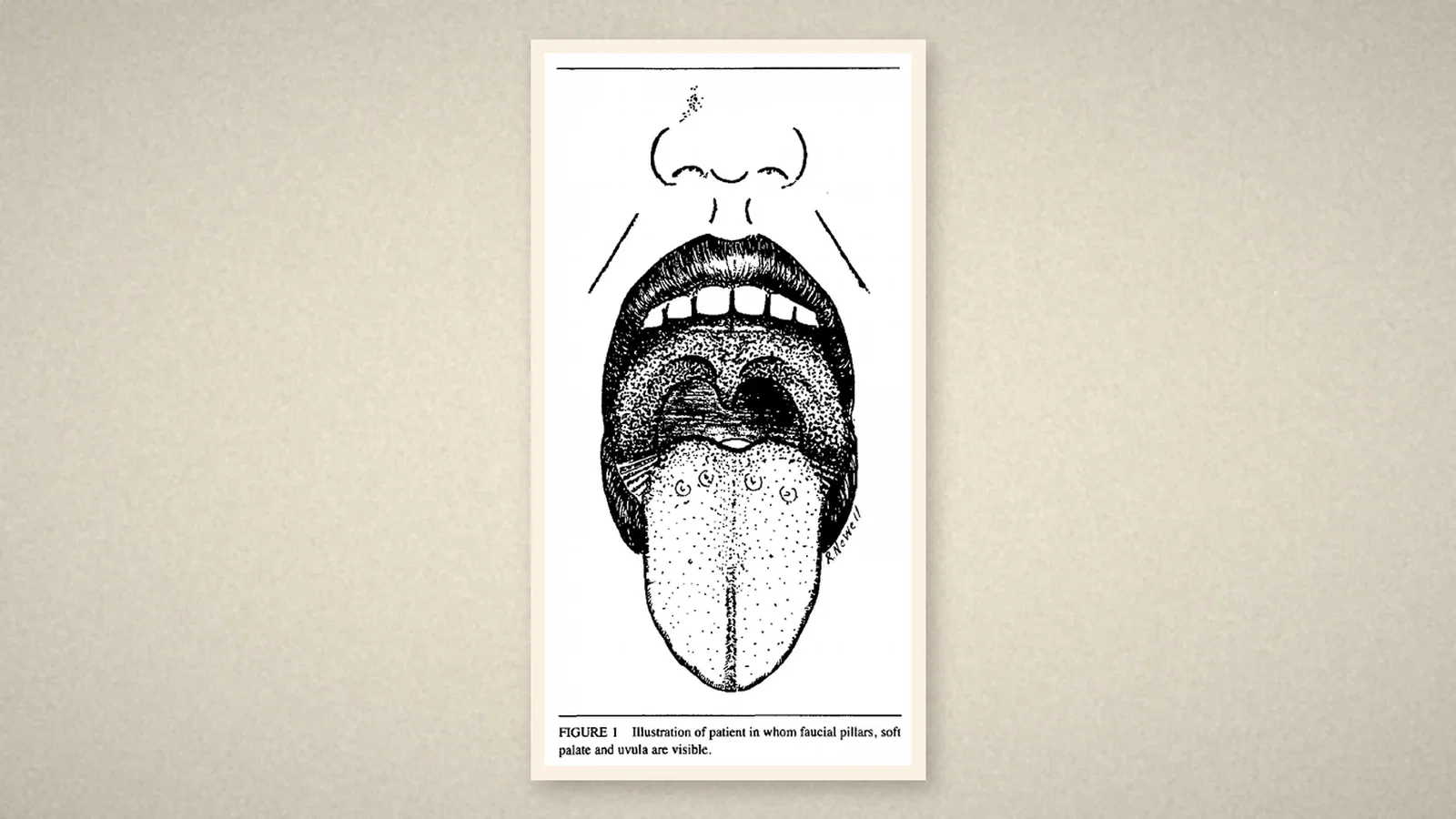
The paper's Figs 1 and 2 are line drawings, not photographs: in Fig. 1 the open mouth shows the pillars, soft palate and uvula; in Fig. 2 all three are hidden by the tongue. The acknowledgements credit Robert Newall with the depiction of the sign. [1, p. 430][1, p. 433]
The same person then induced anaesthesia by a standard protocol, with the patient supine in the "sniffing position", and graded the laryngoscopic view from 1 to 4, grades 1 and 2 counting as adequate exposure and grades 3 and 4 as inadequate. The chi-square test was used. [1, p. 430][1, p. 431]
Results
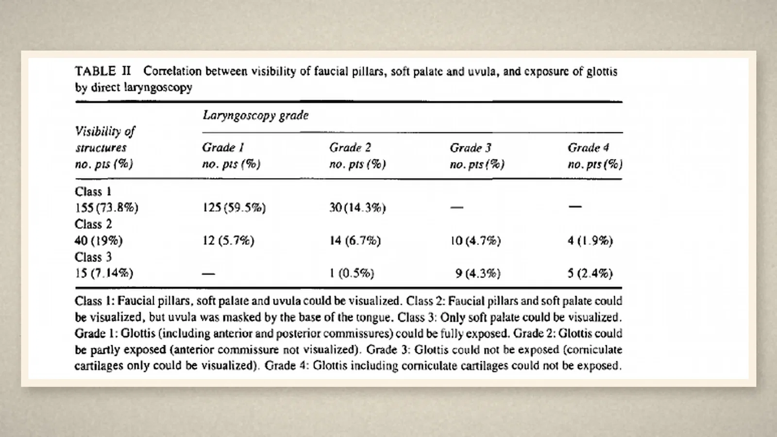
Visibility of the pharyngeal structures correlated significantly with the ease of laryngoscopy (p < 0.001). All 155 patients in class 1 had a grade 1 or 2 view, with no difficult exposures. Of 40 in class 2, 26 were easy and 14 difficult. Of 15 in class 3, none had a full view and only one an easy one; all the rest were grade 3 or 4. All edentulous patients had adequate exposure; 3 of 12 with overbite and 2 of 4 with moderately restricted neck movement did not; all four obese patients having gastroplasty were adequate. [1, p. 431][1, p. 432]
The authors' own reservations
The discussion reviewed earlier anatomical signs and radiographic measurements and noted that such features vary between people and over a lifetime, so that the same feature may seem normal to one observer and abnormal to another. The goal was a clinical method that was "simple, easy to apply, and amenable to objectivity". The tongue being the largest single structure in the mouth affecting access to the larynx, the method assessed the size of its base indirectly, by its relation to the pharyngeal structures. [1, p. 432][1, p. 433]
The authors were also frank about the study's weaknesses. Only the principal author had used the method before; since the other participants did not know whether it was useful, "observer blindness was not thought to be warranted". The person who assessed the airway also performed the laryngoscopy, because a second anaesthetist could not be provided for so many patients. They concluded that the results were highly significant and supported the view that difficult laryngeal visualisation "can be predicted in most cases" by the visibility of the faucial pillars and uvula. [1, p. 433]
What the record shows
The study turned a hypothesis about the size of the tongue into a bedside check that needs no equipment and takes seconds, and tested it against what the laryngoscope showed. Its design also had clear limits: no blinding, the same person assessing and intubating, and relatively healthy patients.
Sources and reading limits
Read: Mallampati, Gatt, Gugino, Desai, Waraksa, Freiberger and Liu, "A clinical sign to predict difficult tracheal intubation: a prospective study", Can Anaesth Soc J 1985;32:429-34 (the whole paper: abstract, hypothesis, methods with Tables I and II and Figs 1 and 2, results, discussion and French résumé). Obtained through institutional access.
Not covered: the principal author's 1983 hypothesis in the same journal; the earlier predictors the authors cite (Cass, James and Lines, 1956; White and Kander, 1975; Nichol and Zuck, 1983); later revisions of the classes and validation studies. The Wood Library-Museum and History of Anaesthesia Society entries were used only as leads.
Limits: as the authors state, the study was not blinded, only the principal author knew the method beforehand, and the same person assessed the airway and performed laryngoscopy; patients were ASA 1 or 2, with an uneven sex ratio. Premedication and induction doses, blade size and patients' height and weight are not repeated, and page crops stop before them. Figs 1 and 2 are drawings signed by Newall, not photographs; patients are not identified.
Three illustrations are crops of Can Anaesth Soc J 1985 pp. 429, 430 and 432 (title and abstract, Fig. 1, Table II). One is a text diagram drawn from the paper, with no people drawn.
- Mallampati SR, Gatt SP, Gugino LD, Desai SP, Waraksa B, Freiberger D, Liu PL. A clinical sign to predict difficult tracheal intubation: a prospective study. Can Anaesth Soc J. 1985;32:429-34. DOI
Continue exploring
Related reading connects shared questions and contexts; it does not by itself establish historical influence.
- 1984: Cormack and Lehane Grade the Laryngoscopic View in Four Levels
- 1978: Finucane and Kupshik Exchange a Leaking Tracheal Tube over a Stilette
Anesthesia Epochs · The Source Room · About & Editorial Method
Published November 3, 2026. Sources and reading limits are stated above. Illustrations are labelled as original source images or AI-made. Historical accounts are educational; they are not current treatment instructions.