Boston anaesthetists threaded a 32-gauge catheter through a 26-gauge needle to give continuous spinal anaesthesia without the headaches of large catheters; within two years, four patients given continuous spinal anaesthesia were reported with cauda equina syndrome.
Reading Library · Anesthesia Epochs · 中文版
Watch the education video (for clinicians)
1990: Spinal Microcatheters — The Cauda Equina Reports · Anesthesia Epochs Education Series
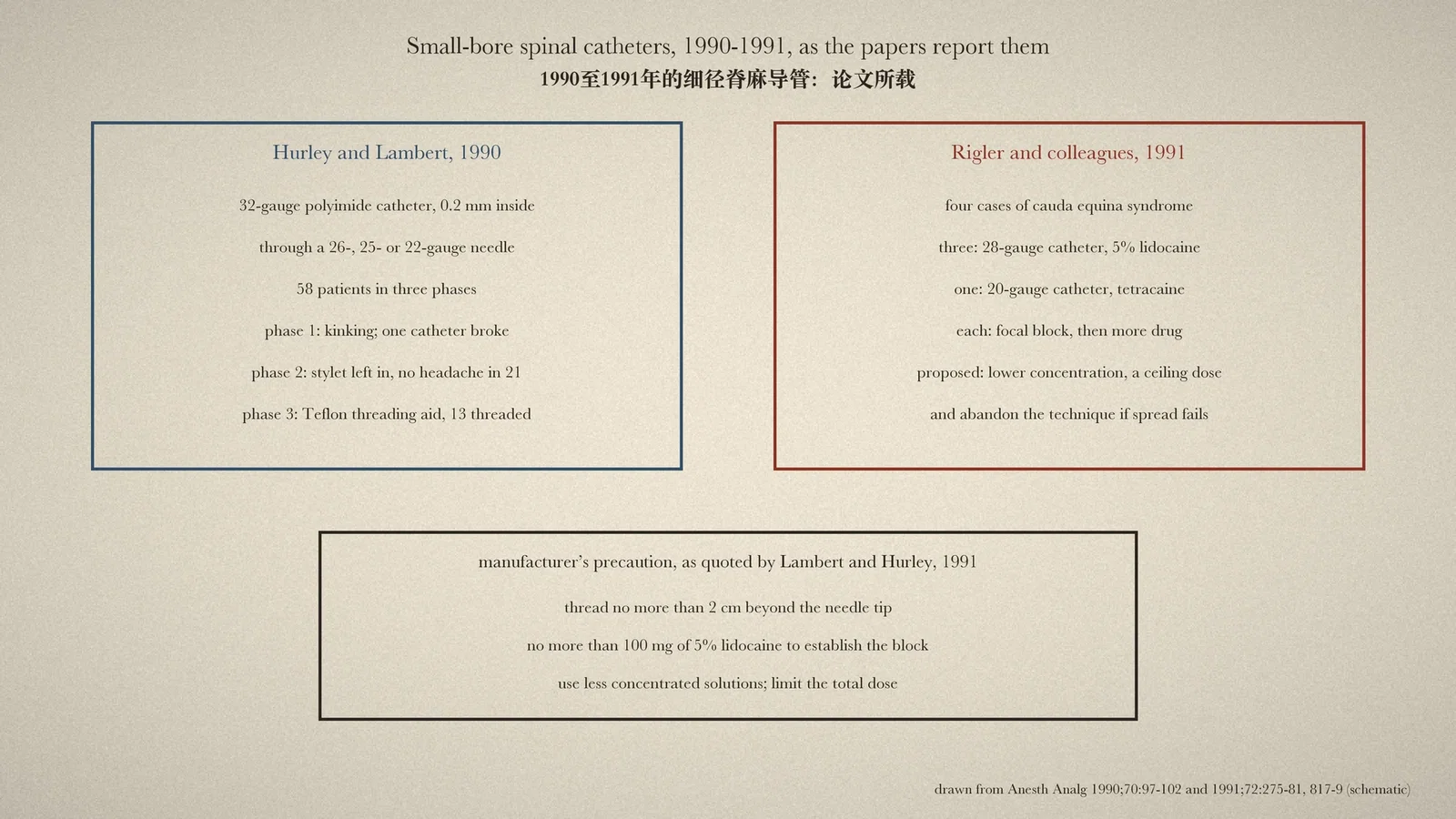
A catheter in the subarachnoid space lets an anaesthetist give spinal anaesthesia in small steps and keep it going as long as surgery lasts. Its price had always been the hole: large needles and catheters meant headaches, so continuous spinal anaesthesia was kept mostly for the elderly. Around 1990 that seemed about to change, when catheters fine enough to pass through spinal needles appeared. Within two years the same journal carried reports of patients left with cauda equina syndrome. This article reads the first microcatheter report and the two 1991 papers on the injuries, all in full. [1][2][3]
A catheter through a 26-gauge needle
Ronald J. Hurley and Donald H. Lambert of Brigham and Women's Hospital, Boston, began their 1990 report with history, citing Dean's description of continuous spinal anaesthesia "at the 75th annual meeting of the British Medical Society in 1907" (an earlier article in this series dates Dean's paper to 1906). The technique, they wrote, was usually reserved for elderly patients and long operations "because of the high incidence of postdural puncture headache (PDPH) when the currently available (large diameter) catheters are used in young patients". Their answer was "a 32-gauge polyimide microcatheter that threads through a 26-gauge spinal needle", 91 cm long, with an inside diameter of 0.2 mm and a fine steel stylet. [1]
Three phases
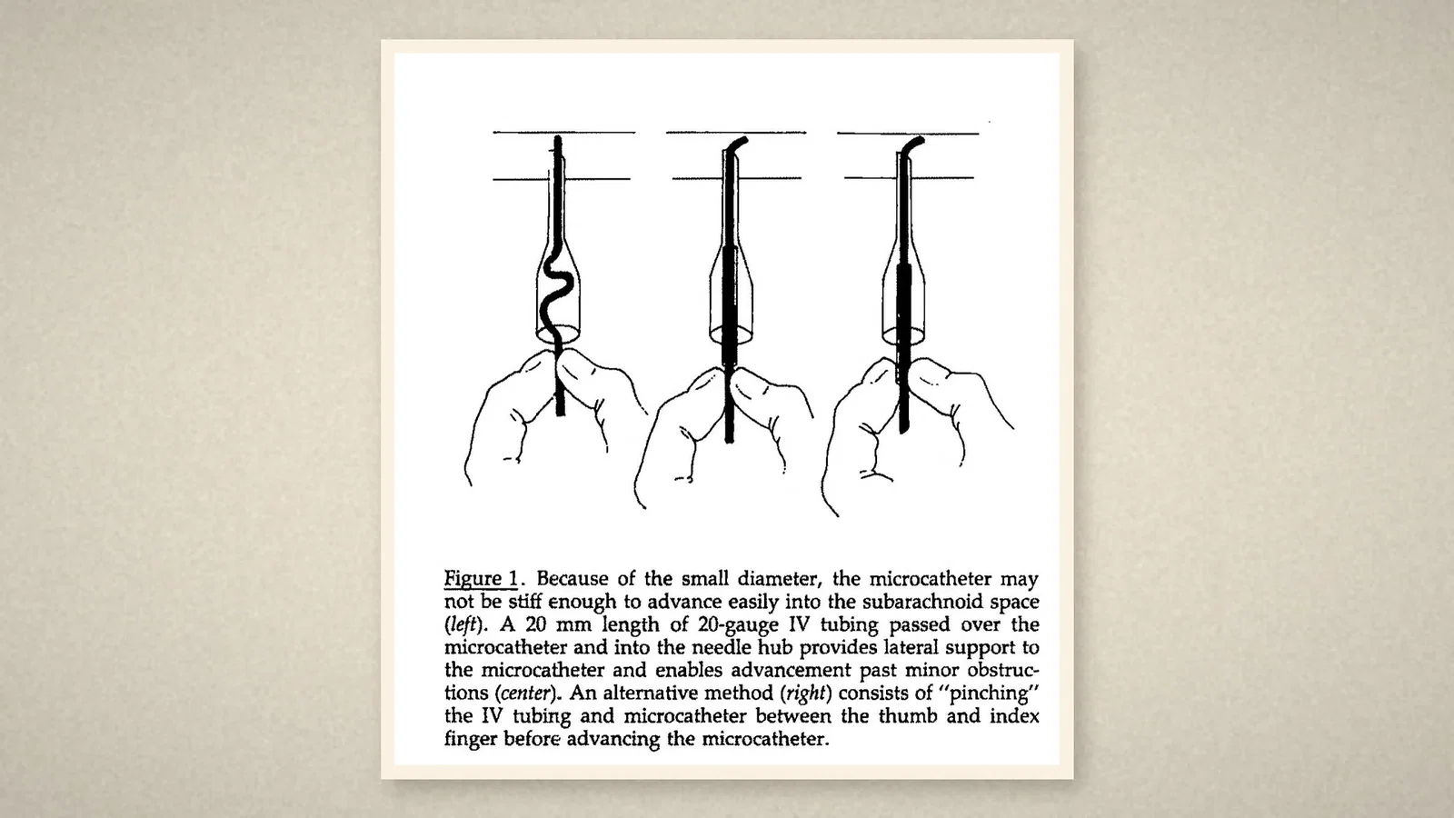
In 58 patients the method went through three phases. In the first, with the stylet removed, catheters kinked under the skin and in the tape, the stylet was hard to withdraw, and one catheter broke during removal, "leaving 5-6 cm of catheter in the patient". In the second, the stylet was left in place for stiffness and radiopacity; no headache occurred in 21 patients. In the third, a 20-mm length of Teflon intravenous tubing slipped over the catheter gave it support in the needle hub, and there were no threading failures in 13 patients. Two blocks in that phase were inadequate, and in trying to improve them "more intrathecal local anesthetic was injected than would have been administered with a single-injection technique"; the authors suspected that the distribution of the drug "may have been restricted". [1]
Four cases
In March 1991 Mark L. Rigler, Kenneth Drasner and colleagues from four institutions reported "four cases of cauda equina syndrome occurring after continuous spinal anesthesia". Three had 28-gauge catheters (CoSpan) and hyperbaric 5 per cent lidocaine; one had a 20-gauge catheter and tetracaine. None had the 32-gauge microcatheter of the Boston report. What the cases shared was a pattern: "there was evidence of a focal sensory block and, to achieve adequate analgesia, a dose of local anesthetic was given that was greater than that usually administered with a single-injection technique". The first patient, for example, received 150 mg of 5 per cent lidocaine in three doses. The authors postulated that "the combination of maldistribution and a relatively high dose of local anesthetic resulted in neurotoxic injury", and proposed a lower concentration, a "ceiling" dose, manoeuvres to spread the drug, and abandoning the technique if those failed. [2]
A model of the injury
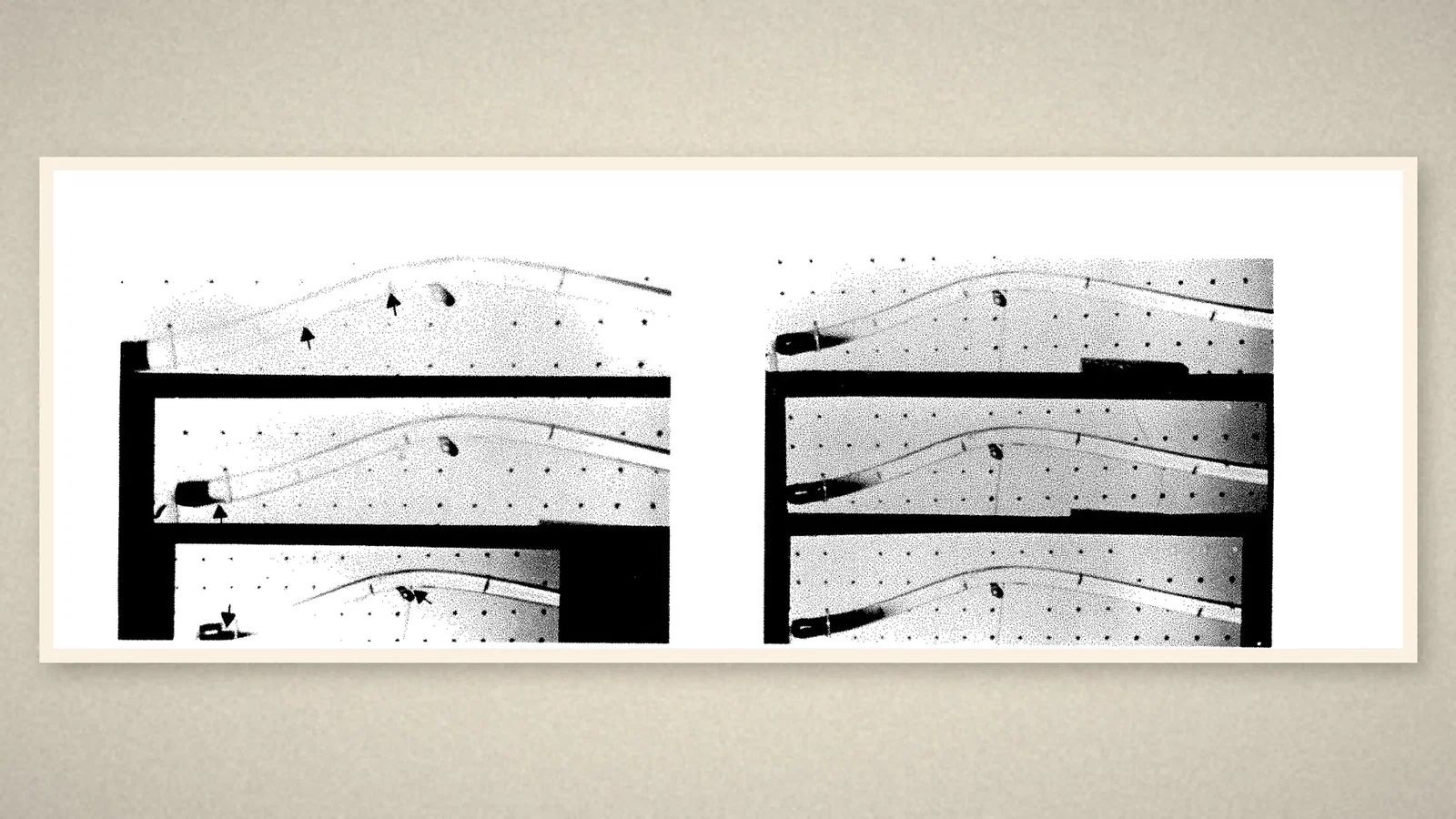
In the same volume Lambert and Hurley responded. The catheter's maker had issued a precaution after reports of perineal sensory loss and sphincter changes: do not thread the catheter more than 2 cm beyond the needle, limit 5 per cent lidocaine in dextrose to 100 mg to establish the block, use less concentrated solutions, and limit the total dose. With a spinal canal model made of tubing and dyed solutions, they showed how a slow stream of hyperbaric lidocaine from a small catheter could pool at the end of the dural sac, where the nerves of the cauda equina lie. They speculated that doses of 5 per cent lidocaine in dextrose "exceeding 200 mg may cause cauda equina syndrome". [3]
What the record shows
The small-bore catheter solved the problem it was designed for: in Boston, headaches were rare. It also created a new one. Injected slowly through a narrow lumen, a concentrated hyperbaric drug could stay where it landed, and when the block looked patchy the natural response, more drug, could injure the nerves. The lesson recorded in 1991 was about dose and distribution, not only about the catheter. What regulators did next is not covered here.
Sources and reading limits
Read: Hurley and Lambert 1990, Rigler and colleagues 1991, and Lambert and Hurley 1991, all in full; the 2024 ASA timeline entry, which dates the microcatheters to 1989.
Not covered: the correspondence that followed; later regulatory action; later trials. Case details are given only as the authors printed them. Doses are as printed.
Three illustrations are page crops from the 1990 and 1991 papers; one is drawn from them.
- Hurley RJ, Lambert DH. Continuous spinal anesthesia with a microcatheter technique: preliminary experience. Anesth Analg. 1990;70:97-102. Internet Archive
- Rigler ML, Drasner K, Krejcie TC, Yelich SJ, Scholnick FT, DeFontes J, Bohner D. Cauda equina syndrome after continuous spinal anesthesia. Anesth Analg. 1991;72:275-81. Internet Archive
- Lambert DH, Hurley RJ. Cauda equina syndrome and continuous spinal anesthesia. Anesth Analg. 1991;72:817-9. Internet Archive
- Australian Society of Anaesthetists. Anaesthesia history timeline. 2024. ASA
Continue exploring
Related reading connects shared questions and contexts; it does not by itself establish historical influence.
- 1906: Dean Leaves the Spinal Cannula In
- 1944: Tuohy Threads a Ureteral Catheter into the Spinal Space
Anesthesia Epochs · The Source Room · About & Editorial Method
Published November 2, 2026. Sources and reading limits are stated above. Illustrations are labelled as original source images or AI-made. Historical accounts are educational; they are not current treatment instructions.