A study of 50 healthy subjects, with the conditions of measurement retained.
Reading Library · Anesthesia Epochs · 中文版
Watch the companion film
1982: Aaslid — Transcranial Doppler · Watch on YouTube
Historical anchor: 1982. Date details and source qualifications are explained below.
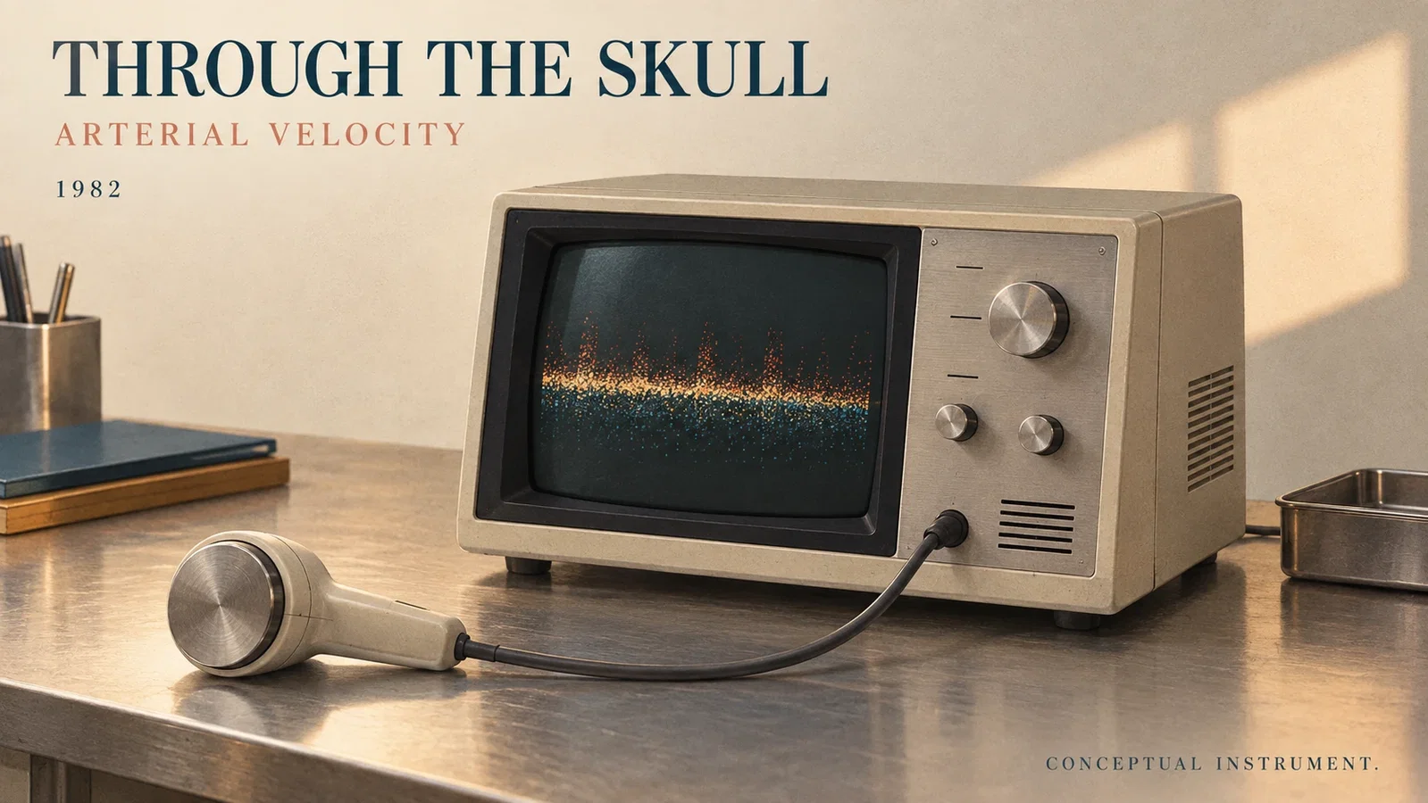
The adult skull presented an acoustic obstacle. In 1982, Rune Aaslid, Thomas-Marc Markwalder and Helge Nornes reported using lower-frequency ultrasound through the temporal region to obtain Doppler signals from basal cerebral arteries.[1, p.769] Their measurement was blood flow velocity. That choice of quantity defines both the achievement and the limits of the report.
The paper acknowledged earlier Doppler observations in extracranial vessels, exposed intracranial arteries during surgery and infants with open fontanelles. An intact adult skull posed a different problem. Bone attenuated ultrasound, while lower frequencies and the thinner temporal region offered a possible route through it.[1, p.769] This article marks publication of the 1982 report; it does not make that year the origin of every form of cerebral ultrasound.
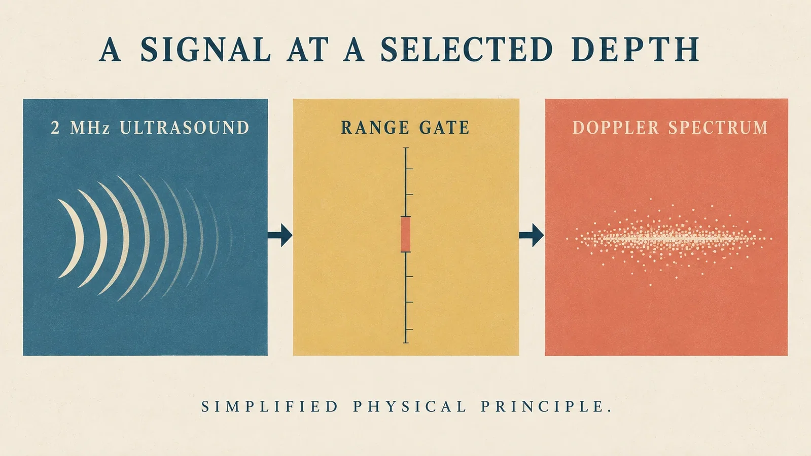
The laboratory prototype used 2 MHz ultrasound and range gating to receive signals at a selected depth. A spectrum analyser distinguished flow direction, and the investigators estimated a time-mean velocity from the spectral envelope.[1, pp.769–770] An acoustic window still had to be found in each person. Converting a frequency shift into velocity also depended on the angle between the beam and the vessel. The output was not an automatic colour map of all cerebral vessels.
Fifty healthy subjects, aged 20 to 65 years, were studied. Bilateral middle cerebral artery (MCA) recordings were obtained in all 50. Other vessels were less accessible: signals were insufficient for velocity measurement in 20% of the anterior cerebral arteries (ACA) and 40% of the posterior cerebral arteries (PCA) examined.[1, pp.769,771] Those percentages describe arteries, not ten and twenty people. The study's account of feasibility includes its incomplete measurements.

Velocity alone does not supply volume flow. In discussing collateral circulation, the authors assumed that the arterial lumen remained relatively constant during testing before using changes in velocity to infer relative changes in volume flow.[1, pp.773–774] They also proposed detecting vasospasm after subarachnoid haemorrhage. The high-velocity observations in spastic arteries, however, were explicitly marked as material still to be published. The healthy-subject study did not validate a diagnostic threshold or demonstrate improved patient outcomes.
Jöbsis's near-infrared work offers a useful comparison of what a signal represents. His 1999 retrospective describes separating haemoglobin and cytochrome oxidase signals, while an unknown optical path length limited conversion to absolute concentrations.[2, p.394] TCD interprets frequency shifts through assumptions about angle and arterial velocity; that optical work interprets spectral components and relative changes. Both seek information from outside the head, but their readings cannot be exchanged as a single measure of cerebral blood supply. The site's related link makes this measurement comparison, without asserting historical transmission.

The report made velocity and direction in some arteries behind an intact skull available for repeated observation. Its conditions remained part of the method: finding a window, considering angle, distinguishing vessels and stating the assumptions used to interpret a change in velocity.[1] The result and its constraints belong in the same historical account.
Image note: all four images are modern editorial concepts, not replicas of the 1982 prototype or journal figures. Spectral marks are symbols without data, and the functional diagram is not a probe-positioning guide. The study illustration does not turn artery-level failures into patient counts. The velocity/flow illustration supplies neither diagnostic thresholds nor clinical decisions.
Sources and reading limits
- Aaslid R, Markwalder TM, Nornes H. Noninvasive transcranial Doppler ultrasound recording of flow velocity in basal cerebral arteries. J Neurosurg. 1982;57(6):769–774. Original article; public original PDF. PMID 7143059. All six original pages and seven figures read. The methods print “2 mHz”; the introduction and discussion establish the intended MHz range. The reference here uses 2 MHz. December 1982 is the issue date; May 13 is manuscript receipt, not publication.
- Jöbsis-vanderVliet FF. Discovery of the near-infrared window into the body and the early development of near-infrared spectroscopy. J Biomed Opt. 1999;4(4):392–396. Original author retrospective. Page 394 reread for optical signal separation and the unknown path-length limitation. The other four pages were not reread for this article; this is not a claim to have read the original 1977 Science paper.
Continue exploring
Related reading connects shared questions and contexts; it does not by itself establish historical influence.
- 1977: Jöbsis Detects Near-Infrared Signals Through an Intact Head
- 2008: The BLUE Protocol Turns Lung Ultrasound into an Algorithm
The Source Room · About & Editorial Method
Sources and reading limits are stated above. Illustrations are labelled as original source images or AI-made. Historical accounts are educational; they are not current treatment instructions.