Continuous amplitude trends, frequency analysis and the limits of each record.
Reading Library · Anesthesia Epochs · 中文版
Watch the companion film
1969–1984: Early Processed EEG — CFM, CFAM and Lifescan · Watch on YouTube
Historical anchor: 1980. Approximate placement; see the essay for the date range and evidence.
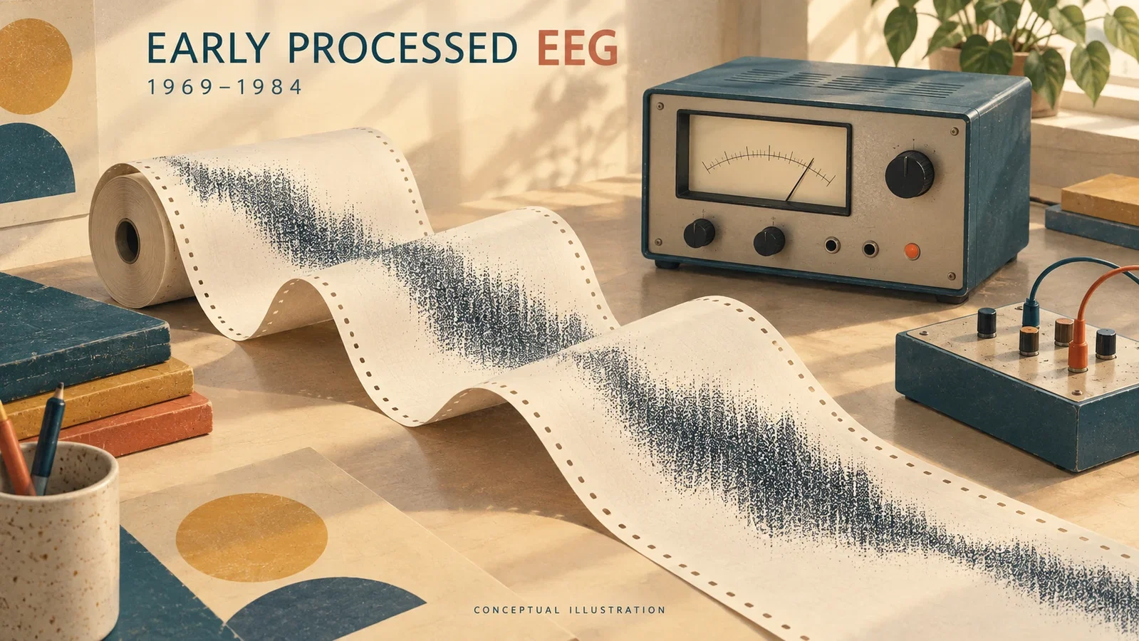
A paper recording, a microprocessor study and a regulatory decision document different parts of a device's history. The sources for this article provide examples from 1969, 1983 and 1984. Together they describe early ways of presenting EEG activity for sustained observation, without establishing a single succession of commercial products.
Maynard, Prior and Scott's 1969 report addressed long-term observation after resuscitation. Their cerebral function monitor (CFM) filtered the signal from a pair of electrodes, compressed its amplitude logarithmically and produced a slow paper trace. An accompanying impedance record helped identify electrode or equipment problems.[1, pp.545–546] Continuous trends complemented intermittent conventional EEG examinations. That combination offers a concrete comparison with the article on bedside assessment of coma. The illustrative cases did not validate a universal anesthetic-depth scale.
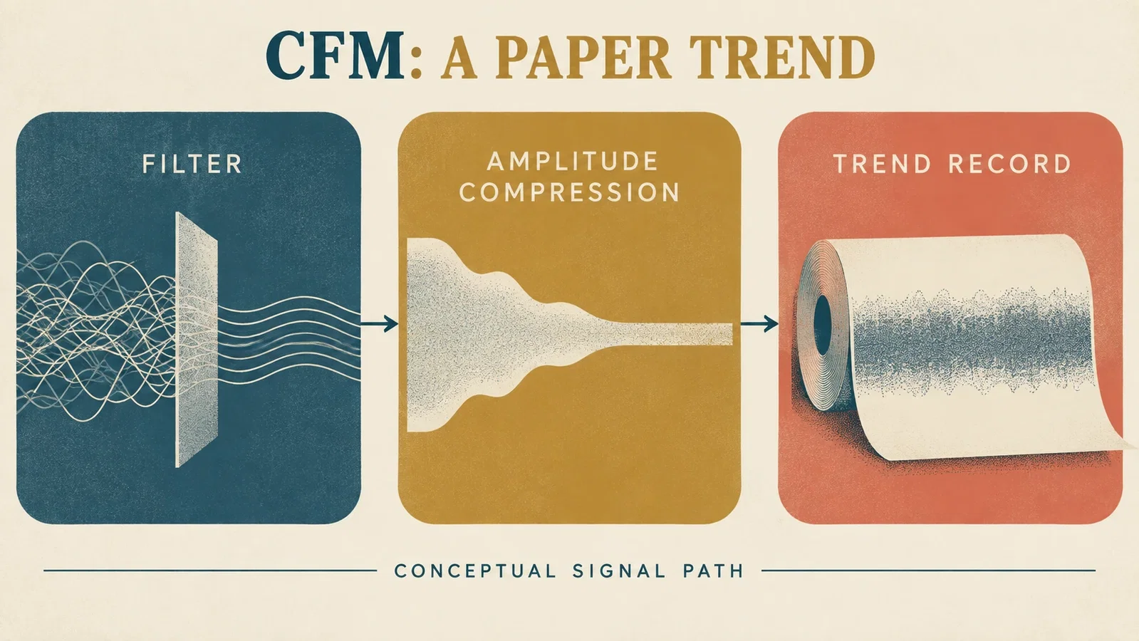
The CFAM paper published in 1983 described a microprocessor-based development of the CFM, adding detailed amplitude analysis and frequency analysis by a modified zero-crossing method. Its citation of a 1979 development report also prevents treating 1983 as an established invention date.[2, pp.1265–1266]
Frequency information could expose changes that amplitude alone obscured. The display nevertheless represented weighted, filtered activity, rather than the power in each frequency band of the original EEG. The clinical evaluation involved only five patients undergoing breast surgery with a combination of anesthetics. Differences between patients and the need for further investigation remained explicit.[2, pp.1267–1270]
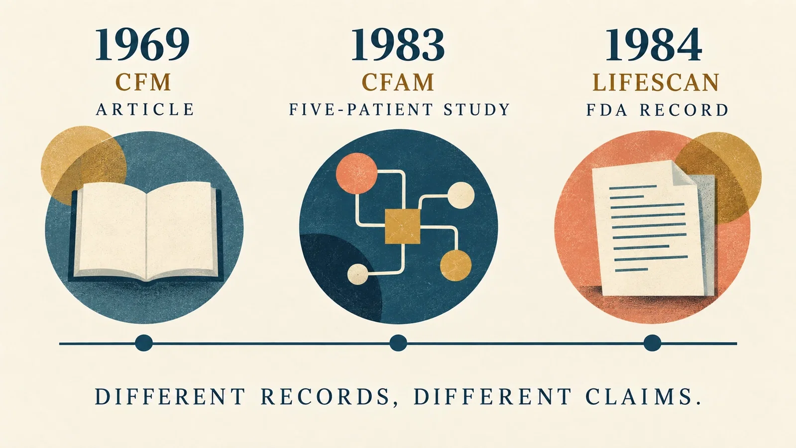
Lifescan supplies a different sort of date. FDA entry K841790 identifies the LIFESCAN EEG MONITOR, names Neurometrics as applicant and records a substantially equivalent decision on July 12, 1984.[3] This establishes a device identity and regulatory event. It does not establish invention, explain the algorithm, or show that Lifescan developed directly from CFAM.
Product names require similar care. Physiometrix's annual report for 2004 identifies PSA 4000 as its own product, describes commercial introduction in 2000, and lists Aspect Medical separately as a competitor.[4] PSA 4000 enters this account only to clarify the identity of a later product, not as a device of the 1970s or 1980s.

The editorial connection to the later BIS article remains useful: early amplitude and frequency trends can be compared with Aspect's later index-generating EEG devices. FDA entry K963644 identifies the A-1000/A-1050 and a 1996 regulatory decision.[5] This comparison distinguishes display approaches and product stages. It does not establish direct transmission between manufacturers, or turn regulatory records into evidence of reduced awareness or improved outcomes.
Image note: all four images are modern editorial illustrations. Devices, paper strips and traces are conceptual, not reproductions of original data, specific models or clinical scenes. The timeline places different evidence types alongside one another. Signal-processing arrows do not describe historical causation between products. The 1980 timeline year is an approximate placement for the 1969–1984 range.
Sources and reading limits
- Maynard D, Prior PF, Scott DF. Device for continuous monitoring of cerebral activity in resuscitated patients. Br Med J. 1969;4:545–546. Original article scans. PMID 5354856. Both original pages, including diagrams and captions, newly read.
- Sebel PS, Maynard DE, Major E, Frank M. The cerebral function analysing monitor (CFAM): A new microprocessor-based device for the on-line analysis of the EEG and evoked potentials. Br J Anaesth. 1983;55:1265–1270. Original publisher article. DOI:10.1093/bja/55.12.1265. All six original pages read, including methods, figures and limitations.
- US Food and Drug Administration. LIFESCAN EEG MONITOR, K841790 · FDA device record. Applicant: Neurometrics. Substantially equivalent decision dated July 12, 1984. Original regulatory database entry read; this is not a clinical study or a record of invention.
- Physiometrix, Inc. Form 10-K for the year ended December 31, 2004, signed March 31, 2005. Item 1, Introduction, Patient State Analyzer, Products and Technology, and Competition sections read for product identity and the company's reported chronology. The entire financial filing was not reviewed. Manufacturer statements about clinical benefits are not adopted.
- US Food and Drug Administration. A-1000 EEG MONITOR AND A-1050 EEG MONITOR, K963644 · FDA device record. Applicant: Aspect Medical Systems. Substantially equivalent decision dated October 8, 1996. Original database entry read; its linked five-page summary is not covered.
The identity of Rampil's 1998 review was verified, but its full text is not covered. A 1991 Lifescan clinical paper was found only in abstract form; its algorithm and clinical findings are not developed here. This is a bounded set of early examples, not an exhaustive device chronology.
Continue exploring
Related reading connects shared questions and contexts; it does not by itself establish historical influence.
- 1966: Plum and Posner Make Coma a Diagnostic Problem
- 1996: The FDA Clears BIS to Aid Anesthetic Monitoring
The Source Room · About & Editorial Method
Sources and reading limits are stated above. Illustrations are labelled as original source images or AI-made. Historical accounts are educational; they are not current treatment instructions.