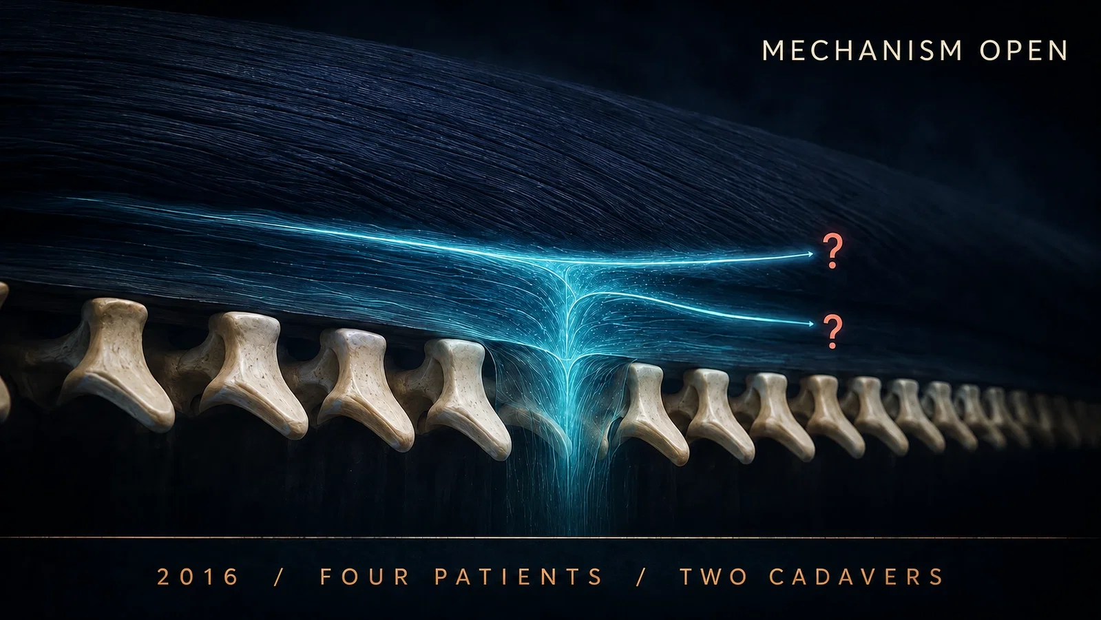
The 2016 report made the transverse process a visible endpoint while preserving a conflict between living sensory observations and cadaveric spread.
Reading Library · Anesthesia Epochs · 中文版
Watch the companion film
2016: Forero — The Erector Spinae Plane Block · Watch on YouTube
Historical anchor: 2016. Date details and source qualifications are explained below.
The abstract tells a two-patient story.
Both patients had severe thoracic neuropathic pain. Both experienced striking relief and broad sensory change after an ultrasound-guided injection. The authors gave the intervention a new name: the erector spinae plane block.
The complete paper contains a larger and less tidy experiment.
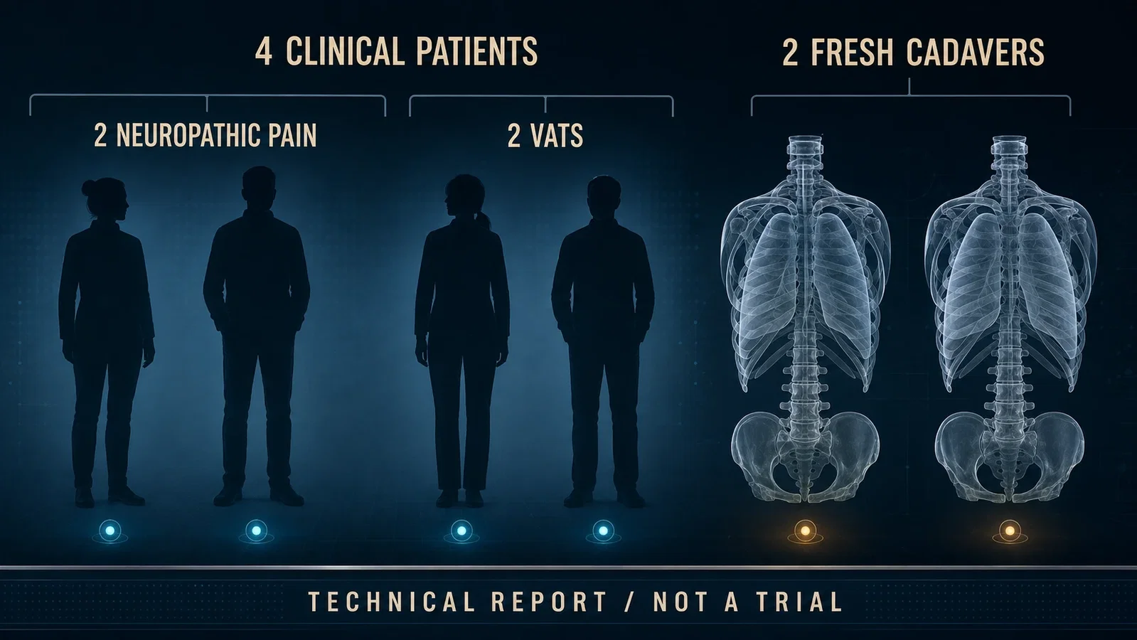
Two more patients received the block for pain after video-assisted thoracic surgery. Two fresh human cadavers were injected and dissected. The full denominator was therefore four clinical patients and two cadavers, not two patients and not a trial.
Across those six subjects, three parts of ESPB appeared at different speeds. The name arrived first. A reproducible landmark came next. A stable mechanism did not arrive with either of them.
A new target inherited an older question
The first patient was a 67-year-old man with severe pain along the left posterior chest wall. The pain was initially understood as postherpetic neuralgia. Later imaging identified metastatic rib disease from bladder cancer.
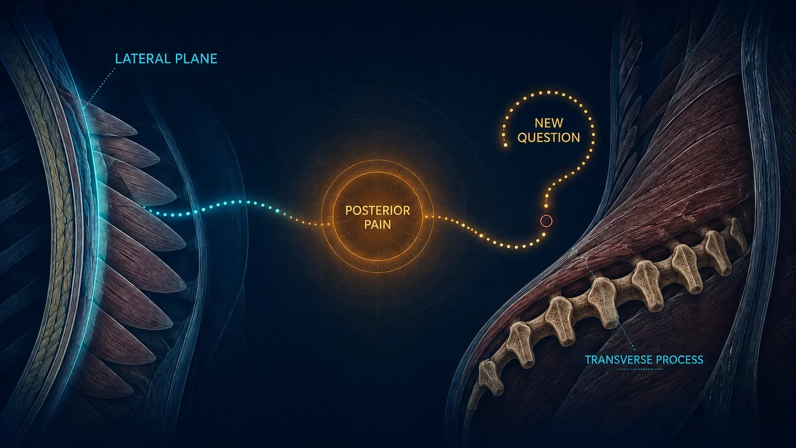
Forero and colleagues considered the serratus plane block, which Blanco and colleagues had described in 2013. They worried that a lateral chest-wall plane would not adequately cover pain close to the back. Instead of abandoning the idea of interfascial spread, they moved it posteriorly.
That decision connects the two historical articles.
ESPB did not appear in isolation from earlier plane-block work. It carried forward the idea that ultrasound could make tissue layers visible and that fluid spreading through a layer could become the intervention. The new question was where that layer should be placed when the painful territory lay close to the spine.
The transverse process gave the technique a physical grammar
On ultrasound, the investigators could identify trapezius, rhomboid major, and erector spinae over the thoracic transverse processes.
Their earliest injection lay superficial to erector spinae, between that muscle and rhomboid major. A later version placed the target deep to erector spinae, where the needle met the transverse process and fluid lifted the muscle from the bone.
The transverse process did more than identify a location. It gave the technique a physical endpoint.
The operator did not need to chase a small nerve. A row of bony landmarks could be seen beneath a broad muscle layer. Contact with bone limited further forward movement, while separation of the fascial plane could be watched in real time. Fluid then traveled longitudinally along the paraspinal gutter.
This was a grammar that another clinician could recognize: the transverse process could be found, erector spinae could be identified, and the opening plane could be observed.
Reproducibility of that action, however, did not prove how the action produced analgesia.
The clinical signal was strong and uncontrolled
After the first superficial injection, the first patient's pain reportedly fell from 10 out of 10 to zero. Two hours later, diminished pinprick sensation extended approximately from T2 to T9, from near the posterior midline toward the midclavicular region, with additional change in the axilla and medial upper arm.
The signal was dramatic. The causal structure was not clean.
Pain returned 36 hours later. Repeated blocks produced about 12 hours of complete relief on average. The team then used a catheter for five days while adding gabapentin and amitriptyline. The longer course cannot be assigned to one injection.
Two weeks later, a contrast study showed longitudinal spread from T1 to T11, with more medial than lateral extension. The same CT exposed a lytic rib lesion and changed the understanding of the patient's pain.
The scan was therefore both a spread observation and a diagnostic event. One living contrast study could show that long paraspinal travel was possible in that patient. It could not establish a universal path.
One patient carried two planes
The second neuropathic-pain patient had chronic symptoms after malunion of multiple ribs and thoracotomy.
A superficial injection relieved pain and allodynia, but the investigators could not detect a clear pinprick sensory block. Three hours later, they injected deep to erector spinae at the T5 transverse process. This time, sensory loss extended roughly from T3 to T9 across the posterior and lateral chest wall, while sparing the axilla.
The sequence helped move the authors toward the deep target.
It was not a randomized comparison of planes. The same person received both injections only hours apart. Residual effects could overlap, assessment was not blinded, and the later one-month improvement remained a single-patient observation.
The case refined an anatomical hypothesis. It did not estimate superiority.
Two surgical cases expanded the question, not the evidence level
The report then described two patients undergoing video-assisted thoracic surgery, one for a wedge resection and one for a lobectomy.
Both received a deep ESP block. Their recovery-room pain and sensory findings encouraged the authors to consider acute postsurgical applications in addition to chronic neuropathic pain.
Neither case demonstrated an opioid-sparing effect.
Both patients also received general anesthesia and systemic opioids. Postoperative hydromorphone remained part of care. Sensory assessment differed between the cases, and one patient had no formal cutaneous map. There was no untreated or alternative-block comparison and no prespecified opioid outcome.
The two cases brought ESPB from the pain clinic into the operating-room narrative. They did not turn a technical report into an efficacy trial.
The cadavers refused one simple route
The first fresh cadaver received bilateral injections superficial to erector spinae, between that muscle and rhomboid major. Dye traveled longitudinally and stained lateral branches of the dorsal rami and the anterior surface of erector spinae. It did not pass beyond the intercostal muscles to show involvement of the ventral rami.
The second cadaver received bilateral injections deep to erector spinae at T5. CT showed long craniocaudal spread, approximately C7 to T8 on one side and T1 to T8 on the other. Dissection found some dye passing through costotransverse and intertransverse regions toward the origins of both dorsal and ventral rami.
Placed side by side, the two specimens made the deep plane look more anatomically persuasive.
They also made certainty impossible. Each target was represented by one cadaver. The observations could not separate plane from individual anatomy, and dye in a nonliving body could not reproduce every feature of local anesthetic in a breathing patient.
The contradiction was published with the invention
The authors did not hide the problem.
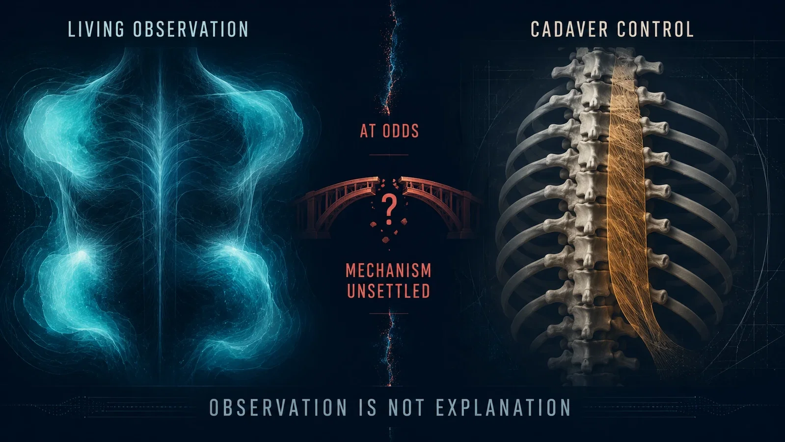
They wrote that the superficial cadaver result was at odds with their initial clinical observations. The living sensory fields appeared to require more than dorsal-ramus involvement, yet dye in the superficial cadaver did not reach the ventral rami.
They proposed that living dynamics might matter. Breathing, tissue tension, and pressure could permit movement that a cadaver did not reproduce. Based on the deep cadaver and their subsequent clinical experience, they recommended targeting the plane between erector spinae and the transverse process rather than the earlier superficial plane.
That recommendation made historical sense within the report. It was still a recommendation derived from four patients and two cadavers, not a contemporary guideline.
The paper also recorded interindividual variation in sensory extent. It called for studies of reproducibility, injection site and volume, the extent of blockade, and systemic local-anesthetic exposure through plasma measurements.
The original report ended with open work, not a closed mechanism.
Simplicity and safety were propositions, not measured outcomes
Forero and colleagues regarded simplicity and safety as the block's major advantage.
Their reasoning was visible. The transverse process was easy to identify and acted as a bony backstop. The intended plane lay posterior to deeper structures such as the pleura. Compared with deliberately entering the paravertebral space, the action appeared forgiving.
The report could not measure that promise.
Four clinical patients provide no denominator for uncommon complications and no comparison of risks. The statement that no structure was at immediate risk described an anatomical rationale. It was not a complication rate.
Historical accuracy therefore requires two clauses: the authors designed and presented ESPB as a simple, potentially safer action; their technical report did not prove safety or superiority.
Twenty later injections made the explanation less automatic
In 2018, Jason Ivanusic, Yasutaka Konishi, and Michael Barrington tested the deep T5 target in ten unembalmed, never-frozen human cadavers. Bilateral injections produced twenty anatomical observations.
Dye again spread widely in craniocaudal and lateral directions around erector spinae. It did not, however, travel consistently into the structures proposed to explain the broad clinical fields. The ventral rami were unstained after nineteen of twenty injections. Only two injections tracked along dorsal rami through the costotransverse foramen toward a dorsal root ganglion.
The authors found no consistent anterior spread into the paravertebral or intercostal spaces. They questioned whether ESPB could be treated as a simpler version of paravertebral block and proposed that lateral cutaneous branches of intercostal nerves might explain part of the observed territory.
Even this larger cadaver study stopped short of a universal mechanism. Intrathoracic pressure, tissue tension, gravity, and position could behave differently in living patients. Dye remained a model for local-anesthetic movement, not the clinical event itself.
The study weakened the assumption that broad fascial spread automatically meant ventral-ramus or paravertebral spread. It did not prove that living anterior spread never occurred.
A name, a landmark, and a research program
The early importance of ESPB does not depend on treating four cases as definitive evidence.
The 2016 report joined a visible bony endpoint, an opening fascial plane, and a set of clinical observations that other investigators could reproduce and challenge. It gave regional anesthesia a stable action before it had a stable explanation.
That asymmetry was productive.
Once the target could be found, researchers could test different planes, map sensation, image contrast, dissect cadavers, compare related blocks, measure systemic exposure, and eventually ask clinical trial questions. The original paper's own conflict made those studies necessary.
Forero and colleagues did not deliver a finished theory and then demonstrate it in four patients. They made a plane over the transverse process into a shared experimental object.
The action could be repeated.
The mechanism remained open.
Sources and reading limits
Sources: Forero M et al., The Erector Spinae Plane Block: A Novel Analgesic Technique in Thoracic Neuropathic Pain, Reg Anesth Pain Med 2016;41(5):621-627, PMID 27501016; Ivanusic J et al., A Cadaveric Study Investigating the Mechanism of Action of Erector Spinae Blockade, Reg Anesth Pain Med 2018;43(6):567-571, PMID 29746445; PubMed and Crossref identity records; Internet Archive acquisition provenance; two 2017 PubMed comment records used only as title-level nomenclature context. All cited sources were read for this article.
Visual interpretations
These illustrations explore the setting and ideas. Depicted people, actions, instruments and documents are reconstructions, not evidence of their exact historical appearance.
Continue exploring
Related reading connects shared questions and contexts; it does not by itself establish historical influence.
Historical connections
- Earlier connection: 2013: The Serratus Plane Block Begins as a Sensory Map
Related reading
- 2013: The Serratus Plane Block Begins as a Sensory Map
- 2019: Chronic Pain Enters ICD-11, Not All as Disease
The Source Room · About & Editorial Method
Sources and reading limits are stated above. Illustrations are labelled as original source images or AI-made. Historical accounts are educational; they are not current treatment instructions.