The 2013 report made two planes around serratus anterior visible and testable, while leaving surgical analgesia, safety, and comparative value unanswered.
Reading Library · Anesthesia Epochs · 中文版
Watch the companion film
2013: Blanco — The Serratus Plane Block · Watch on YouTube
Historical anchor: 2013. Date details and source qualifications are explained below.
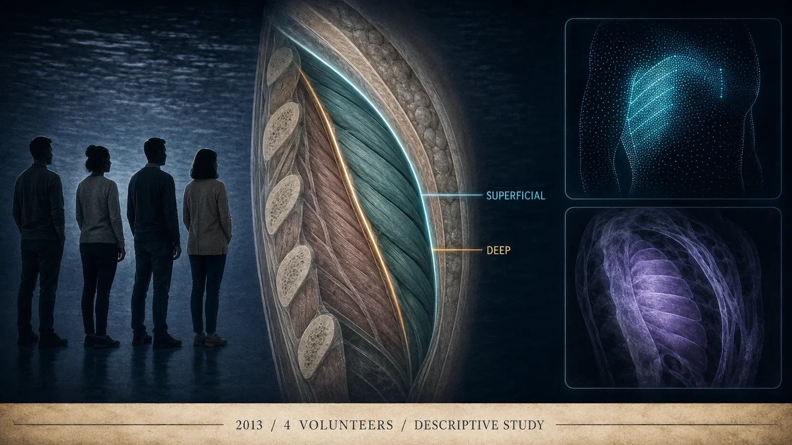
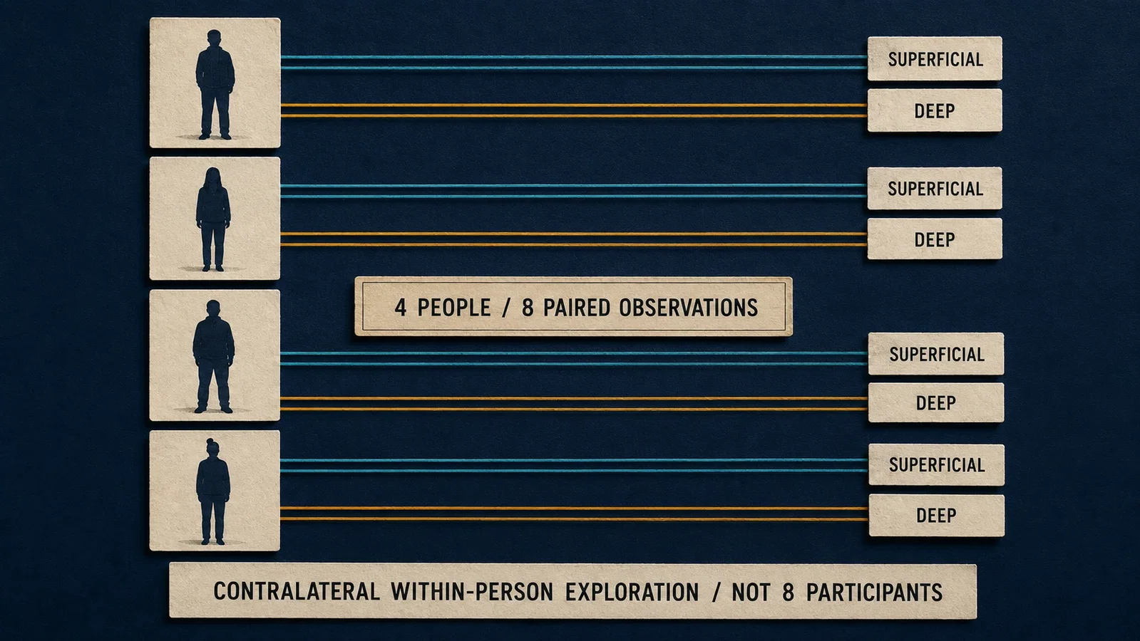
The 2013 serratus plane block paper contains eight blocks but only four independent people.
That distinction is the best place to begin. It reveals both the ingenuity of the experiment and the limits of the claims that can be built from it.
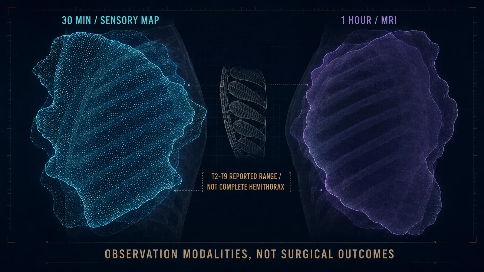
Each female volunteer contributed a pair of contralateral observations. On one side, the investigators studied a plane superficial to serratus anterior. On the other, they studied a plane deep to the muscle. Thirty minutes later they mapped sensory change with pinprick. After one hour they used MRI to examine where the experimental injectate appeared to have spread.
The design turned two barely visible tissue spaces into a comparison that could be seen, felt, and published. It did not turn four volunteers into a clinical trial of postoperative analgesia.
What began in 2013 was not a finished answer. It was a research question with anatomy.
The denominator is part of the discovery
Blanco, Parras, McDonnell, and Prats-Galino reported four female volunteers aged 23, 23, 32, and 46. The article states that they were unrelated to the investigators, that local ethics approval had been obtained, and that written informed consent was collected. The visible text does not formally label the volunteers as healthy, so this account does not add that adjective.
Every person received two blocks, one on each side. This paired arrangement allowed the investigators to place superficial and deep observations within the same body rather than compare two unrelated groups.
That is a useful exploratory design. It can reduce some between-person variation and make an anatomical contrast legible in a very small study. But its apparent symmetry can mislead. Eight block observations do not provide the independence of eight participants. The sides were not two large randomized groups. The experiment could reveal patterns worth pursuing, not stable population estimates.
This denominator matters whenever the paper's numerical precision becomes seductive. A mean reported to the nearest minute can still rest on four people. A broad sensory territory can still vary from person to person. The absence of an observed complication can still be almost uninformative about uncommon harm.
Small exploratory studies are not weak because they fail to be large trials. They become misleading only when later storytellers ask them to answer questions their design never contained.
A sensory experiment can be clinically suggestive without being a surgical outcome study
The formal title was Serratus plane block: a novel ultrasound-guided thoracic wall nerve block. The 2013 article did not use the later standardized title serratus anterior plane block.
Its methods were built around observation. The investigators used ultrasound to distinguish a plane superficial to serratus anterior from one deep to it. The experimental mixture included a local anaesthetic and gadolinium so that spread could be examined with MRI. Those historical drug and imaging details explain the study's architecture; they are not a current recipe.
At 30 minutes, the team delineated sensory loss to pinprick. They divided the chest wall into anterior, lateral, and posterior regions and recorded the extent separately. MRI followed at one hour. The project therefore produced two kinds of map: a map of reported and tested sensation, and a map of image signal.
It did not enroll anyone undergoing breast, thoracic, or upper-abdominal surgery. There was no incision, control technique, postoperative pain trajectory, opioid outcome, recovery measure, or discharge endpoint. There was no pharmacokinetic analysis and no sample designed to estimate safety.
The paper's enthusiasm about possible perioperative use was a hypothesis growing out of sensory and imaging observations. It was not itself evidence that surgery had become less painful.
The T2-T9 headline hides several different maps
The paper summarized its result as dermatomal paraesthesia from T2 to T9, with numbness in all four volunteers. That statement is accurate at the level of the reported overall pattern. It is not a description of uniform, complete hemithorax anaesthesia.
Table 2 makes the difference visible. Superficial observations ranged from T2-T6 to T2-T9 depending on the volunteer and whether the anterior, lateral, or posterior chest wall was being considered. Deep observations also differed by person and region, commonly ending between T5 and T9. The map was broad, but it was not a single identical field laid over four bodies.
The distinction was recognized immediately. In an editorial printed in the same issue, Tighe and Karmakar challenged the suggestion of complete hemithorax paraesthesia. They pointed to posterior primary rami, anterior cutaneous intercostal branches near the sternum, and supraclavicular nerves as anatomical reasons that important territory would remain outside the proposed block.
Their criticism did not deny that the volunteers experienced sensory change. It denied the conversion of that change into a more comprehensive claim about the whole hemithorax.
That same-issue timing is important. The limit was not invented years later after the technique had been scrutinized by a larger field. It was visible on publication day.
The superficial plane looked more durable, but the authors kept the conclusion provisional
The numerical contrast between the two planes was striking.
For the superficial observations, the reported mean duration was 752 minutes with a standard deviation of 21 for intercostal sensory effects, and 778 minutes with a standard deviation of 43 for the motor-nerve measure. For the deep observations, the corresponding values were 386 with a standard deviation of 160 and 502 with a standard deviation of 150 minutes.
The investigators therefore favored the superficial plane. They also wrote that the study was descriptive and that randomized controlled trials would be needed for a definitive assessment.
Both parts of that conclusion belong together. Omitting the preference would make the experiment seem directionless. Omitting the qualification would turn an exploratory within-person contrast into established superiority.
The larger standard deviations in the deep observations also matter. They remind the reader that individual responses did not collapse into one clean result. The exact means describe these four volunteers; they do not establish how long a surgical patient should expect analgesia to last.
The study created a reason to test the superficial plane more rigorously. It did not complete that test.
MRI made the hypothesis visible without becoming its final judge
The MRI reconstructions are among the paper's most memorable features. They appear to transform an ultrasound-guided injection into a three-dimensional anatomical object.
Yet the investigators documented a problem with that apparent clarity. Gadolinium signal could resemble surrounding fat on MRI. Image thresholds and processing were needed to isolate the presumed contrast distribution. More importantly, the clinically observed sensory territory extended beyond what the MRI seemed to show.
This mismatch should not be treated as an embarrassment to be edited out of the history. It is scientifically productive.
Pinprick mapping depends on stimulus, reporting, and interpretation. MRI depends on tracer behavior, tissue contrast, acquisition, and post-processing. Partial agreement can support an anatomical hypothesis while disagreement identifies what neither method can settle alone.
The paper therefore did more than provide a compelling image. It exposed a measurement problem that later fascial-plane research would repeatedly encounter: the route taken by a visible injectate is related to, but not identical with, the territory of nerve function or clinical analgesia.
An image can show where signal appears. It cannot by itself tell us how a patient will experience surgery.
The editorial kept four unanswered clinical questions in view
Tighe and Karmakar's editorial admired the innovation while refusing to let possibility become proof.
First, the block had not yet been described for analgesia during or after surgery. A sensory map in volunteers could not establish its place beside techniques already used in clinical practice.
Second, the proposed coverage was anatomically incomplete. Serratus plane block would not reproduce the broad somatic and autonomic effects of thoracic paravertebral or epidural blockade.
Third, Blanco and colleagues had provided no pharmacokinetic data. The idea that placement in a less vascular area might lower local-anaesthetic toxicity risk remained an assumption to be tested.
Fourth, comparative role was unresolved. The editorial expressed particular doubt about equivalence to paravertebral block for major breast surgery with axillary dissection, as well as use as a sole anaesthetic. It also left chronic pain and other possible applications open.
The editorial's tone is historically useful because it was neither dismissive nor promotional. It recognized a promising anatomical technique and then listed the evidence that promise still owed.
In effect, the two papers divided the work. The original report made the planes and sensory signal visible. The editorial translated that visibility into a clinical research agenda.
Four uneventful observations cannot certify safety
The original report noted no side effects in the four volunteers. That is a factual observation. It is not a general safety result.
Rare complications require a denominator large enough to reveal them. Comparative safety requires a comparator. Local-anaesthetic toxicity requires exposure and pharmacokinetic information. None was available here.
The same-issue editorial treated lower toxicity risk as an unproven assumption. Years later, the 2021 breast-surgery meta-analysis reported no procedure-related complications among its included randomized trials, but also cited a case report of pneumothorax and called for higher-quality trials to establish safety.
Those later facts do not prove that the 2013 volunteers were unsafe. They show why none observed must not be rewritten as safe.
Evidence language should preserve the size of the window through which an event was observed. In 2013, that window contained four people.
A cadaver study later showed why spread and effect must remain separate
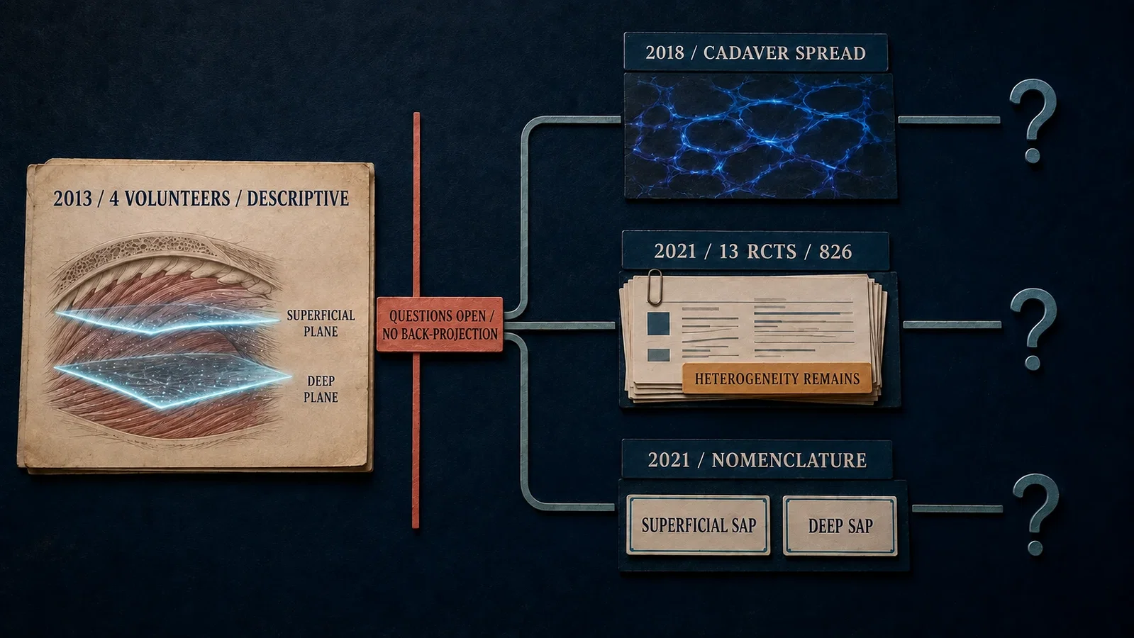
Kunigo and colleagues returned to the anatomical problem in 2018. They used nine adult Thiel-embalmed cadavers, all older than 80, to compare 20 mL on one side with 40 mL on the other. One 40 mL hemithoracic wall was excluded because of a surgical scar, leaving 17 evaluable sides.
The larger volume produced wider craniocaudal staining. The mean number of stained intercostal nerves was 5 +/- 0.7 with 40 mL and 4 +/- 1.1 with 20 mL, p=0.02. All T2-T5 nerves were stained in the 40 mL group, while all T3-T4 nerves were stained in the 20 mL group. T1, pectoral, long thoracic, and thoracodorsal nerves were seldom stained, and dye did not reach the axillary or dorsal regions.
That study sharpened the anatomical picture, but its authors explicitly warned that clinical effect cannot be predicted from cadaveric research. No anatomist verified the dissections, donor height and weight were not recorded, and embalmed tissue cannot reproduce living physiology.
The historical lesson is not that one volume should replace another. It is that even a more direct anatomical observation produced a distribution narrower and different from the original sensory report.
Dye, MRI signal, pinprick sensation, and postoperative analgesia occupy related layers of evidence. They are not interchangeable endpoints.
By 2021, trials existed, but uncertainty had not disappeared
Hu and colleagues' 2021 systematic review and meta-analysis moved the question into breast surgery. It included 13 randomized controlled trials and 826 participants.
The synthesis suggested reduced postoperative opioid use and lower pain scores in several postoperative periods. That was later clinical evidence, not a hidden result of the four-volunteer report.
Its uncertainty was substantial. Heterogeneity reached I2=100% for postoperative opioid consumption and I2=94% for intraoperative opioid consumption. Several pooled pain-score outcomes had I2 values between 81% and 95%. The GRADE table rated most outcomes as low-quality evidence, with postoperative nausea and vomiting rated moderate.
The authors also acknowledged a limited number of participants, clinical heterogeneity arising from different general and local anaesthetic regimens, and too few randomized trials to compare serratus anterior plane block adequately with other regional techniques.
The later literature therefore did not produce a simple retrospective verdict that the 2013 authors had been right about everything. It did something more valuable. It began to test the clinical questions that the original design could not contain, while making new sources of variation visible.
The name matured after the research question had already spread
A second 2021 paper addressed language rather than analgesic effect.
An ASRA-ESRA Delphi consensus recommended superficial serratus anterior plane (SAP) block with 80% agreement and deep serratus anterior plane block (SAP) with 91% agreement. Consensus on the corresponding anatomical descriptions was 52% for the superficial approach and 76% for the deep approach.
Those figures show that naming and anatomy do not always stabilize at the same speed. The labels achieved stronger agreement than one of the proposed descriptions.
The later terminology helps separate the two planes that Blanco and colleagues had explored. It should not be back-projected into the original article's title. In 2013 the published name was serratus plane block; in 2021 the field supplied a more standardized vocabulary for its superficial and deep descendants.
This is another way a technique can evolve. First the anatomy becomes experimentally interesting. Later the community negotiates how to speak about it consistently.
The real milestone was making an unfinished question reproducible
It would be easy to narrate the serratus plane block as a clean sequence: invention, proof, adoption.
The archived sources describe something less linear and more credible.
Four volunteers allowed two tissue planes to be compared within the same bodies. Sensory testing suggested broad but variable territories. Duration observations favored the superficial plane. MRI made spread visible but did not fully match sensation. A same-issue editorial challenged complete coverage, pharmacokinetic assumptions, safety implications, and surgical equivalence. Later cadaveric work complicated the anatomical map. Randomized trials moved the question into patients but introduced heterogeneity and evidence-quality limits. A consensus then standardized the names.
The 2013 report matters because it gave later researchers something specific to disagree with, measure differently, test clinically, and rename.
That is a meaningful form of innovation. It does not require pretending that four volunteers established a complete thoracic-wall anaesthetic, proved postoperative benefit, or certified safety. It requires recognizing the point at which an anatomical possibility became a shared experimental object.
Eight block observations did not make eight independent people.
They made two tissue planes difficult to ignore.
Sources and reading limits
Sources: Blanco R, Parras T, McDonnell JG, Prats-Galino A. Anaesthesia. 2013;68(11):1107-1113. PMID 23923989. DOI 10.1111/anae.12344; Tighe SQM, Karmakar MK. Anaesthesia. 2013;68(11):1103-1106. PMID 24032664. DOI 10.1111/anae.12423; Kunigo T et al. JA Clin Rep. 2018;4:10. PMID 29457120. PMCID PMC5804672. DOI 10.1186/s40981-018-0147-4; Hu NQ et al. Pain Res Manag. 2021;2021:7849623. PMID 34733377. PMCID PMC8560299. DOI 10.1155/2021/7849623; El-Boghdadly K et al. Reg Anesth Pain Med. 2021;46(7):571-580. PMID 34145070. DOI 10.1136/rapm-2020-102451. All cited sources were read for this article.
Visual interpretations
These illustrations explore the setting and ideas. Depicted people, actions, instruments and documents are reconstructions, not evidence of their exact historical appearance.
Continue exploring
Historical connections
These links follow specific development, adoption or citation connections recorded in the underlying research. They do not imply that history had one inevitable path.
Related reading connects shared questions and contexts; it does not by itself establish historical influence.
- 1994: A Brachial Plexus Study Watches the Needle and Local Anesthetic
- 2016: The Erector Spinae Plane Block, from Four Patients and Two Cadavers
The Source Room · About & Editorial Method
Sources and reading limits are stated above. Illustrations are labelled as original source images or AI-made. Historical accounts are educational; they are not current treatment instructions.