Early cases, the monitor view and tube placement.
Reading Library · Anesthesia Epochs · 中文版
Watch the companion film
2001: GlideScope — The Video Laryngoscope · Watch on YouTube
Historical anchor: 2001. Date details and source qualifications are explained below.
2001 · Reading the early evidence behind a new airway device
The year attached to GlideScope needs an attributed source. Verathon's anniversary release, published in April 2011, distinguished Jack Pacey's work beginning in late 1999 from introduction of the device in 2001. The manufacturer's current website instead describes an introduction in 1999. This article uses the dated retrospective account to anchor the event to 2001, without assigning an exact first-sale date or claiming the world's first video laryngoscope. The early clinical literature supplies a separate observation: the later case series included use beginning in November 2001.[1;2;4, pp.194–195]
In 2003, Richard M. Cooper described a 74-year-old man returning for elective surgery. Previous records documented repeated difficulty or failure with direct laryngoscopy, and an episode of regurgitation and aspiration during management with a laryngeal mask. He declined the recommended awake fibreoptic intubation. After induction on this occasion, GlideScope provided a Cormack–Lehane grade I view, and intubation was completed using a malleable stylet. The report showed what an alternative view could make possible in one patient. It did not establish management for every difficult airway, and Cooper explicitly called for larger clinical series.[3, pp.611–613]
The device in those pages should be distinguished from later models. Manufactured by Saturn Biomedical Systems, it had a sharply angled black plastic blade, a camera beneath the blade and a cable running from the handle to a separate seven-inch LCD monitor. Early images were monochrome; red and blue light-emitting diodes provided illumination. The camera transferred the view to a screen without requiring a direct line of sight from the operator's eye to the glottis. Figure 2 in the 2003 report showed a typical monitor image, explicitly not an image of the reported patient. It cannot serve as a photograph of that particular encounter.[3, pp.612–613;4, p.192]
The subsequent multicentre series examined use on a broader scale. Published in 2005, it described 728 uses involving 133 operators at five hospitals between November 2001 and March 2003. Six incomplete records were excluded, leaving 722 for analysis. Clinicians selected patients, availability affected use, training was not standardized and there was no formalized study protocol. This was early clinical experience, rather than a trial randomly assigning comparable patients to competing laryngoscopes.[4, pp.191,193–196]
The location of failure was especially instructive. Among 26 cases in which GlideScope was abandoned for another method, 14 had already yielded a grade I laryngeal view. Failure here meant abandoning the technique, sometimes after a single attempt. It did not mean that the airway ultimately remained unsecured: the paper reported successful subsequent intubation with other devices in all these cases. Seeing the glottis and delivering a tube to it required separate assessment. Experience, abandonment criteria and patient selection also limited comparisons with failure rates in other studies.[4, pp.194,196–197]
The authors' relationships are part of that evidence. The 2005 paper disclosed that John A. Pacey developed the device and was chief executive of its manufacturer, Saturn. Cooper was a consultant to and investor in the company and had received travel support. The paper also stated that there was no research funding. That statement did not remove the disclosed relationships; the relationships alone do not invalidate the observations either. Both belong in the account.[4, p.191]
A specific comparison is available in the 2013: An Old Airway Algorithm Makes Room for Video Laryngoscopy. Its algorithm included video-assisted laryngoscopy among initial choices while retaining the distinction between awake and postinduction approaches and attention to oxygen delivery throughout management. The technology had acquired an explicit place within a larger strategy. This comparison concerns the later placement of a class of devices, without claiming that a particular 2001 product directly caused the guideline update.[5, PDF pp.5,7]
The official appendix to the 2016: 1,018 Difficult Intubations Make 'Try Again' a Measurable Risk provides another comparison. GlideScope appeared as a separate device group, alongside groups that differed in age, weight, airway examination and anticipated difficulty. Recording the device across a network made its context of use available for comparison and made selection differences visible. The baseline table itself did not establish a superior device. This account uses the available peer-reviewed appendix and does not reconstruct results from the unavailable main article.[6, appendix 1]
GlideScope's early history centres on a change in how the laryngeal view was obtained, a change that still had to be judged against tube placement and clinical context. The 2003 case established a possibility; the 2005 series placed that possibility alongside failures, training and selection. Preserving those layers gives the device its historical significance without making a clear screen a guarantee that every difficulty has been solved.[3, p.613;4, pp.196–197]
Images
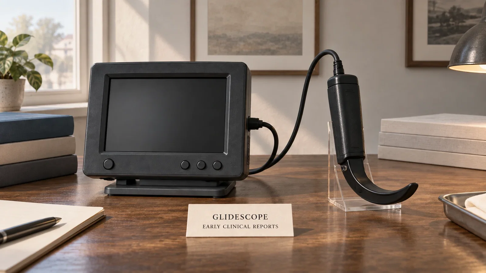
Modern editorial reconstruction informed by the early description of a separate monitor and black blade; not a 2001 photograph or an exact model replica.
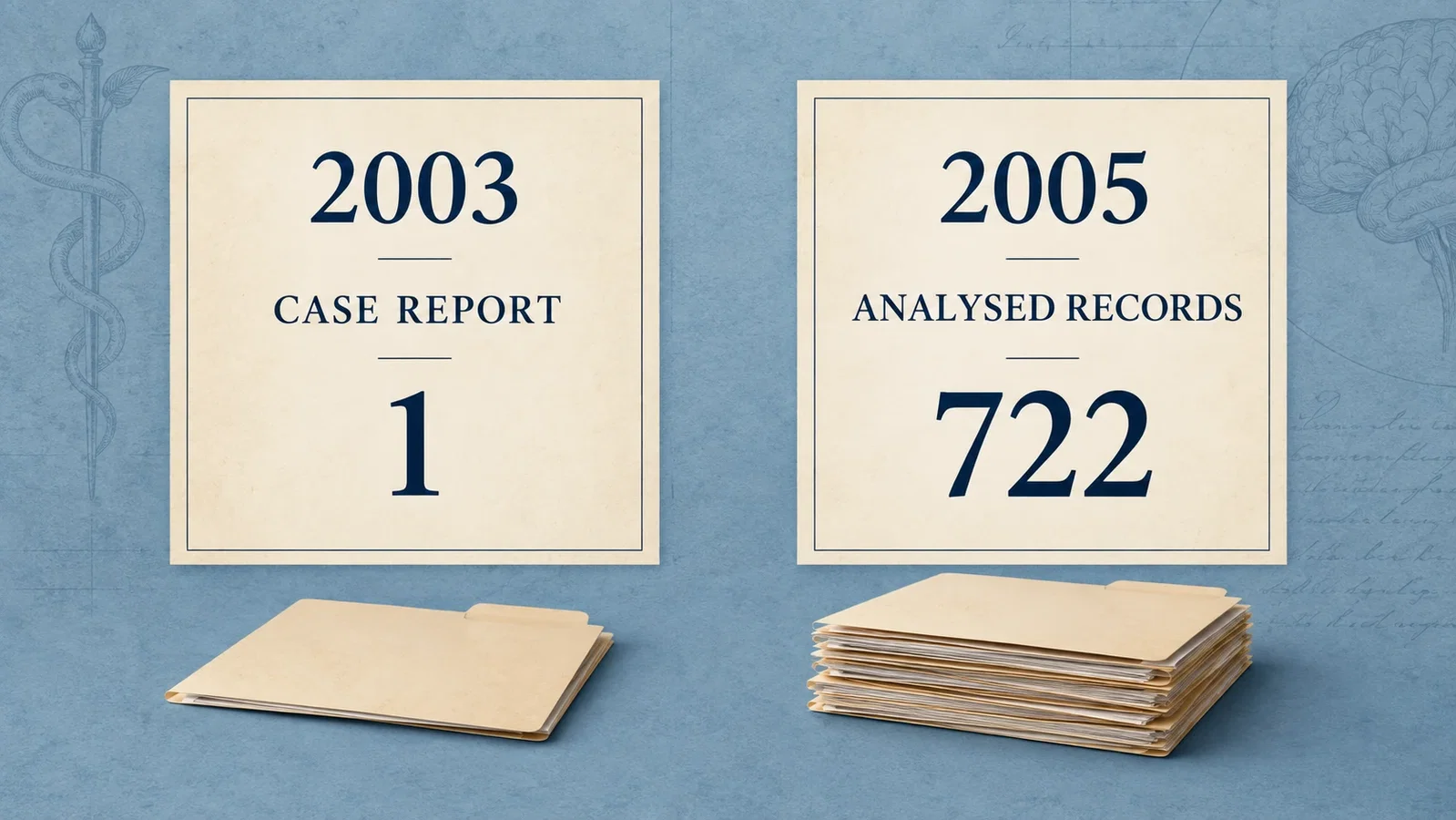
The 2003 single case and the 722 analysed records in the 2005 series represent different levels of evidence.

Fourteen of the 26 abandoned uses had achieved a grade I view. The inner panel is a subset, not a second independent cohort.

Editorial comparison with the 2013 guideline and PeDI appendix, not a causal diagram or a clinical procedure.
Sources and reading limits
- Verathon Inc. GlideScope video laryngoscope…celebrates 10th anniversary. April 18, 2011. Original company release. Full article read; retrospective corporate account, with promotional outcome claims excluded.
- Current Verathon product page. History paragraph and early reference entry read. Its 1999 wording differs from the 2001 introduction in reference 1.
- Cooper RM. Use of a new videolaryngoscope (GlideScope) in the management of a difficult airway. Can J Anaesth. 2003;50:611–613. DOI:10.1007/BF03018651. Publisher original PDF. All three pages and figures read; correct PMID 12826557.
- Cooper RM, Pacey JA, Bishop MJ, McCluskey SA. Early clinical experience with a new videolaryngoscope (GlideScope) in 728 patients. Can J Anaesth. 2005;52:191–198. DOI:10.1007/BF03027728. Publisher original PDF. All eight pages, tables and figures read; PMID 15684262.
- Apfelbaum JL et al. Practice guidelines for management of the difficult airway: an updated report. Anesthesiology. 2013;118:251–270. DOI:10.1097/ALN.0b013e31827773b2. Archived ASA PDF. From an archived copy. This version has placeholder journal pagination.
- Fiadjoe JE et al. PeDI registry prospective cohort analysis, supplementary appendix. Parent article DOI. The complete two-page peer-reviewed appendix was read; it is not the main article.
The 2005 paper contains inconsistencies between counts, some percentages and table totals. This account retains explicitly attributed counts without silently correcting the article or constructing a superiority comparison from those percentages. NLM comment links were checked, but comment bodies were not read; absence of an observed erratum link is not proof that no correction exists. All four images are modern editorial illustrations in the original landscape format.
Sources and reading limits are stated above. Illustrations are labelled as original source images or AI-made. Historical accounts are educational; they are not current treatment instructions.
Continue exploring
Related reading
Related reading connects shared questions and contexts; it does not by itself establish historical influence.