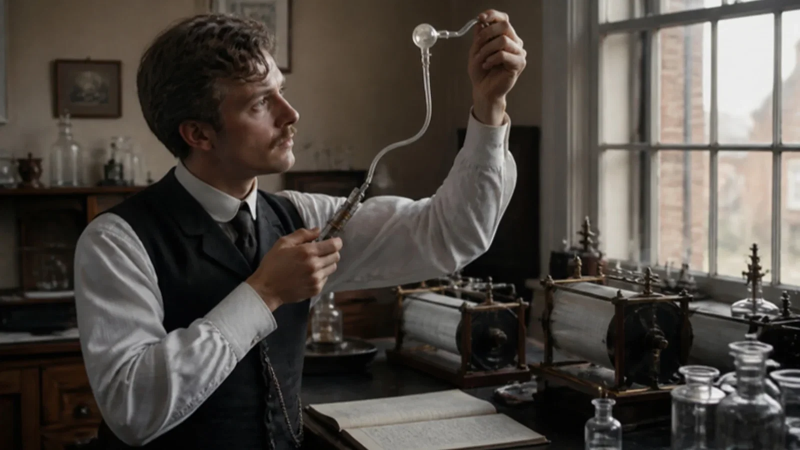
In 1889 Henry Head described a bent cannula whose thin rubber chamber, filled with glycerine, sealed a rabbit’s right bronchus so that one lung stayed still while the other was ventilated.
Reading Library · Anesthesia Epochs · 中文版
Watch the education video (for clinicians)
1889: Head's Bronchial Cannula — A Rubber Chamber in the Bronchus · Anesthesia Epochs Education Series
Every cuffed tracheal tube and every bronchial blocker works the same way: a thin-walled chamber around the tube is inflated through a small channel until it seals the airway. The Australian Society of Anaesthetists' timeline credits the "invention of cuffed tubes (these being for endobronchial tubes)" to Henry Head in 1889, in the course of physiological lung studies. This article reads Head's paper, "On the regulation of respiration", in volume 10 of the Journal of Physiology, where the device is described and drawn. The paper turns out to differ from the timeline on where the work was done and in which animals. [1][2]
A problem in physiology
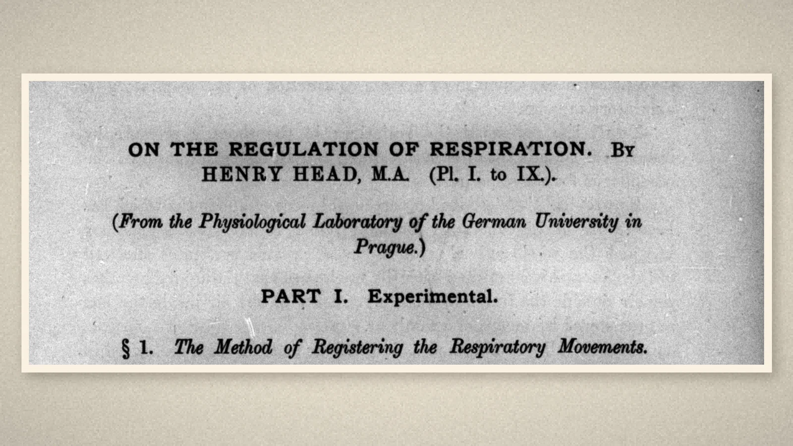
Head, signing as "Henry Head, M.A.", wrote from the Physiological Laboratory of the German University in Prague. The paper is a long study of how the lungs, through the vagus nerves, regulate breathing, carried out on rabbits narcotised with chloral. One question needed a special apparatus: what happens to breathing "when one or both lungs remain permanently at the same volume". Inflating both lungs meant closing the trachea, so that the air stagnated and the animal became dyspnoeic, and "the results were always complicated by dyspnoea". To study one lung held at a constant volume, Head had to find "some means of eliminating the dyspnoea": the other lung had to keep breathing. [1]
The cannula
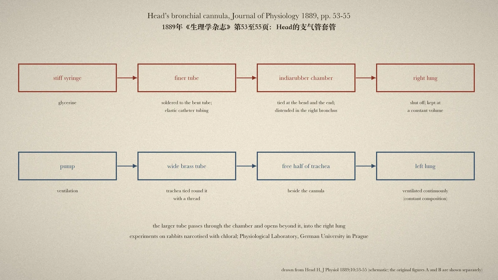
The answer was "a cannula ... constructed on the principle of Marey's Cardiac sounds". A fine tube was bent so that it could be passed down the trachea into the right bronchus. A still finer, slightly shorter tube was soldered to it, opening just below the bend. "A piece of thin indiarubber tubing was passed over the far end of both tubes and tied firmly at the bend and at the extreme end of the larger tube." The smaller tube now opened into a chamber formed by the rubber, and "fluid forced down the smaller tube will distend the walls of this chamber". The larger tube passed right through the chamber and opened beyond it. [1]
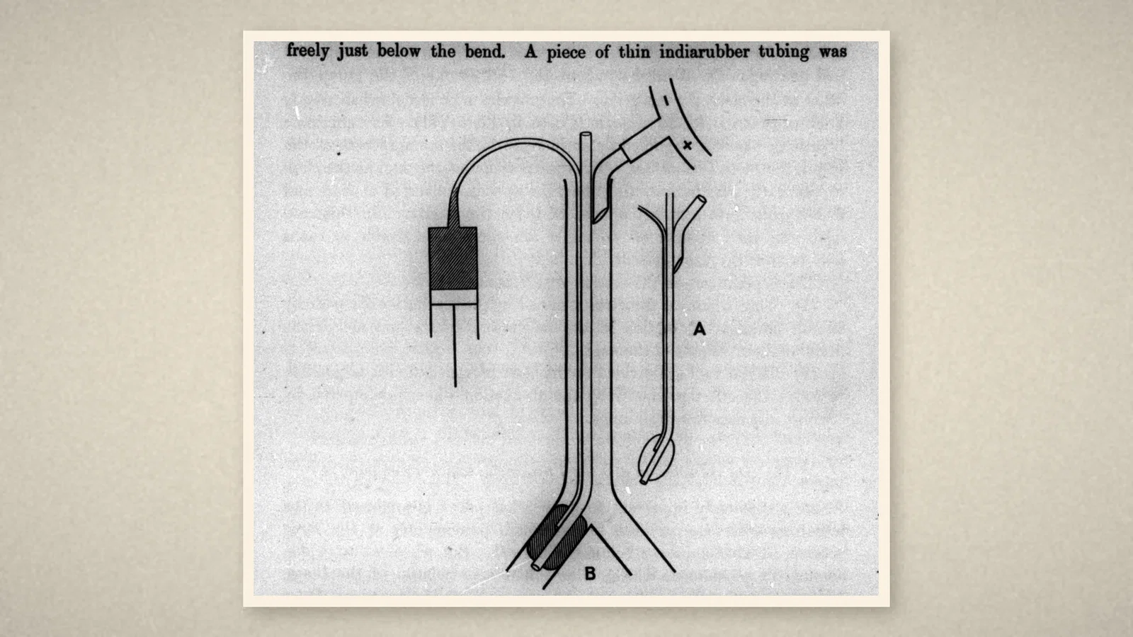
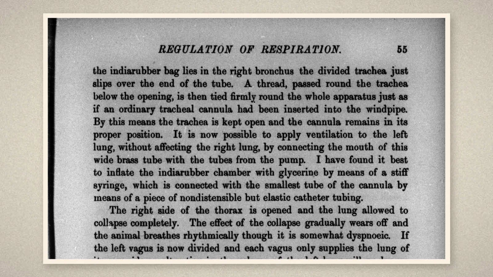
Passed into the right bronchus and distended, the chamber "will shut off the right lung entirely except for the opening afforded by the larger tube of the cannula". The cannula was sized so that "at least half of the trachea remains perfectly free", and the left lung could breathe or be ventilated past it. A wide brass tube was fitted so that the divided trachea slipped over its end, and a thread round the trachea held the whole apparatus "just as if an ordinary tracheal cannula had been inserted into the windpipe". Ventilation could then be applied to the left lung alone from the pump. For the cuff itself, Head wrote, "I have found it best to inflate the indiarubber chamber with glycerine by means of a stiff syringe", connected to the smallest tube through "nondistensible but elastic catheter tubing". [1]
How it was used
The experiment was not gentle. The right side of the chest was opened and the right lung allowed to collapse; the left vagus was cut, so that each lung's nerve ran only to its own side; then the cuff was inflated in the right bronchus and the right lung distended. Because the right chest was open, that lung stayed at the same volume whatever the animal did, while the left lung was ventilated continuously: "Thus one lung remains at a constant volume whilst the air in the other is kept of a constant composition." Head then had to check, by closing the trachea or watching for the rhythm of the pump, that the two vagi did not cross; animals in which they did were useless for the experiment. [1]
What the record shows
Head's cannula contains, in a few lines of text and one drawing, the parts of every modern bronchial blocker and cuffed tube: a thin rubber sleeve tied to the tube at both ends, a separate fine inflation channel, a stiff syringe, an inflation tube that does not stretch, a lumen that passes through the cuff to the lung beyond, and a size chosen so that the other lung's airway stays open. Its purpose was lung isolation: one lung held still, the other ventilated. That is the same problem the thoracic anaesthetist solves when one lung must be quiet for surgery.
Sources and reading limits
Read: Head, “On the regulation of respiration”, Journal of Physiology 1889;10, the first part of the paper, including pp. 1 and 53–55; the ASA timeline entries for 1889.
Not covered: the second part of the paper; Marey’s apparatus for cardiac sounds; later cuffed tubes. The ASA places the work at University College Hospital or the London Hospital and in dogs, and calls the device a double-lumen endobronchial tube; the paper is headed from the Physiological Laboratory of the German University in Prague, the experiments described are on rabbits, and the device is a single bent tube with a fine inflation tube, the other lung breathing through the trachea beside it. The article follows the paper. The animal experiments are recorded, not endorsed.
Three illustrations are crops of the 1889 journal, including its original figures; one is drawn from the paper.
- Head H. On the regulation of respiration. J Physiol. 1889;10:1-70. Internet Archive
- Australian Society of Anaesthetists. Anaesthesia history timeline. 2024. ASA
Continue exploring
Related reading connects shared questions and contexts; it does not by itself establish historical influence.
- 1931: Guedel and Waters' Cuffed Endotracheal Tube
- 1949: Carlens's Double-Lumen Catheter Separates the Two Lungs
Anesthesia Epochs · The Source Room · About & Editorial Method
Published November 2, 2026. Sources and reading limits are stated above. Illustrations are labelled as original source images or AI-made. Historical accounts are educational; they are not current treatment instructions.