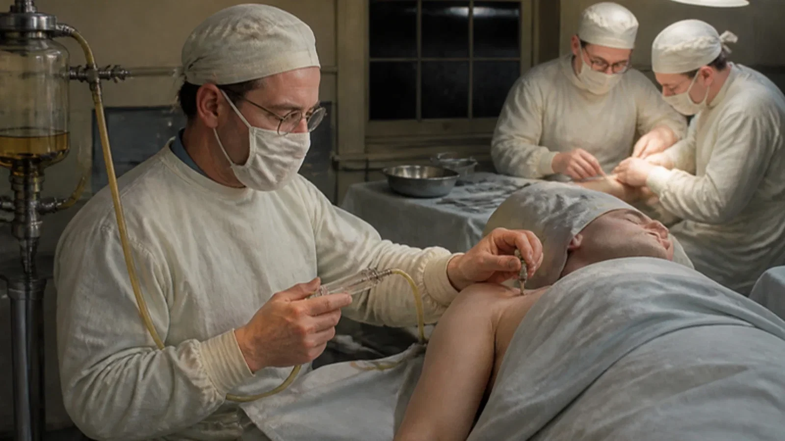
A Brooklyn anaesthesiologist fixed a blunt malleable needle beside the subclavian artery with a cork from an ether can, connected it through tubing and a two-way valve to a syringe, and topped up procaine for operations of up to four hours.
Reading Library · Anesthesia Epochs · 中文版
Watch the education video (for clinicians)
1946: Ansbro's Continuous Brachial Plexus Block — A Corked Needle · Anesthesia Epochs Education Series
A nerve block lasts only as long as its drug. In the spine and the caudal canal, one answer had been to leave something in place and keep dosing. On 4 April 1946 F. Paul Ansbro, director of anaesthesiology at Kings County Hospital in Brooklyn, told the Brooklyn Surgical Society how to do the same for the brachial plexus. The paper, "A method of continuous brachial plexus block", appeared in the American Journal of Surgery that June; this article reads it in full. [1]
The problem
Ansbro's argument started from arithmetic. Fifty cubic centimetres of 1 per cent procaine "will lose its anesthetic effect within forty to sixty minutes", and the long-acting drugs of the day were too toxic in the volumes a plexus block needed. When an operation outlasted the block, a general anaesthetic had to be added, which was "usually not desired and oftentimes deliterious", and the surgeon hurried. For poor-risk patients, Ansbro wrote, a nerve block was often the method of choice. [1]
The brachial plexus block itself had a reputation "for its many failures". Ansbro gave its history briefly: Hirschel's axillary block in 1911, and Kulenkampff's supraclavicular method "the next year", the most used and safer than other routes. (Kulenkampff's own paper, read for another article in this series, appeared in October 1911.) Ansbro had come to trust a supraclavicular method proposed by Macintosh and Mushin, and it "suggested to the writer the possibility of continuing the injections with the needle remaining in situ". [1]
A needle held by a cork
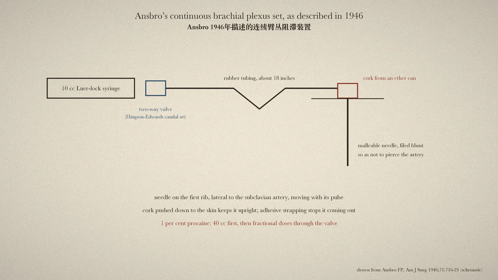
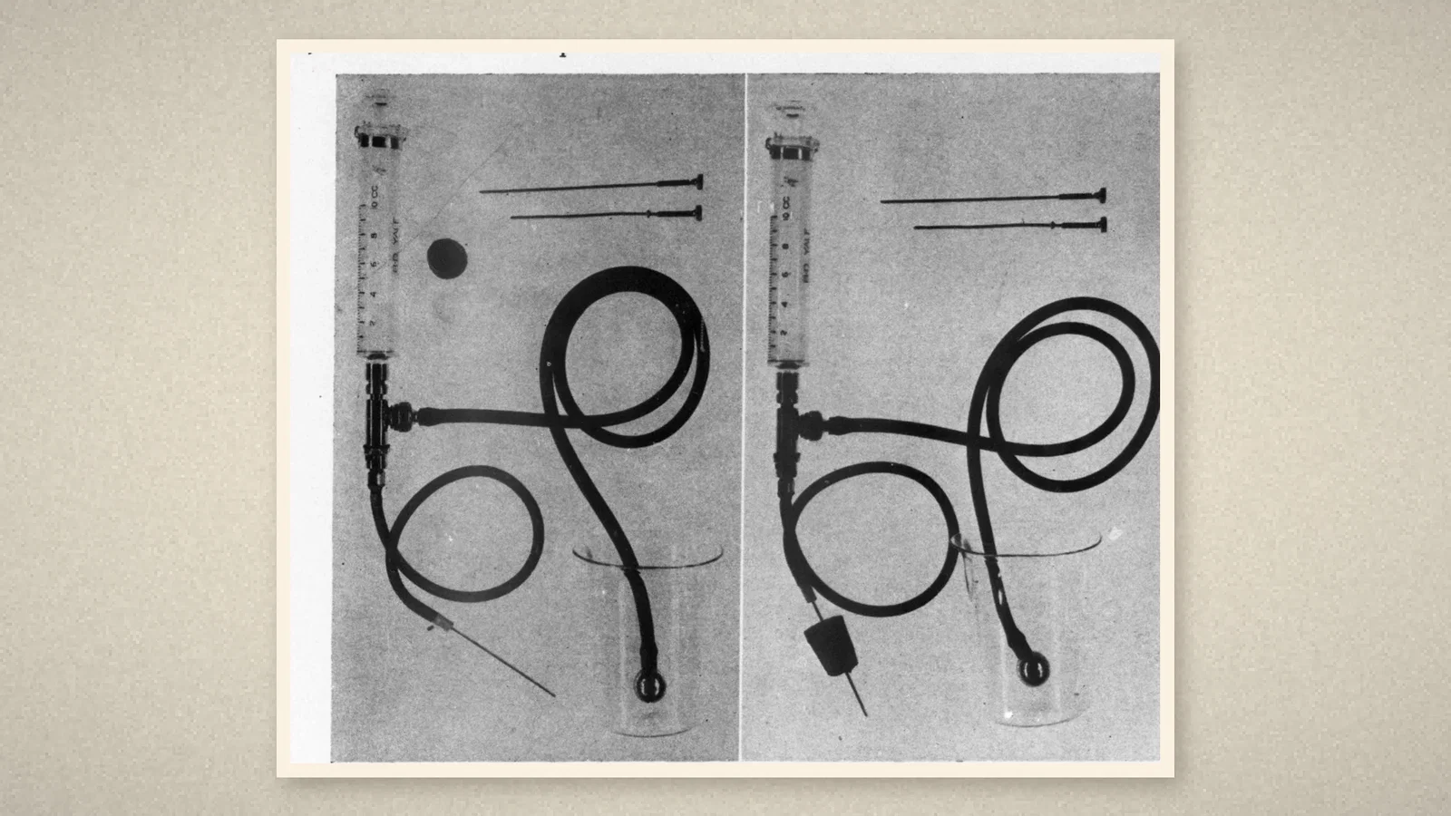
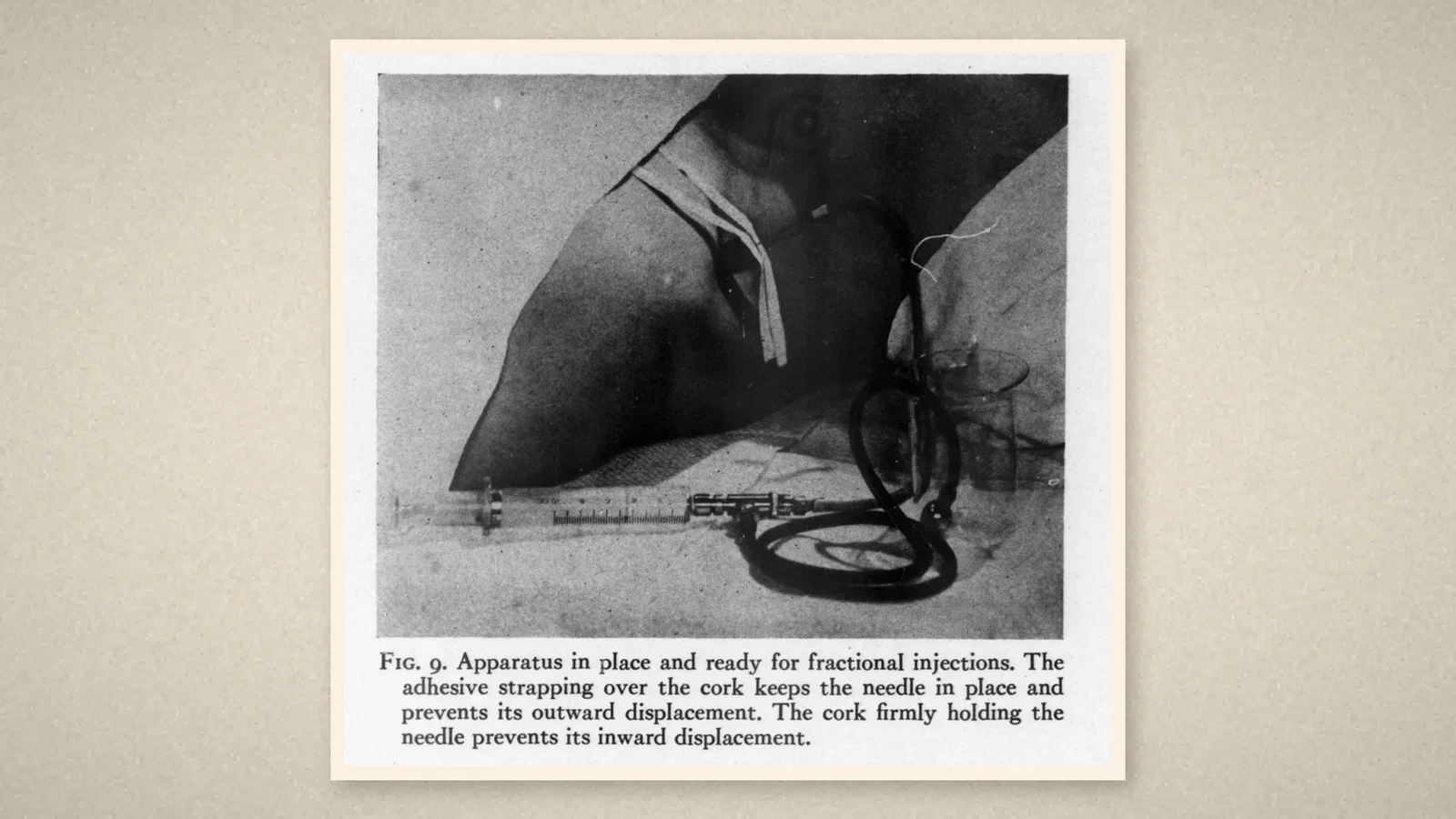
The apparatus was a 10 cc Luer-lock syringe with the two-way valve "as used in the Hingson-Edwards continuous caudal method", about 18 inches of tubing, a malleable needle "filed to a blunt end to prevent perforation of blood vessels", and "a cork stopper from an ether can". The landmark was the subclavian artery crossing the first rib. With a finger on the artery, the needle was passed through a wheal above the clavicle until it touched the rib, lying lateral to the artery: if the needle "is pulsating with the subclavian artery, then it, of necessity, lies in contact with the brachial plexus". Paraesthesiae were helpful but "not essential". The cork was then pushed down to the skin to hold the needle upright and stop it going deeper, and adhesive strapping over the cork stopped it coming out. [1]
After careful aspiration, 40 cc of 1 per cent procaine without adrenaline was injected; anaesthesia usually followed within fifteen minutes, and further doses were given through the tubing as needed. A sharp, large needle was used first to pierce the skin, and the blunt one followed its track. [1]
Twenty-seven cases
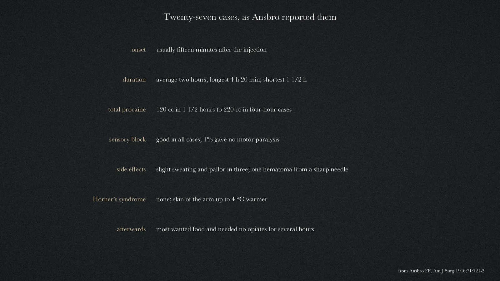
In twenty-seven cases, operations from open reductions of the forearm, arm and shoulder to repair of the median nerve, anaesthesia lasted on average two hours, the longest four hours twenty minutes. Sensory block was good in all; 1 per cent procaine did not paralyse the muscles. Total doses ran from 120 cc in an hour and a half to 220 cc in four-hour cases. Three patients had slight sweating and pallor; one early hematoma came from a sharp needle. No patient showed Horner's syndrome, and the blocked arm warmed by up to 4 °C. Most patients wanted food on return to bed and needed no opiates for several hours. [1]
Ansbro also used the block to relieve vascular spasm. In a patient with brachial artery embolism, a "painful, swollen, cold, wet and cyanotic arm" became "painless, warm, dry and pink" within two hours; the patient died of a pulmonary embolus within another hour. [1]
What the record shows
Ansbro's 1946 method kept a peripheral block going for hours with equipment borrowed from obstetric caudal analgesia and a cork from an ether can. What stayed in the body was a blunt needle, not a catheter; the arterial pulse, not a nerve stimulator or ultrasound, told the operator where the needle was. The paper does not claim to be the first continuous peripheral block, and this article does not either.
Sources and reading limits
Read: Ansbro's 1946 paper in full.
Not covered: the papers of Macintosh and Mushin and of Hingson and Edwards that Ansbro drew on; later continuous peripheral catheter techniques. Ansbro dates Kulenkampff's method a year after Hirschel's; the original Kulenkampff paper is dated 1911. The case results are the author's own. Doses are as printed.
Two illustrations are page crops from the 1946 paper; two are drawn from it.
- Ansbro FP. A method of continuous brachial plexus block. Am J Surg. 1946;71(6):716-22. Internet Archive
Continue exploring
Related reading connects shared questions and contexts; it does not by itself establish historical influence.
- 1911: Two Needles for the Brachial Plexus
- 1944: Tuohy Threads a Ureteral Catheter into the Spinal Space
- 1942: Hingson and Edwards Sustain Caudal Analgesia Through Labor
Anesthesia Epochs · The Source Room · About & Editorial Method
Published November 2, 2026. Sources and reading limits are stated above. Illustrations are labelled as original source images or AI-made. Historical accounts are educational; they are not current treatment instructions.