
In May 1969 the British Journal of Anaesthesia (vol. 41, pp. 453-4) printed a case report by Raymond Vale of Guy's Hospital, London, 'Selective bronchial blocking in a small child'. A 14-month-old child needed removal of the left lower lobe for an abscess that communicated with the bronchial tree, so the lobe had to be isolated, but Thompson and Magill blockers were clearly too large. Vale used a small Fogarty embolectomy catheter instead, passed through an infant bronchoscope into the left main bronchus and inflated beyond its tip, before a nasotracheal tube was placed. At thoracotomy the lower lobe was purple and not inflating while the upper lobe inflated; the catheter later became dislodged and the bronchus was snared from outside. The child did well. Vale commented that blocker design had hardly changed because double-lumen tubes were almost ubiquitous, yet blockers still had a place. In June 1982 Inoue and colleagues of Tokai University, Japan, described in the Journal of Thoracic and Cardiovascular Surgery a tracheal tube with a channel carrying a movable blocker, used in 34 thoracotomies, and called it a refinement of the technique using a Fogarty embolectomy catheter, citing Ginsberg in 1981 and Vale in 1969.
Reading Library · Anesthesia Epochs · 中文版
Watch the education video (for clinicians)
1969: Fogarty Bronchial Blockers — One Lobe, One Balloon · Anesthesia Epochs Education Series
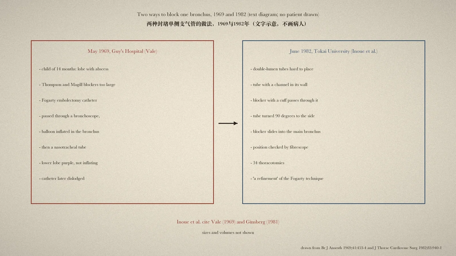
In surgery for a lung abscess, the great fear is that pus will spill into other lobes during handling. To wall the diseased lobe off, a blocker has to sit in its bronchus, but for a child just over a year old the blockers then in use were all too large. A two-page case report in May 1969 solved the problem with a fine catheter made for removing blood clots. Thirteen years later a surgical team in Japan called its own new tube a refinement of that idea. [1, p. 453][2, p. 941]
One child's operation
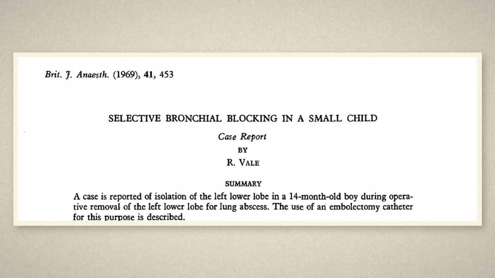
The report, "Selective bronchial blocking in a small child", was by Raymond Vale of Guy's Hospital, London. Its summary records the isolation of the left lower lobe in a 14-month-old child during removal of that lobe for lung abscess, and describes the use of an embolectomy catheter for the purpose. [1, p. 453]
The child coughed up pus and the radiograph showed a fluid level in the abscess cavity, suggesting a connection with the bronchial tree, so an anaesthetic technique that blocked off the affected lobe was desirable. But Thompson and Magill blockers "would clearly be too large", and it was decided to use a small Fogarty embolectomy catheter. [1, p. 454]
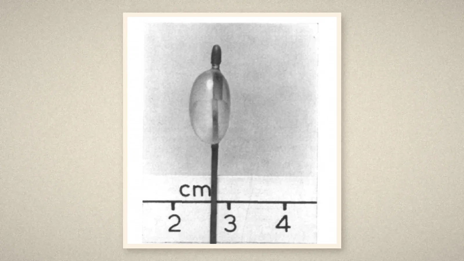
After induction, an infant bronchoscope was passed into the left main bronchus. The left upper lobe opening could not be seen, but the Fogarty catheter was pushed beyond the bronchoscope's tip and its balloon inflated. The catheter's end was clamped and sealed with bone wax, the bronchoscope was withdrawn with the catheter left in place, and a nasotracheal tube was passed. Auscultation suggested that the left lower lobe was no longer aerating, the child was turned onto the right side, and surgery began. [1, p. 454]
When the chest was opened no suction was needed: the lower lobe was purple and not inflating, while the upper lobe was pink and inflating. But after the lower lobe had been handled, suction was needed several times and brought up a good deal of pus, showing that the catheter had become dislodged. The lower lobe bronchus was then held shut from outside with a rubber snare until the bronchus clamp could go on. The child did well afterwards. [1, p. 454]
A neglected device
In the closing comment Vale wrote that the design of bronchial blockers had hardly changed since their introduction, largely through lack of interest and "the almost ubiquitous use of double-lumen" tubes. Yet "there is still a place, however, for their use" in the definitive management of lung surgery, as in this case. The acknowledgement thanks Lord Brock, under whose care the child was treated. [1, p. 454]
A new tube thirteen years later
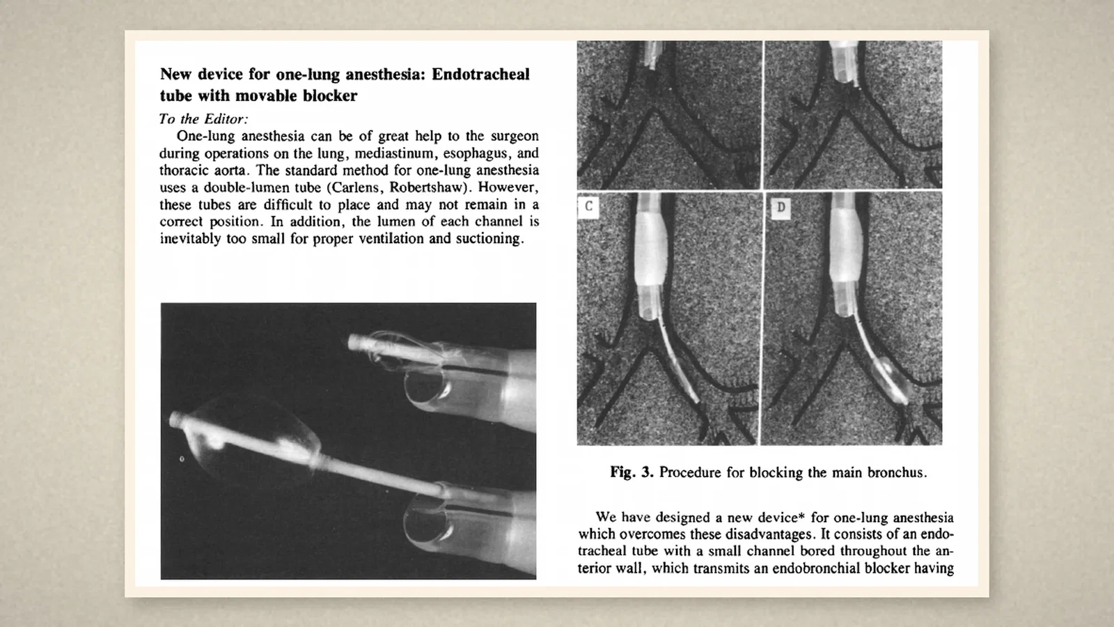
In June 1982 the Journal of Thoracic and Cardiovascular Surgery printed a letter from Hiroshi Inoue and four colleagues of the First Department of Surgery, Tokai University School of Medicine: "New device for one-lung anesthesia: endotracheal tube with movable blocker". The standard method of one-lung anaesthesia, they wrote, was the double-lumen tube, but such tubes were difficult to place, might not stay in position, and had channels "inevitably too small for proper ventilation and suctioning". [2, p. 940]
Their device was a tracheal tube with a small channel bored through its front wall, carrying a bronchial blocker with a low-pressure, high-volume cuff. After ordinary intubation the tube was turned 90 degrees toward the side of the thoracotomy. The blocker was pushed down so that it slid along the tracheal wall into the target main bronchus, and its position was fine-tuned with a flexible bronchofiberscope or a radiograph. Once the patient had been turned and the chest opened, the blocker's cuff was inflated for one-lung anaesthesia. [2, p. 940][2, p. 941]
The device had been used for 34 thoracotomies with satisfactory results. The letter ends by saying that one-lung anaesthesia with it "appears to represent a refinement of the technique using a Fogarty arterial embolectomy catheter". Its two references are Ginsberg's paper of 1981 and Vale's case report of 1969. [2, p. 941]
What the record shows
A borrowed embolectomy catheter, one child, a blocker that slipped during surgery: Vale's report was small, but it named a need that double-lumen tubes had hidden. Thirteen years later Japanese surgeons built the blocker into the wall of the tracheal tube, and placed their device explicitly in this line.
Sources and reading limits
Read: Vale, "Selective bronchial blocking in a small child", Br J Anaesth 1969;41:453-4 (the whole report, with page images checked); Inoue, Shohtsu, Ogawa, Kawada and Koide, "New device for one-lung anesthesia: endotracheal tube with movable blocker", J Thorac Cardiovasc Surg 1982;83:940-1 (the whole letter with its three figures). Both are publisher PDFs of the published articles, obtained through institutional access.
Not covered: Ginsberg's 1981 paper cited by Inoue; Lines's 1969 report on bronchial blocking; the original descriptions of the Thompson and Magill blockers; later blockers, including a 2010 timeline entry. The ASA timeline entries were not used as evidence.
Limits: some timelines name Fogarty blockers in 1969. The sources show one case report using a Fogarty embolectomy catheter as a blocker and, thirteen years later, a different device whose authors call it a refinement of that technique. Vale's case is a single child, and the catheter dislodged during surgery. Inoue's letter reports 34 thoracotomies without detailed results. Drug doses, catheter sizes and balloon volumes are not repeated, and page crops stop before them; the child's radiographs are not shown. Patients are not identified.
Three illustrations are crops of Br J Anaesth 1969 pp. 453 and 454 (the photograph of Fig. 2, without the caption giving the volume) and J Thorac Cardiovasc Surg 1982 p. 940. One is a text diagram drawn from these sources, with no patient drawn.
- Vale R. Selective bronchial blocking in a small child. Br J Anaesth. 1969;41:453-4. DOI
- Inoue H, Shohtsu A, Ogawa J, Kawada S, Koide S. New device for one-lung anesthesia: endotracheal tube with movable blocker. J Thorac Cardiovasc Surg. 1982;83:940-1. ScienceDirect
Continue exploring
Related reading connects shared questions and contexts; it does not by itself establish historical influence.
- 1957: Macintosh and Richards Describe an Illuminated Introducer for Tracheal Tubes
- 1949: Macintosh Threads the Tracheal Tube over a Gum-Elastic Catheter
Anesthesia Epochs · The Source Room · About & Editorial Method
Published November 3, 2026. Sources and reading limits are stated above. Illustrations are labelled as original source images or AI-made. Historical accounts are educational; they are not current treatment instructions.