In October 1983 the Clinical Reports of Anesthesiology (vol. 59, no. 4) printed 'Evaluation of pulse oximetry' by Mark Yelderman and William New Jr. of the Department of Anesthesia, Stanford University School of Medicine, accepted on 24 March 1983. Blood gases, they wrote, are reliable but invasive and intermittent; transcutaneous oxygen needs local heating and still fails to reflect arterial oxygenation perfectly; ear oximeters 'arterialize' the ear with heat and chemicals and struggle to separate arterial from tissue and venous absorbance. Pulse oximetry places any pulsating arterial bed between a two-wavelength light source and a detector and uses only the pulsatile part of the signal, which comes solely from arterial blood, to calculate saturation beat by beat with Beer's law. A finger pulse oximeter had recently become commercially available, run by a microprocessor, with no heating and no user calibration. In five healthy non-smoking student volunteers, whose inspired oxygen was lowered and then raised in steps through a modified anaesthesia machine, readings were compared with arterial samples on a CO-Oximeter: 79 paired points gave a correlation coefficient of 0.98, and the authors judged the instrument linearly accurate and precise over the range tested.
Reading Library · Anesthesia Epochs · 中文版
Watch the education video (for clinicians)
1983: Pulse Oximetry — Only What Pulses · Anesthesia Epochs Education Series

In today's operating room a small clip on a fingertip and a number on a screen are taken for granted. In 1983 such an instrument had only just come on the market, and two anaesthetists at Stanford set out to answer a plain question: was the number right? Their method was to have healthy volunteers breathe less and less oxygen, step by step, while arterial blood was drawn for comparison. [1, p. 349][1, p. 350]
Nothing else was good enough
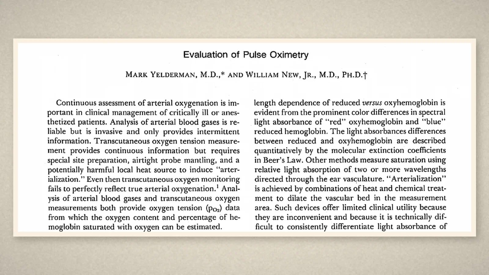
In October 1983 the Clinical Reports of Anesthesiology (vol. 59, no. 4) printed "Evaluation of pulse oximetry" by Mark Yelderman and William New Jr. of the Department of Anesthesia, Stanford University School of Medicine. It had been accepted on 24 March 1983. [1, p. 349]
Continuous assessment of arterial oxygenation, the authors began, matters in the care of critically ill and anaesthetised patients. Yet each existing method had a flaw. Arterial blood gas analysis is reliable, but invasive, and gives only intermittent information. Transcutaneous oxygen tension is continuous, but it needs special site preparation, an airtight probe and "a potentially harmful local heat source" to arterialize the skin, and even then it fails to reflect true arterial oxygenation perfectly. Other methods shine two or more wavelengths through the ear, its vessels dilated by heat and chemicals; such devices, the authors wrote, are inconvenient and cannot consistently separate the absorbance of arterial blood from that of tissue and venous blood, so their clinical use is limited. [1, p. 349]
Watching only what pulses
The deficiency, they went on, could be circumvented by measuring absorbance changes that are time-coherent with arterial pulsation. Pulse oximetry places any pulsating arterial bed between a two-wavelength light source and a detector. As the bed expands and relaxes it changes the path the light takes and so the amount detected, producing "the familiar plethysmograph waveform". [1, p. 349]
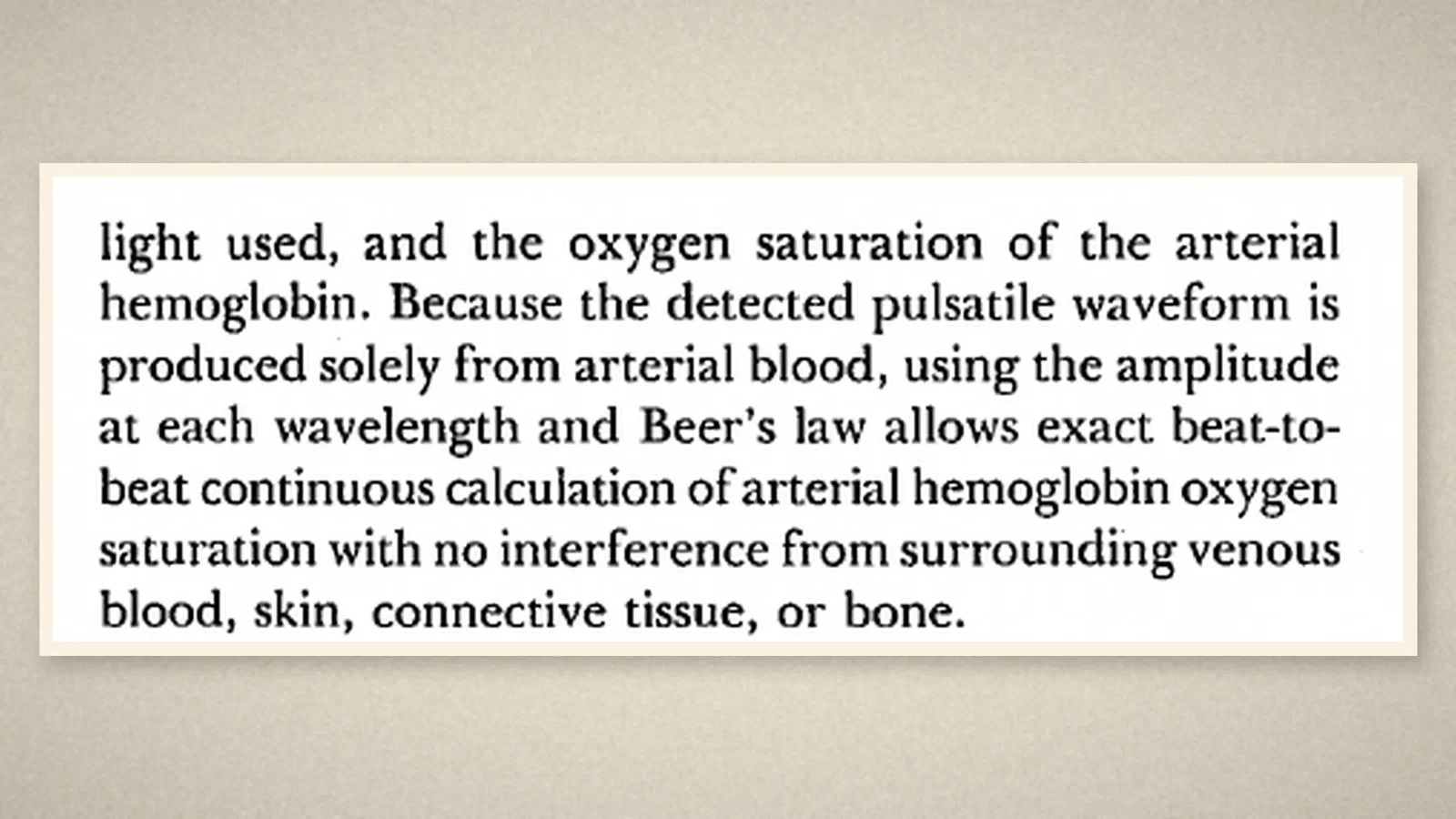
The key is that this pulsatile waveform comes solely from arterial blood. Using its amplitude at each wavelength and Beer's law, arterial haemoglobin saturation can be calculated beat by beat, continuously, with no interference from the surrounding venous blood, skin, connective tissue or bone. [1, p. 350]
A pulse oximeter, the authors noted, had recently become commercially available; a footnote names it as the Nellcor N-100 of Hayward, California. Two light-emitting diodes and a photodiode sat in a finger receptacle. No heating or arterialization was needed, a microprocessor handled control, calculation and display, and no calibration by the user was required. [1, p. 350]
Making volunteers hypoxic
To know whether it was accurate across a wide range of saturations, people had to be made genuinely short of oxygen. The protocol, the authors wrote, followed guidelines for the ethical evaluation of devices, which require clinically induced hypoxaemia for a meaningful assessment. [1, p. 350]
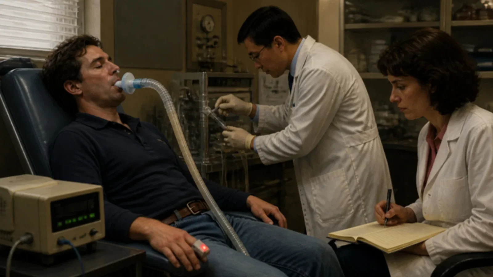
They modified an Ohio recirculating anaesthesia machine so that inspired oxygen could be held exactly steady. Under guidelines approved by the local institutional review board, five healthy, non-smoking college students were chosen. Each was interviewed, filled in a detailed medical questionnaire, had a full examination and laboratory tests, was told the whole procedure and its risks, was asked to sign consent only on the day of the study, and was paid. Intravenous and radial arterial lines were placed, and the oximeter was put on the index finger of the same side. Inspired oxygen was lowered in steps; at each plateau, once the readings were stable, arterial blood was drawn and analysed at once on an IL282 CO-Oximeter. The subjects were kept talking throughout and reminded that they could stop the procedure themselves at any time by removing the mask; none did. Oxygen was then raised in steps back to room air, and a few points were taken on high oxygen. Carboxyhaemoglobin and methaemoglobin were treated as dysfunctional and left out of the comparison. [1, p. 350]
Seventy-nine points on a line
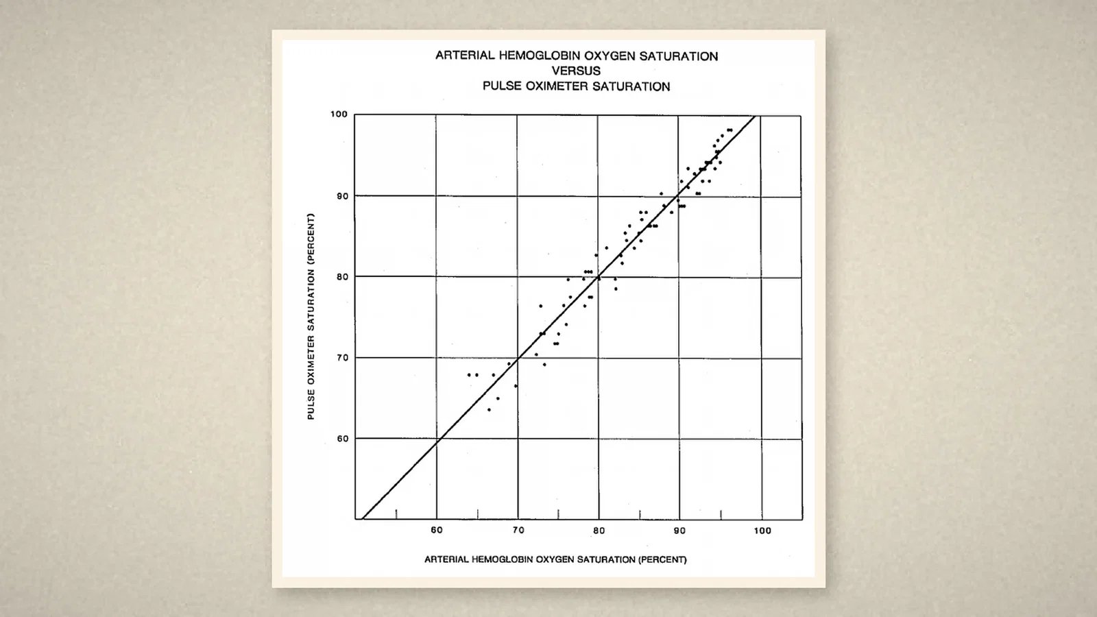
The five subjects gave 79 pooled data points, with a correlation coefficient of 0.98, and all tolerated the procedure without adverse sequelae. [1, p. 350] In discussion the authors said that the pulse technique eliminates artifact from the light absorption of tissue and venous blood, "something not possible with traditional ear oximetry techniques", and that their data showed the instrument to be linearly accurate and precise over the range evaluated. [1, p. 351]
They also set out its limits. Anything that markedly reduces vascular pulsation, such as hypothermia, hypotension or an infusion of vasoconstrictor drugs, weakens the instrument's ability to calculate saturation; a special nasal probe could then be used. With only two wavelengths, some error is inherent when dyshaemoglobins are present. Accuracy at lower saturations was desirable too, but ethically it could not be tested by deliberately making people that hypoxic. [1, p. 351][1, p. 352]
What the record shows
This short report explained the principle of a newly marketed instrument clearly: instead of heating the ear or the skin, look only at the part of the light that beats with the pulse. It tested the readings by inducing hypoxaemia in five volunteers, and it stated honestly the boundaries set by weak pulses, abnormal haemoglobins and ethics.
Sources and reading limits
Read: Yelderman and New, "Evaluation of pulse oximetry", Anesthesiology 1983;59:349-52 (introduction, principle, device, methods, results, discussion and Fig. 1). The end of the preceding clinical report and the start of the following one, printed on the same pages, are not used. Obtained through institutional access.
Not covered: the earlier oximetry literature the authors cite (Wood; Nilsson; Yoshiya and colleagues, 1980); the maker's records on the device's development and release; later clinical studies. The History of Anaesthesia Society and NZSA timeline entries were used only as leads.
Limits: some timelines date commercial pulse oximetry to 1981; the report read here appeared in October 1983 and says only that a pulse oximeter "recently has become commercially available", naming the model in a footnote without a release year. The subjects were five healthy volunteers, not patients. Inspired oxygen levels, gas flows, sample volumes, wavelengths and the saturation range are not repeated in the text, and page crops stop before the methods and device parameters; Fig. 1 is shown as the authors' result. Volunteers are not identified.
Three illustrations are crops of Anesthesiology 1983 pp. 349, 350 and 351 (title and opening, the principle paragraph, Fig. 1). One is a schematic of the principle drawn from the text, with no people and no wavelengths or saturation values.
- Yelderman M, New W Jr. Evaluation of pulse oximetry. Anesthesiology. 1983;59:349-52. DOI
Continue exploring
Related reading connects shared questions and contexts; it does not by itself establish historical influence.
- 1981: Sullivan and Colleagues Treat Sleep Apnoea with Nasal CPAP
- 1958: Severinghaus and Bradley Build Blood pO2 and pCO2 Electrodes
- 1974: Takuo Aoyagi Makes Hypoxia Visible
Anesthesia Epochs · The Source Room · About & Editorial Method
Published November 3, 2026. Sources and reading limits are stated above. Illustrations are labelled as original source images or AI-made. Historical accounts are educational; they are not current treatment instructions.