From pulse 'noise' to continuous oxygenation monitoring — and into the system of anesthesia safety.
Reading Library · Anesthesia Epochs · 中文版
Watch the companion film
1974: Aoyagi and Pulse Oximetry — Making Oxygenation Visible · Watch on YouTube
Historical anchor: 1974. Date details and source qualifications are explained below.
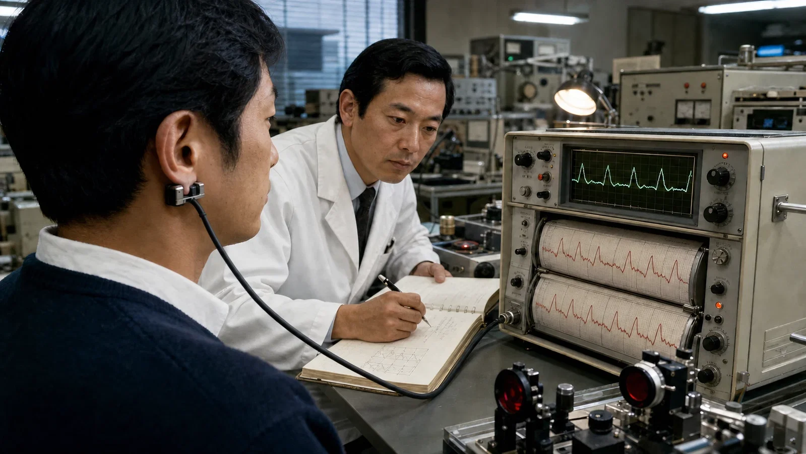
In December 1972, inside Nihon Kohden's development department, Takuo Aoyagi (1936–2020) was trying to remove a troublesome waveform. He was using an ear oximeter to improve dye-dilution measurement of cardiac output, but each heartbeat distorted the trace. His first idea was to cancel this pulse “noise.” Then he recognized that the noise carried the information he needed. The portion of the optical signal that changed with each heartbeat arose chiefly from changing arterial blood volume. If that pulsation could be separated from the surrounding tissue and venous background, it could reveal arterial oxygenation continuously.
I. 1972: When Noise Became Signal
Blood is not a single, fixed shade of red. Oxygenated and deoxygenated hemoglobin absorb red and near-infrared light differently, which makes optical measurement possible. An ear probe, however, does not see arterial blood alone. It also sees skin, soft tissue, bone, and venous blood. Most of that material forms a large optical background around the much smaller arterial change.
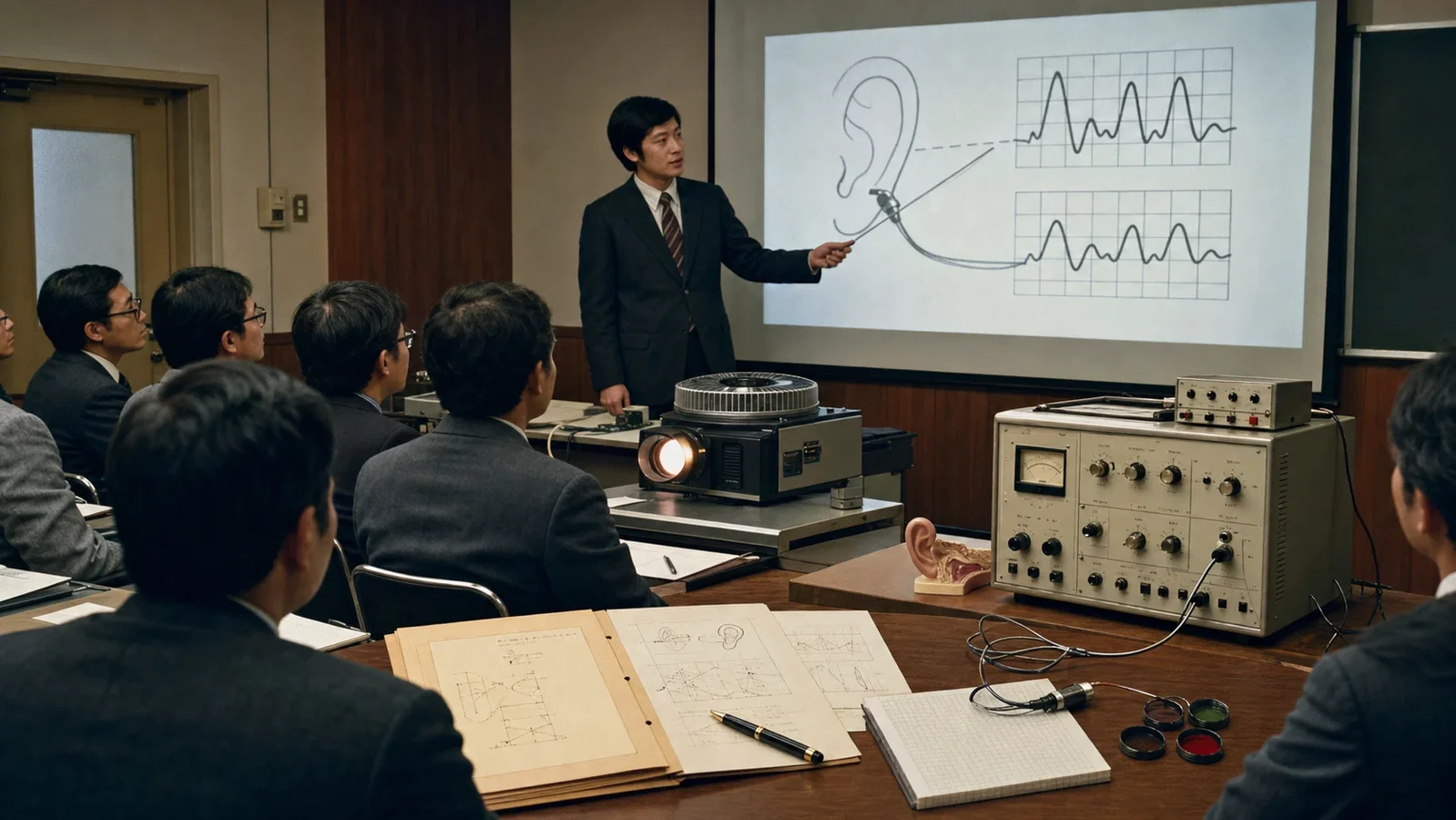
Aoyagi used the heartbeat itself to isolate that change. During systole, arterial blood volume at the measurement site rises slightly, altering the amount of transmitted light. Engineers call this pulsatile component AC and the relatively stable background DC. The instrument normalizes AC to DC at each of two wavelengths, then divides one normalized result by the other. This “ratio of ratios” is mapped through an empirical calibration curve to produce SpO₂.
The pulse oximeter therefore does not see oxygen directly, nor does it measure PaO₂ without an arterial sample. It continuously estimates peripheral arterial oxygen saturation from light and pulsation. The distinction matters. A precise-looking number can still be affected by low perfusion, motion, dyshemoglobins, and other measurement limits. It also cannot determine by itself whether a patient is ventilating adequately.
II. 1974: Two Traceable Acts
This article is anchored in 1974, but that year should not be compressed into a vague claim that the invention suddenly appeared. On 29 March, Aoyagi and his colleague Michio Kishi filed a Japanese patent application titled Optical Type Blood Measuring Equipment. On 26 April, Aoyagi publicly presented the ear-based pulse-oximetry principle at the 13th annual meeting of the Japanese Society of Medical Electronics and Biological Engineering in Osaka. His report was titled Improvement of an Earpiece Oximeter.
Filing a patent application did not mean the patent was granted that day, and a conference report was not a clinical product launch. Keeping the two events separate reveals the actual sequence. The central insight came in 1972. The filing and presentation made it traceable in 1974. Years of device development followed before the method became routine clinical monitoring.
III. 1975–1983: Turning a Principle into a Clinical Device
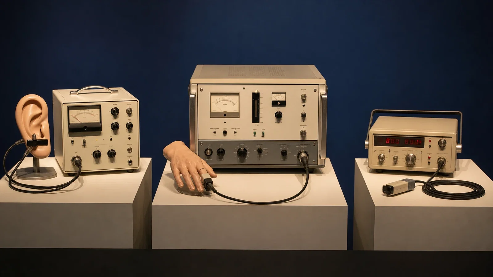
Nihon Kohden introduced the OLV-5100 in 1975. It paired an earlobe probe with a miniature incandescent lamp, while heavy fiber-optic cables carried the light. It was the first ear pulse oximeter, but it was not a commercial success. Sensor performance, usability, and clinical demand had not yet aligned, and the development program was later suspended. A working principle had become a product, but not yet a clinical habit.
Minolta moved the measurement site to the fingertip with the OXIMET MET-1471 in 1977. “Finger-measurement type” did not mean the integrated plastic clip familiar today. The instrument weighed about 7.7 kg, consumed about 130 W, and connected its light source to the probe through fiber-optic paths. The advance was fingertip measurement and instrument design, not modern portability.
The next phase of product development took place in North America. Nellcor produced a prototype in 1982 and marketed the N-100 in 1983. It required no bedside calibration, its sensor was easy to apply, its numerical display was clear, and the pitch of its pulse tone changed with oxygen saturation. Promotion directed toward anesthesiologists also accelerated adoption. The N-100 was not the first pulse oximeter in the United States, but it helped make continuous oximetry routine across North American operating rooms. Clinical adoption required more than the original equation. It also required human-factors design, clinical feedback, manufacturing, and a system ready to use the information.
IV. Visibility Was One Part of Safety
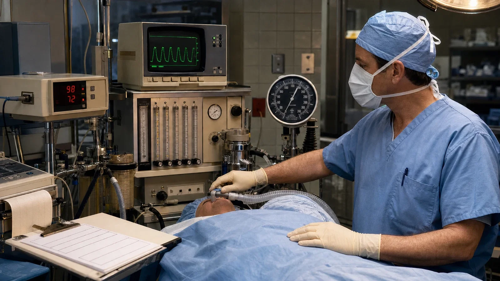
Pulse oximetry changed what an anesthesia team could observe. Before continuous SpO₂ monitoring, recognition of hypoxemia depended more heavily on late clinical signs, circulatory changes, and intermittent blood sampling. A changing number and an audible alarm made a developing episode of hypoxemia visible while there was still time to respond. Oxygenation, however, is not ventilation. A patient receiving supplemental oxygen may maintain SpO₂ for a time while carbon dioxide continues to rise. Capnography supplies information that pulse oximetry cannot replace.
Monitoring standards evolved in stages as well. The Harvard minimum monitoring standards adopted in 1985 established an institutional precedent but did not mandate pulse oximetry for every anesthetic. The original ASA standards adopted in 1986 still encouraged quantitative oxygenation monitoring rather than requiring pulse oximetry in every case. The 1989 revision made a quantitative method of assessing oxygenation, such as pulse oximetry, a formal requirement. Equipment availability, standards, training, alarm response, and safety culture developed together into a monitoring system.
The outcome evidence requires the same restraint. A 2014 Cochrane review included five randomized trials with 22,992 participants. Pulse oximetry improved the detection of hypoxemia and prompted changes in care, but the trials did not show a reduction in perioperative complications or mortality. These results support earlier detection and changes in care, not a claim that one monitor independently reduced rare outcomes. System-wide safety gains cannot be assigned to one device alone.
Within The Anesthesia Epochs, Griffith and Johnson's curare separated muscular relaxation from anesthetic depth and made active management of ventilation increasingly central to anesthesia. Aoyagi added a continuously visible physiological feedback signal. Pulse oximetry did not single-handedly end anesthesia mortality. Its more specific and supportable contribution was to turn oxygenation hidden within skin color and tissue background into a number that a clinical team could see, share, and act upon.
Sources and reading limits
Sources: Severinghaus JW, Anesth Analg. 2007;105(6 Suppl):S1–S4 (PMID 18048890); Miyasaka K et al., J Anesth. 2021;35:671–709 (PMCID PMC8327306); Quaresima V et al., J Biomed Opt. 2024;29(S3) (PMCID PMC11330276); Eichhorn JH et al., JAMA. 1986;256:1017–1020; Pedersen T et al., Cochrane Database Syst Rev. 2014:CD002013 (PMCID PMC6464860); Jubran A, Crit Care. 1999;3:R11–R17; Nitzan M et al., Med Devices. 2014;7:231–239; Becker DE, Casabianca AB. Anesth Prog. 2009;56:14–22 (PMCID PMC2662504).
Visual interpretations
These illustrations explore the setting and ideas. Depicted people, actions, instruments and documents are reconstructions, not evidence of their exact historical appearance.
Continue exploring
Related reading connects shared questions and contexts; it does not by itself establish historical influence.
- Later connection: 2011: Lifebox Works Toward Global Pulse-Oximetry Access
- 1953: Virginia Apgar Gives the Newborn a Common Language
- 1977: Jöbsis Detects Near-Infrared Signals Through an Intact Head
- 1985: Harvard's Monitoring Standards Make Continuous Observation a Shared Requirement
- 1986: The ASA Adopts Monitoring Standards with Four Areas of Assessment
- 2018: Prediction Enters the Monitor, Not the Treatment Order
- 2023: The FDA Creates a Device Type for the NOL Index
- 2024: ASA Revises PACU Handoff to 'Stay with the Patient'
The Source Room · About & Editorial Method
Sources and reading limits are stated above. Illustrations are labelled as original source images or AI-made. Historical accounts are educational; they are not current treatment instructions.
Continue this reading route
Stop 4 of 7 · Back to route · Making Anesthesia Safer: From Observation to Continuous Monitoring
Previous · c. 1964: Block-Aid Makes Muscle-Relaxant Action Repeatedly Observable
Next · 1985: Harvard's Monitoring Standards Make Continuous Observation a Shared Requirement
A signal can be available before its use becomes a requirement. The Harvard standards specified who should observe the patient and what that observation should include.