How an anesthesiologist made obstetric, anesthetic, and resuscitation practice comparable—and where the score stops.
Reading Library · Anesthesia Epochs · 中文版
Watch the companion film
1953: Virginia Apgar — The First Minute · Watch on YouTube
Historical anchor: 1953. Date details and source qualifications are explained below.
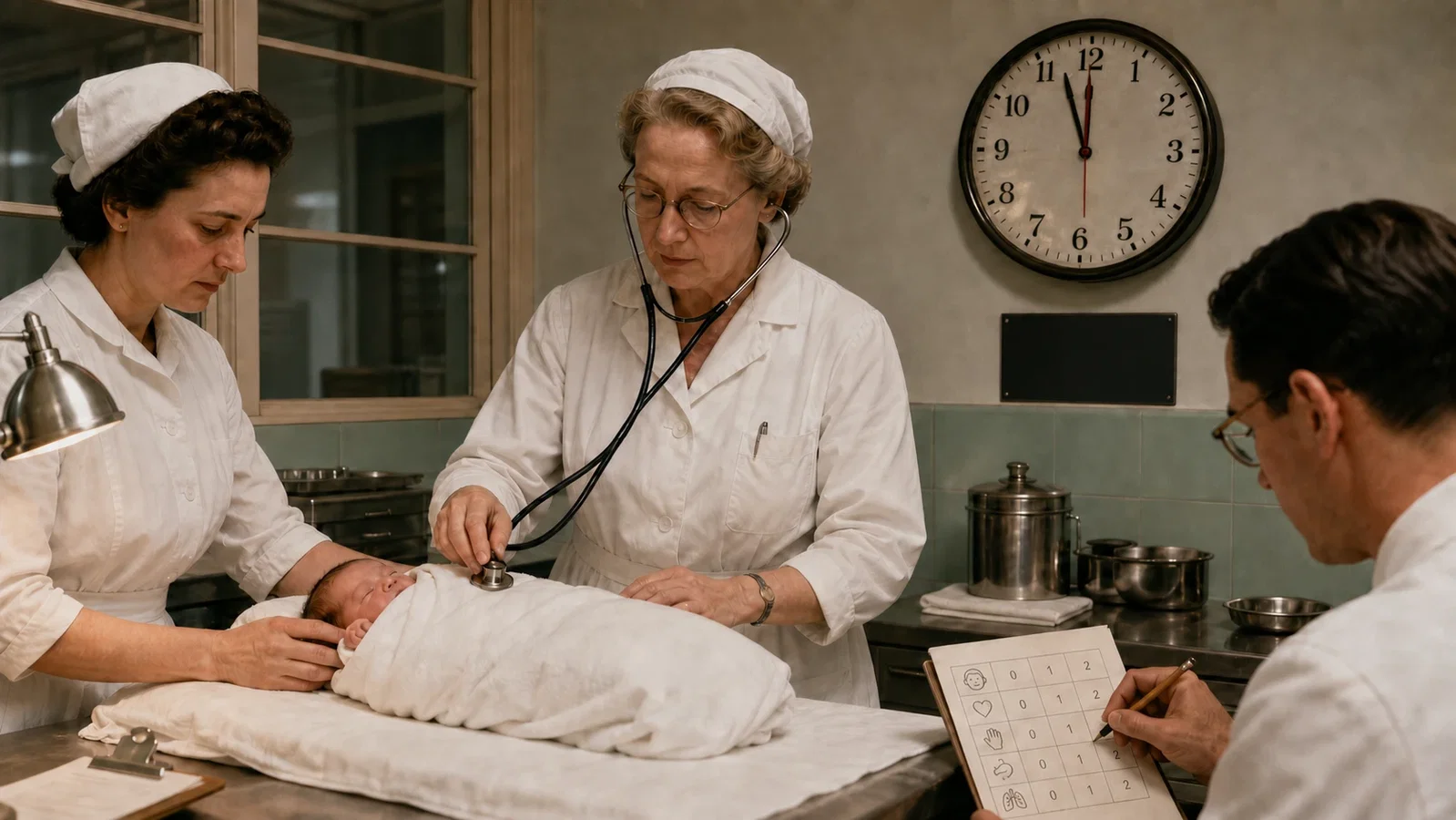
A delivery room had no shortage of observers in 1952. What it lacked was a common language for what they saw. Virginia Apgar presented a new method of newborn evaluation at an anesthesia meeting in Virginia Beach that September, then published it the following year. Her proposal did not begin with the idea of reducing an infant to a number. It began with the recognition that familiar descriptions could not be compared reliably.
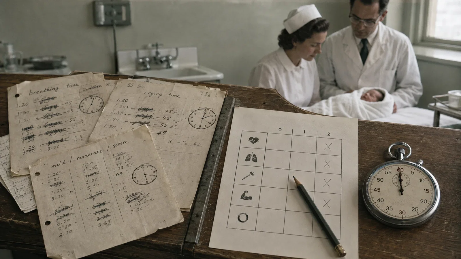
Clinicians recorded “breathing time,” “crying time,” or mild, moderate, and severe depression. Each term concealed a problem. An infant exposed to maternal medication or general anesthesia might take one breath and then remain apneic for several minutes. Some infants never produced what an observer considered a satisfactory cry. The categories of depression left substantial room for individual interpretation. Results from different obstetric practices, anesthetic techniques, and resuscitation methods therefore did not share a stable endpoint.
I. Five Signs at the Same Minute
Apgar selected five signs that could be assessed easily without interrupting care: heart rate, respiratory effort, reflex irritability, muscle tone, and color. Each received 0, 1, or 2 points, for a maximum score of 10. The assessment was fixed at sixty seconds after the infant had been completely delivered.
The fixed time was as important as the total. A measurement taken at the first gasp could not be compared with one taken after a sustained cry or several minutes of observation. By holding the time constant, the score gave clinicians a shared way to discuss obstetric intervention, maternal analgesia and anesthesia, resuscitation, and differences among hospitals.
The 1953 paper did not define the later mnemonic words Appearance, Pulse, Grimace, Activity, and Respiration. That expansion is useful for remembering the score, but it should not be projected backward as Apgar's original terminology. Her paper described the five clinical signs directly and emphasized a uniform method and a precise observation time.
The individual signs also had to reflect the infant's condition at that moment. Heart rate could be assessed by watching the precordium or palpating the umbilical cord near the abdomen. Respiratory effort at sixty seconds mattered more than an isolated earlier gasp. Reflex response, muscle tone, and color were recorded at the same time. As the series developed, many scores were assigned by the anesthesia resident at the delivery. Apgar later advised that the observer should be someone other than the person conducting the delivery, who might be occupied with care or emotionally invested in the immediate outcome.
II. What the First Numbers Could Show
The paper covered 2,096 births over seven and a half months, but that figure was not the denominator for the scoring analysis. The reported score groups contained 1,021 infants:
- Of 65 infants with scores of 0-2, 9 died, or 14%.
- Of 182 infants with scores of 3-7, 2 died, or 1.1%.
- Of 774 infants with scores of 8-10, 1 died, or 0.13%.
The gradient was striking. In this initial hospital series, a lower score was associated with a higher proportion of neonatal deaths. The score organized scattered observations into condition groups that could be discussed and compared.
The design did not test whether using the score reduced mortality. There was no randomized comparison between scored and unscored care, and the data came from an early single-center experience. The supportable conclusion is narrower: among the first 1,021 scored infants, lower scores were associated with higher mortality. That finding supports description and risk stratification. It does not establish how many deaths the score prevented or allow the form itself to be credited with a later decline in infant mortality.
III. A Direct Link to the Curare Era
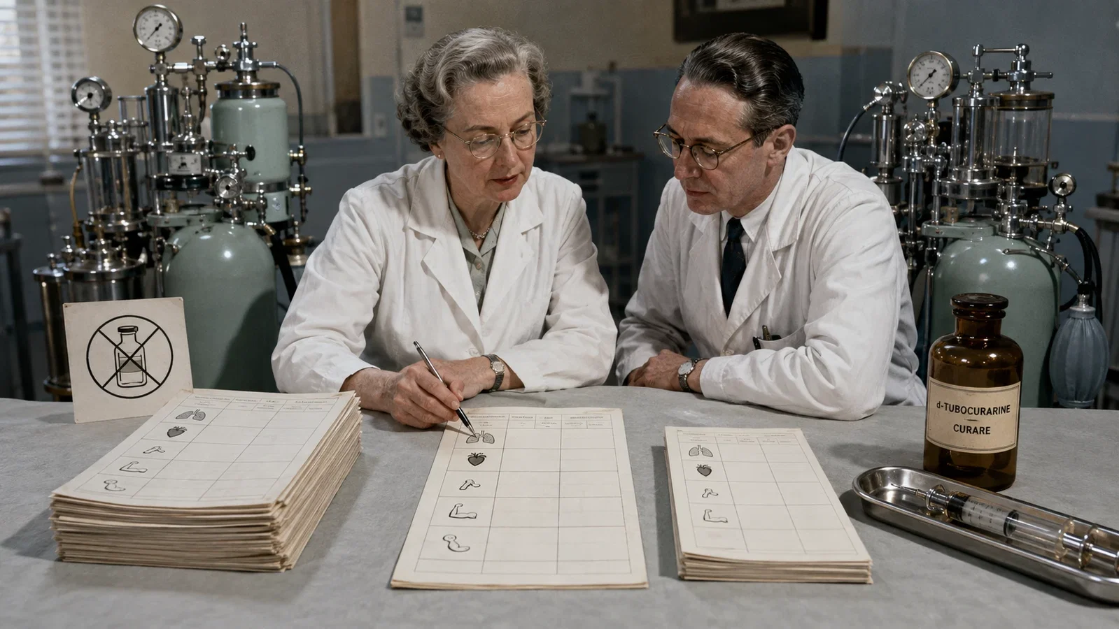
Only a little more than a decade had passed since Harold Griffith and Enid Johnson introduced curare into general anesthesia. Apgar's paper was already asking how newborn condition varied with maternal anesthetic practice. The connection is not a retrospective invention. The original report discussed d-tubocurarine, flaxedil, succinylcholine, and decamethonium.
Twenty women received an intravenous muscle relaxant with cyclopropane anesthesia. Seventeen received d-tubocurarine, while one each received the other three agents. Only 13 of their infants were scored. The paper compared 41 cesarean deliveries using cyclopropane without a relaxant with 13 using cyclopropane plus a relaxant. The mean score was 5.0 in both groups, and about 70% of infants in each group received some form of oxygen administration.
Within that series, Apgar found no evidence that adding a relaxant left the infants in better condition, and she identified maternal respiratory depression as a disadvantage of the technique. These observations were not a modern drug trial. The groups were small and nonrandomized, several relaxants were combined in one category, and obstetric risk, anesthetic management, and contemporary resuscitation practice could all affect the result. The comparison cannot establish that curare-like drugs did not cross the placenta, nor can it establish causal harm.
Its historical value lies in comparability. A common newborn score allowed anesthesiologists to place maternal drug choices and neonatal condition within the same discussion. The Griffith article showed anesthesia being separated into components that could be controlled independently. The Apgar article shows how the effects accompanying those choices could begin to be recorded with a common clinical measure.
IV. The Score Is Not the Signal to Start Resuscitation
A standard can become dangerous when its timing is misunderstood. In her 1966 reflections, Apgar corrected a practice in which some teams withheld active resuscitation until the one-minute score had been assigned. She called that practice wrong. When an infant was known to require help, resuscitation was to begin as soon as a free airway had been assured. The first score was still assigned at sixty seconds, even if resuscitation was already in progress.
That distinction remains central to the score's proper use. The Apgar score describes the infant's condition at a specified time and can help document the response to resuscitation. It does not determine whether initial resuscitation is needed, which step should be taken, or when treatment should begin. The five-minute score, and the change between one and five minutes, provide additional population-level and response information. They still do not foretell an individual infant's death or future neurologic function.
Modern guidance from the American College of Obstetricians and Gynecologists and the American Academy of Pediatrics states the boundary plainly. The Apgar score alone cannot diagnose asphyxia and should not be used to predict individual mortality or neurologic outcome. Low scores can be associated with increased risk in populations while remaining unable to determine one infant's future. Most infants with low scores do not develop cerebral palsy. Description, population association, and individual prediction are different claims.
V. The Least Stable Sign Was There from the Beginning
Apgar described color as the most unsatisfactory of the five signs and the one that caused the most disagreement among observers. Even infants who scored well on the other signs often did not receive full points for color at one minute. She also recognized that natural skin pigmentation could interfere with the observation. A standard that equates the best appearance with being “pink all over” does not provide an equally reliable visual judgment across all skin tones.
A 2025 systematic review included 10 studies examining Apgar scoring or visual cyanosis assessment across Black, Asian, and other minority ethnic newborns. It found possible differences in scoring practice and confirmed that cyanosis is difficult to assess visually. The evidence base was limited. Only two of the seven Apgar studies were rated at low risk of bias across all five quality domains. Definitions and outcomes were heterogeneous, and all but one study classified infants by race or ethnicity rather than measuring skin tone. The heterogeneity prevented meta-analysis.
The careful conclusion is not that every Apgar score has been proven to contain the same systematic bias. It is that a subjective component may not perform with equal reliability across newborns with different skin tones. Pulse oximetry later converted oxygenation into a continuous numerical estimate, but it is not an inherently bias-free substitute. It also cannot replace the clinical information supplied by heart rate, breathing, reflex response, and muscle tone.
VI. A Common Language, Not a Number for Destiny
The durable contribution of the Apgar score is not the compression of a newborn into a number from 0 to 10. It is the discipline of having different observers examine the same five signs at the same minute and record them in the same form. That common language made it easier for obstetric, anesthesia, and resuscitation teams to communicate, compare practice, and study groups of patients.
The language remains useful because its limits can be stated clearly. The score can describe, communicate, compare, and stratify. It cannot make care wait, diagnose asphyxia by itself, or turn a population association into an individual prognosis. Its color component also shows that standardization does not make every observation objective.
This history leads to a related question. When visual observation is unstable, can physiology be converted into a continuous and readable signal? Two decades later, Takuo Aoyagi would find the principle of pulse oximetry in the “noise” of an ear oximeter. Pulse oximetry did not simply replace the Apgar score. It represented another form of the same measurement impulse: giving clinical teams a shared signal while preserving the limits of what that signal can say.
Sources and reading limits
Apgar V. Curr Res Anesth Analg. 1953;32:260-267 (PMID 13083014); Apgar V. Pediatr Clin North Am. 1966;13:645-650; Hammer DJ, Portu R, Santiago A, Colon-Morales MA. Accurate Apgar Scoring. 1968 NLM scientific exhibit; ACOG/AAP Committee Opinion No. 644. Obstet Gynecol. 2015;126:e52-e55, reaffirmed 2025; Fair FJ et al. Pediatr Res. 2025;97:939-952 (PMID 39277651; PMCID PMC12055595).
Visual interpretations
These illustrations explore the setting and ideas. Depicted people, actions, instruments and documents are reconstructions, not evidence of their exact historical appearance.

Continue exploring
Related reading connects shared questions and contexts; it does not by itself establish historical influence.
- 1847: Simpson Separates Labour Pain from Uterine Action
- 1974: Takuo Aoyagi Makes Hypoxia Visible
- 1987: NRP Builds a Neonatal Resuscitation Course That Could Travel
- 2001: COMET Evaluates Labour Analgesia for Mother and Newborn
- 1945–2009: Cuffed Pediatric Tracheal Tubes Rethink Size, Position and Evidence
- 2016–2019: GAS, PANDA and MASK Narrow the Pediatric Neurotoxicity Question
- 2024: ASA Revises PACU Handoff to 'Stay with the Patient'
The Source Room · About & Editorial Method
Sources and reading limits are stated above. Illustrations are labelled as original source images or AI-made. Historical accounts are educational; they are not current treatment instructions.